Dear Editor,
We present our study aimed to detect myofibroblasts (MFs) in salivary mucoceles, and chronic sialadenitis (CS) of minor salivary glands (MSGs) for the better understanding of their pathogenesis and the processes involved in fibrosis, respectively.
Archival specimens of 20 cases of extravasation mucoceles of 10-12 month duration, 6 cases of mucous retention cysts with 1 week-6 months duration and adjacent MSGs (26 cases) together 5 normal lower labial MSgs were used as controls. Immunohistochemical analysis for α-smooth muscle actin (α-SMA) and desmin was performed using the streptavidin-biotin methodin conjuction with morphological analysis (spindle shaped and sometimes stellate) used for MFs identification.
Histologic examination of the adjacent to mucoceles MSGs showed that the degree of fibrosis was normal, low, moderate and severe in 9, 6, 6, and 5 cases, respectively. In 12 cases the adjacent to mucoceles excretory ducts were distended. MFs positive for α-SMA but not for desmin were detected.
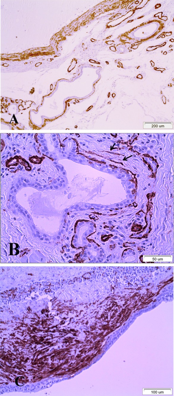
In extravasation mucoceles, MFs were found only in one case (1/20) with duration 4 months. In this case, MFs were present in the wall of granulation tissue that was partially substituted by dense connective tissue (Fig.1A). These results indicate that MFs are not involved in pathogenesis of extravasation mucoceles.
Figure 1. A. Extravasation mucocele where part of granulation tissue was substituted by dense connective tissue with large number of MFs. Also many MFs are presented around two distended excretory ducts adjacent to lesion. B. Small number of MFs around an excretory duct (arrows) in CS with severe degree of fibrosis. C. Large number of MFs is presented around the lining epithelium of mucous retention cyst (a-SMA Immunohistochemistry).

MFs were not found in normal salivary glands and this finding agrees with the results for normal submandibular glands1. Small number of MFs was seen only in one case (1/5) of CS with severe degree of fibrosis but not in cases with normal (0/9), low (0/6) and moderate degree (0/5) of fibrosis (Fig.1B). These results concur with a previous study in CS of submandibular glands and support the suggestion that MFs are not significantly involved in the processes of fibrosis1.
MFs were found around the lining epithelium of 2/6 cases of mucous retention cysts, and in 3/12 cases of the adjacent to mucoceles distended excretory ducts (Fig.1C). Myoepithelial cells give to salivary glands parenchyma sufficient support but are absent from the lining epithelium of excretory ducts. Therefore, our results possibly indicate a muscular supportive role of MFs around the cystic wall of mucous retention cysts and distended excretory ducts.
References
- 1.Harrison JD, Epivatianos A. Immunoprofile of Kuttner tumor ( chronic sclerosing sialadenitis) Inter J Surg Pathol. 2010;18(5):444. doi: 10.1177/1066896910376768. [DOI] [PubMed] [Google Scholar]


