Abstract
Depression is a common comorbidity in temporal lobe epilepsy (TLE) that is thought to have a neurobiological basis. This study investigated the functional connectivity (FC) of medial temporal networks in depression symptomatology of TLE and the relative contribution of structural versus FC measures. Volumetric and functional connectivity MRI (fcMRI) were performed on nineteen patients with TLE and 20 controls. The hippocampi and amygdalae were selected as seeds, and five prefrontal and five cingulate regions of interest (ROIs) were selected as targets. Low-frequency blood-oxygen-level-dependent signals were isolated from fcMRI data, and ROIs with synchronous signal fluctuations with the seeds were identified. Depressive symptoms were measured by the Beck Depression Inventory-II. Patients with TLE showed greater ipsilateral hippocampal atrophy (HA) and reduced FC between the ipsilateral hippocampus and ventral posterior cingulate cortex (vPCC). Neither HA nor hippocampal-vPCC FC asymmetry was a robust contributor to depressive symptoms. Rather, hippocampal-anterior prefrontal FC was a stronger contributor to depressive symptoms in left TLE (LTLE). Conversely, right amygdala FC was correlated with depressive symptoms in both patient groups, with a positive and negative correlation in LTLE and right TLE (RTLE), respectively. Frontolimbic network dysfunction is a strong contributor to levels of depressive symptoms in TLE and a better contributor than HA in LTLE. In addition, the right amygdala may play a role in depression symptomatology regardless of side of the epileptogenic focus. These findings may inform the treatment of depressive symptoms in TLE and inspire future research to help guide surgical planning.
Keywords: Temporal Lobe Epilepsy, Functional Connectivity, Hippocampus, Amygdala, Asymmetry
1. INTRODUCTION
One of the most common comorbidities suffered by patients with temporal lobe epilepsy (TLE) is depression, with the prevalence of depressive disorders estimated at up to 50% [1]. Although depression in TLE was traditionally thought to be secondary to psychosocial variables (e.g., lack of independence, stigma), there is strong evidence of a neurobiological component to depression in TLE that may involve similar mechanisms to those that generate seizures [1]. Obtaining a better understanding of the neurobiological underpinnings of depression in TLE is important because depressive symptoms have been linked to both poor quality of life [2, 3] and poor surgical outcomes [4, 5].
Previous structural neuroimaging studies in individuals with TLE traditionally focused on the contribution of single medial temporal structures, including the hippocampus and amygdala, to depressive symptoms [6]. These studies have shown altered morphometry in depressed patients with TLE relative to those without depression, including bilaterally reduced hippocampal volumes [7] and enlarged amygdala volumes [8, 9]. However, other studies have yielded conflicting results [10–14].
Given the often contradictory findings related to individual structures, a broader network of brain structures may better explain depression in TLE. Recent data have revealed widespread brain structural alterations [15–18] in patients with TLE that extend well beyond the medial temporal lobe (MTL). Importantly, these widespread abnormalities are linked with cognitive and psychiatric symptom manifestations in TLE [19]. Therefore, it is likely that brain networks involved in temporal lobe seizures overlap with those that contribute to depression. In particular, frontolimbic and cingulate networks have been implicated both in TLE [20, 21] and in depression [22, 23].
The current investigation utilized functional connectivity (FC) magnetic resonance imaging (fcMRI) to examine whether connectivity of MTL structures to prefrontal and cingulate cortices relate to self-reported depressive symptoms in patients with TLE. FcMRI analyses probe regions of the brain whose low-frequency blood oxygen level dependent (BOLD) signal fluctuations are synchronous, implying network connectivity among regions. We isolated low-frequency BOLD fluctuations from task performances (i.e., task-regressed fMRI)- a method successfully utilized in investigations of healthy controls and other clinical disorders to study cooperation among regions [24].
To date, altered FC of the hippocampus [25, 26] and the amygdala [27, 28] has been reported in studies of patients with TLE, but only one study has examined limbic network alterations in depression in TLE [29]. This study revealed regions of decreased and increased FC in depressed relative to non-depressed TLE patients, including decreased FC between the limbic system and prefrontal lobe and increased FC between the limbic system and the angular gyrus. The current study extends the existing literature by (1) investigating how structural volumes and FC of these limbic networks differentially contribute to depression in TLE, and (2) exploring differences in these relationships between left TLE (LTLE) and right TLE (RTLE). We hypothesized that alterations in FC of MTL networks would be associated with depression symptomatology in patients with TLE. Specifically, we predicted that decreased frontolimbic functional cooperation would be associated with higher self-reported symptoms of depression. We also predicted that frontolimbic FC would be a stronger contributor to depressive symptoms than structural volumes of the hippocampus or the amygdala. Due to potential differences in disease mechanisms between LTLE and RTLE, as indicated by difference in neuroanatomical compromise [30, 31] and studies suggesting greater depressive symptoms in LTLE compared to RTLE [20], we predicted that LTLE would show greater reductions of frontolimbic FC than RTLE.
2. MATERIAL AND METHODS
2.1 Participants
Nineteen patients with a diagnosis of medically refractory TLE and 20 healthy control individuals participated. This study received prior approval by the institutional review board, and informed consent was obtained from each participant. All patients who met inclusion criteria were consecutively recruited from the UCSD Epilepsy Center where they were under evaluation for surgical treatment. They were diagnosed by experienced epileptologists (E.S.T and V.J.I.), according to the International League Against Epilepsy criteria [32]. Patients were classified into LTLE (n = 11) or RTLE (n = 8) based on seizure onsets recorded by video-EEG including electrographic ictal onset, seizure semiology, and neuroimaging results. Where clinically indicated, patients underwent Phase II video-EEG monitoring using 5-contact foramen ovale electrodes to exclude bilateral independent seizure onsets. Clinical MRI scans were available on all patients, and were visually inspected by a board-certified neuroradiologist for detection of hippocampal sclerosis (HS) and the exclusion of contralateral temporal lobe structural abnormalities. In 12 patients (7 LTLEs, 5 RTLEs), MRI findings suggested the presence of ipsilateral HS. No patients showed evidence of contralateral HS or extrahippocampal pathology on clinical MRI. Five patients had a history of febrile seizures (3 LTLE, 2 RTLE). All patients were treated with antiepileptic medications (AEDs) at the time of the study (see supplemental table). Control participants were recruited from the greater San Diego area via study flyers and word of mouth. They were screened for neurological or psychiatric conditions per their self-report.
2.2 Procedure
2.2.1 Measurement of Depression
All participants completed the Beck Depression Inventory-II (BDI-II), a widely used 21-item multiple-choice measure that assesses emotional, cognitive, and vegetative symptoms of depression [33]. A value of 0 to 3 is assigned to each item, with 3 being the most severe, yielding a possible total score ranging from 0 to 63. The total score served as a dependent variable in the current study. BDI-II was part of a larger neuropsychological battery and usually was administered within one week, if not on the same day, of the MRI scans.
2.2.2 Image Acquisition
2.2.2.1 Structural MRI
Magnetic resonance imaging was performed on a General Electric Discovery MR750 3T scanner with an 8-channel phased-array head coil. Image acquisitions included a conventional 3-plane localizer and a T1-weighted 3D structural sequence (TR = 8.08 msec, TE = 3.16 msec, TI = 600 msec, flip angle = 8°, FOV = 256 mm, matrix = 256 × 192, slice thickness = 1.2mm). All patients were seizure-free for a minimum of 24-hours prior to the MRI scan.
2.2.2.2 Functional Data Acquisition
Functional T2*-sensitive echo planar imaging (EPI) sequence (TR = 3000 msec, TE = 30 msec, flip angle = 90°, FOV = 220 mm, 64 × 64, slice thickness = 2.5 mm) was performed. Forty-seven axial slices were obtained during each TR, covering the entire cortex. The first five volumes were discarded, and a total of 172 volumes were obtained per each run. A total of two runs were acquired per participant, with two different phase encoding directions (forward and reverse) to correct for geometric distortions in the EPI images [34]. The order of the phase encoding directions, and combination of the phase encoding directions and word lists, were counterbalanced across the participants to control for order effects.
2.2.3 Image Processing
2.2.3.1 Surface Reconstruction, Segmentation, and Parcellation
Individual T1-weighted images were used to construct models of each participant’s cortical surfaces using FreeSurfer software 4.5.0 (http://surfer.nmr.mgh.harvard.edu). Volumes of the hippocampi and amygdalae were obtained using FreeSurfer’s automated atlas-based segmentation [35]. All segmentations and cortical parcellations were visually inspected by a trained image analyst to ensure accuracy of the results.
2.2.3.2 Functional Connectivity Analysis
Functional imaging data were processed and analyzed using the Analysis of Functional Neuroimages (AFNI) [36], Surface Mapping (SUMA) software [37], and MatLab (MathWorks, Natick, MA). We isolated low-frequency BOLD fluctuations (0.008-0.08 Hz) from a task fMRI dataset and performed a seed-based approach for FC analysis [38]. We chose the hippocampus and the amygdala as the “seed” regions because of their prominent roles in TLE and mood [10, 39], and the frontal and cingulate cortices as target regions of interest (ROIs) due to the past literature in depression indicating altered FC between the limbic system and these regions [23, 40, 41]. The task performed during the functional runs was an event-related, semantic judgment task where participants pressed a button with their left index finger whenever a low-frequency animal word, interspersed with other words and false font sequences, appeared on the screen. The block-design implementation of this task is detailed in McDonald et al. [42].
DICOM images from the two functional runs were reconstructed into two separate 3d+time files, with the first volume used for correction of geometric distortions, resulting in 171 volumes per run. Each functional volume was registered to the first volume of each run using a 3D co-registration algorithm (the program 3dvolreg of AFNI), and slice time correction was applied (the program 3dtshift of AFNI). Two fMRI runs were concatenated into a total of 342 time points. The signal intensities were normalized. Images were resampled to 2.5 mm3 isotropic voxels. Then, hemodynamic responses to each stimulus type of the task were estimated using AFNI’s 3dDeconvolve using a cubic-spline (“csplin”) basis function that covered a 15-second period after each stimulus onset, in order to remove the task-related signal fluctuations. To reduce noise, six motion parameters and baseline drifts were also modeled. The residuals obtained from this task regression were then fed forward to the FC analysis and were treated as analogous to resting state data.
Task-regressed data and the motion files were filtered using a bandpass filter of 0.008-0.08 Hz. Cerebral parcellations according to the Destrieux cortical atlas [43] and subcortical volume segmentation were accomplished using FreeSurfer, and converted to volume data using the program @SUMA_Make_Spec_FS of SUMA. Binary masks for the whole brain, white matter, and ventricles obtained from this segmentation were used to extract mean signals to be included as regressors in subsequent analysis. Binary masks of the hippocampus and amygdala were projected to the functional images in the native space to extract the average time-series from each seed. Each of the averaged time-series was correlated with every voxel in the brain at the individual-subject level, to obtain the intrinsic connectivity maps, where motion parameters, global signal level and scanner drift, measured via CSF and white matter signal fluctuations, were regressed out as nuisance variables. Voxel-wise correlation coefficients were then converted into Fisher’s Z.
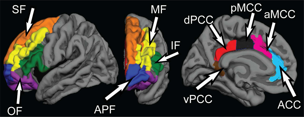
To conduct ROI analysis, five cingulate ROIs were used; anterior part (ACC), middle-anterior part, middle-posterior part, posterior-dorsal part, and posterior-ventral part (vPCC) of the cingulate cortex, see Figure 1. Multiple parcelled Destrieux regions were combined to create meaningful ROIs for prefrontal ROIs. Specifically, anterior prefrontal (APF), superior frontal, orbital frontal, middle frontal, and inferior frontal ROIs were created, as shown by Figure 1. The means of Fisher’s Z of these ROIs were obtained bilaterally for the connectivity with the bilateral hippocampal and amygdalar seeds.
Figure 1.
Regions of interest in the cingulate and prefrontal cortices. SF = superior frontal; MF = middle frontal; IF = inferior frontal; OF = orbitofrontal; APF = anterior prefrontal; ACC = anterior cingulate cortex; aMCC = anterior part of middle cingulate cortex, dPCC = dorsal part of the posterior cingulate cortex, pMCC = posterior part of the middle cingulate cortex vPCC = ventral part of posterior cingulate cortex
2.3 Statistical analysis
An alpha of .01 was used in all analyses in order to strike a balance between Type I and Type II errors. Age, years of education attained, left/right volume asymmetry of the hippocampi and amygdalae calculated as ([left volume – right volume]/([left volume + right volume]/2), and BDI-II scores of the healthy controls and patients with LTLE and RLTE, were compared by analyses of variance (ANOVAs). Structural and functional asymmetries were included because asymmetry is often a more robust measure of TLE-related pathology relative to measures of ipsilateral pathology alone [44]. Clinical variables (i.e., age of disease onset and duration of illness) were compared between LTLE and RTLE groups using t-tests. Correlation coefficients were calculated between these clinical variables, structural volume asymmetries, and BDI-II score.
Group differences in the frontolimbic connectivity strength were investigated with repeated-measures ANOVAs (RMANOVAs) performed for each ROI, with the side of the seed (left/right) and of the target ROI (left /right) as within-subject factors, and group membership (control, LTLE, and RTLE) as a between-subject factor. To examine asymmetry of FC in patients, RMANOVAs were run for each of the ROIs, with group (LTLE/RTLE) as a between-subject factor, and the side of the seed (ipsilateral/contralateral to the seizure focus) and of the target ROI (within/cross hemisphere connection) as within-subject factors. Then, asymmetry indices (ipsilateral Fisher’s Z – contralateral Fisher’s Z) were created for ROIs that showed significant asymmetry in the above analysis. These indices were then entered into a correlational analysis to examine the relationship between FC asymmetry and depressive symptoms. For ROIs without significant asymmetry, ipsilateral and contralateral FC values were separately correlated with BDI-II scores to investigate the relationship. Finally, to investigate the relative contribution of structural volumes versus FC for predicting BDI-II scores, hierarchical regression analysis was performed, with the structural volume asymmetry at step 1, and FC strengths at step 2.
3. RESULTS
There were no statistically significant differences among the controls, LTLE, or RTLE, in age or years of education (F[2,38] = 1.17, p = .37, F[2,38] = 2.84, p = .07, respectively). The distribution of the two genders across the groups was comparable (χ2[2] = 2.47, p = .29). Table 1 presents demographic, clinical, and structural volume information of our participants. The BDI-II scores ranged from minimal (0) to moderate (22) level of self-reported depressive symptoms in patients. There was a trend for a difference in BDI-II scores among the three groups (F[2,37] = 3.014, p = .062), with a tendency for patients to present with greater levels of depressive symptomatology. There was no difference in the BDI-II score between patients with LTLE and RTLE. BDI-II scores were not correlated with disease duration or age of seizure onset in patients. Between LTLE and RTLE, there was no statistically significant difference in age of seizure onset or duration of seizure disorder.
Table 1.
Demographic, clinical, and volumetric information of the participants
| Healthy Control (n = 20) |
LTLE (n = 11) | RTLE (n = 8) | |
|---|---|---|---|
| Age | 42.26 (15.80) | 39.93 (11.54) | 33.00 (14.56) |
| Education | 15.85 (2.25) | 14.27 (1.74) | 14.00 (2.73) |
| BDI-II | 5.1 (6.05) Range: 0 – 21 |
10.82 (7.36) Range: 1 – 21 |
9.86 (7.71) Range: 0 – 18 |
| Age of Onset | - | 17.50 (15.17) | 20.00 (11.24) |
| Disease Duration | - | 21.86 (17.01) | 12.75 (13.60) |
| Left Hippocampus Volume | 3947.10 (368.88) | 3359.09 (789.98)* | 3975.25 (611.36) |
| Right Hippocampus Volume | 3987.40 (366.37) | 3980.55 (335.42) | 3418.38 (1183.00) |
| Left Amygdala Volume | 1526.25 (247.17) | 1443.27 (358.29) | 1669.88 (256.47) |
| Right Amygdala Volume | 1720.15 (247.63) | 1774.36 (266.75) | 1743.50 (488.20) |
| Hippocampal Asymmetry | −.01 (.05) | −.18 (.19)*^ | .20 (.26)^ |
| Amygdala Asymmetry | −.12 (.13) | −.22 (. 17)^ | −.02 (.20)^ |
Note:
denotes a patient group showing statistically significant difference from the control group at alpha of .05.
denotes LTLE and RTLE significantly different from each other at alpha of .05 Although the RTLE group show larger left hippocampal volume than the healthy control, the difference did not reach statistical significance, and by removing one outlier from RTLE group, their left hippocampal volume is smaller than healthy controls.
3.1 Structural Asymmetry of the Hippocampus and Amygdala
Hippocampal asymmetry (HA) was significantly different among the three groups (F[2,38] = 6.289, p = .005), with both LTLE and RTLE groups showing smaller ipsilateral relative to contralateral hippocampal volumes. When LTLE and RTLE groups were combined, longer disease duration was associated with greater ipsilateral-to-contralateral HA (r = −.575, p = .01). There was a trend between LTLE and RTLE to show different amygdala asymmetry (F[2,38] = 5.408, p = .029), with LTLEs showing smaller ipsilateral relative to contralateral amygdala volume while RTLE showed the opposite asymmetry. This was due to one patient with RTLE showing an asymmetry in the opposite direction (left < right). Without this patient, ANOVA indicated that LTLE and RTLE both showed smaller ipsilateral volume (F[2,37] = 6.767, p = .003).
3.2 Group Differences in Functional Connectivity
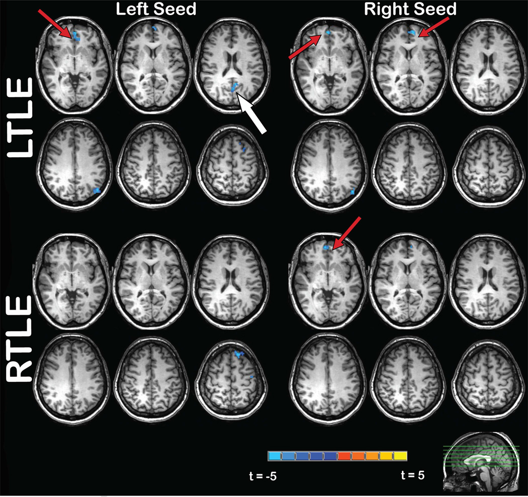
Figure 2 portrays group maps of the average FC differences between patients with LTLE and controls and patients with RTLE and controls. Reductions in FC were predominately seen in patients between the hippocampi and anterior cingulate (LTLE and RTLE), and between the hippocampus and posterior cingulate (LTLE only). At the whole brain level, other small areas of decreased FC were observed in LTLE and RTLE [see 45 for details], but fell outside of the theory-driven ROIs of this paper. Table 2 shows the mean FC strengths of the hippocampus and the amygdala within these ROIs. There was a tendency for reduced FC between the hippocampus and the ACC and vPCC ROIs in patients with TLE, F(2,36) = 4.138, p = .024, and F(2,36) = 3.928, p = .029, respectively. Reduced FC did not depend on the side of the seed or of the target ROI. No effects were significant in the amygdala FC ROI analyses (See supplemental figure).
Figure 2.
T-statistic maps of hippocampal functional connectivity group difference between controls and each of the patient groups based on the whole brain analysis (voxel-wise threshold p = .005, with cluster correction, p < .05). Cold colors indicate areas where a patient group showed a relative decreased in connectivity, while warm colors indicate areas where a patient group showed a relative increase in connectivity, relative to healthy controls. The red arrows point to the anterior cingulate region, while the white arrow points to the posterior cingulate region.
Table 2.
Average functional connectivity strength between ipsilateral and contralateral seed regions and target ROIs in the within-hemisphere and cross-hemisphere connection
| Ipsilateral Amygdala |
Contralateral Amygdala |
Ipsilateral Hippocampus |
Contralateral Hippocampus |
|||||
|---|---|---|---|---|---|---|---|---|
| Within | Cross | Within | Cross | Within | Cross | Within | Cross | |
| Anterior Prefrontal | −.007 | −.012 | −.016 | −.014 | −.025 | −.012 | −.027 | −.023 |
| Inferior Frontal | −.019 | −.028 | −.035 | −.035 | −.042 | −.051 | −.512 | −.035 |
| Middle Frontal | −.044 | −.047 | −.047 | −.052 | −.043 | −.062 | −.050 | −.055 |
| Superior Frontal | −.029 | −.033 | −.026 | −.029 | −.015 | −.040 | −.005 | −.024 |
| Orbitofrontal | .013 | .002 | .008 | .012 | −.006 | −.006 | −.010 | −.015 |
| Within | Cross | Within | Cross | Within | Cross | Within | Cross | |
| ACC | −.018 | −.018 | −.015 | −.024 | −.031 | −.031 | −.001 | −.014 |
| aMCC | −.029 | −.024 | −.024 | −.032 | −.038 | −.042 | −.037 | −.040 |
| dPCC | −.050 | −.038 | −.034 | −.045 | −.023 | −.024 | .004 | .001 |
| pMCC | −.030 | −.036 | −.035 | −.031 | −.030 | −.034 | −.041 | −.038 |
| vPCC | −.026 | −.023 | −.012 | −.009 | .024* | .028^ | .089* | .090^ |
Note: ACC = anterior cingulate cortex, aMCC = anterior part of middle cingulate cortex, dPCC = dorsal part of the posterior cingulate cortex, pMCC = posterior part of the middle cingulate cortex, vPCC = ventral part of the posterior cingulate cortex
3.3 Functional Connectivity Asymmetry of the Hippocampus and Amygdala
Next, in order to investigate whether FC values show asymmetry similar to the hippocampal volume, RMANOVAs with group membership as a between-subject factor, and the side of the seed and of the target ROI as within-subject factors, were performed. The analyses showed a significant main effect of the side of the seed (F = 9.864, p = .006) in the vPCC region, suggesting that FC between the ipsilateral hippocampus and the vPCC was lower than FC between the contralateral hippocampus and the vPCC. This suggests that FC of the hippocampi to this target ROI was asymmetrical in patients with TLE, therefore asymmetry indices were created for inclusion in the FC-BDI analysis.
3.4 Relationship Between Volumetric and FC Asymmetry, and BDI-II Score
There was a tendency for patients with LTLE, but not RTLE, to show a relationship between HA with BDI-II score (r = −.64, p = .034), with a greater ipsilateral hippocampal volume loss associated with higher BDI-II scores. Amygdalar volume asymmetry was not statistically associated with BDI-II scores in patients.
As for FC, the asymmetry of the hippocampus-vPCC connection was marginally associated with the BDI-II score in patients with TLE (r = −.47, p = .049). Specifically, greater ipsilateral reductions in FC between the hippocampus and vPCC tended to be associated with higher levels of self-reported depressive symptoms. However, when LTLE and RTLE were examined separately, FC asymmetry did not significantly correlate with BDI-II scores. No other asymmetry indices (cross-hemisphere or combined) were associated with BDI-II scores.
3.5 Relationship Between Prefrontal and Cingulate FC, and BDI-II Score
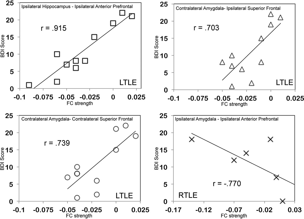
Because a lack of FC asymmetry in TLE does not necessarily mean a lack of contribution of FC to depression symptomatology, the relationship between the BDI-II scores and the hippocampal and amygdalar connectivity with the prefrontal and remaining cingulate ROIs was investigated (Figure 3). In patients with LTLE, FC of the contralateral amygdala to the contralateral and ipsilateral superior frontal ROI, and FC of the ipsilateral hippocampus to the ipsilateral APF ROI were positively correlated with BDI-II score (r = .739, p = .009, r = .703, p = .006, and r = .915, p < .001, respectively) indicating that increased FC between the hippocampus/amygdala seeds and these frontal regions was associated with higher depression scores. In patients with RTLE, FC of the ipsilateral amygdala to ipsilateral APF ROI was negatively correlated with BDI-II score (r = −.770, p = .043), indicating that decreased ipsilateral (right) amygdala-prefrontal FC was associated with higher depression scores.
Figure 3.
Scatterplots showing the relationship between depression (BDI-II score) and FC of the ipsilateral hippocampus to ipsilateral anterior prefrontal ROI (top left), the contralateral amygdala to contralateral superior frontal ROI (bottom left), and the contralateral amygdala to ipsilateral superior frontal ROI (top right) in LTLE, and FC of the ipsilateral amygdala to ipsilateral anterior prefrontal ROI (bottom right). Note that in the bottom left panel, 2 patients with the same BDI score of 8 had very similar functional connectivity values thus they are overlapping.
Finally, hierarchical regression was performed within the LTLE group to determine the relative contribution of HA and FC strengths to APF ROI, as both were associated with depressive symptoms in LTLE. The analysis indicated that FC between the ipsilateral hippocampus and the ipsilateral APF ROI explained a significant amount of variance in the depression score (R2 change = .496, p < .001) above and beyond the amount of variance that HA explained (R2 = .409, p = .034). When hippocampus-APF FC was entered at step 1, it explained 83.7% of the variance (p < .001). These analyses indicate that FC was a stronger contributor than HA, and, it explained a significant amount of unique variance above and beyond HA, with a total of 90.5% of the variance accounted for by the model. FC strengths in these regions did not show statistically significant associations with disease duration or age of onset.
4. DISCUSSION
The main finding of the current study is that FC between the hippocampus and vPCC was reduced ipsilaterally in patients with TLE, but this asymmetry was only a weak contributor to self-reported depressive symptoms. Rather, FC of the hippocampus to the APF cortex predicted depressive symptoms in LTLE, and it was a stronger contributor than hippocampal atrophy. These data suggest the presence of neurobiological factors contributing to depressive symptoms in TLE, but the network showing the greatest ipsilateral reductions in TLE (i.e., hippocampal-vPCC) is not necessarily driving depression. This may indicate that although TLE and depressive symptoms share common pathogenic mechanisms that include the hippocampus and PCC network, dysregulation of other brain regions (i.e., anterior and superior prefrontal cortex) [46] are involved in modulating this association.
4.1 Role of Frontolimbic Network in Co-morbid Depression in TLE
Our finding that frontolimbic dysregulation contributes to depressive symptoms in TLE is consistent with Chen et al. [29] who found reduced limbic connectivity with inferior frontal cortex and Brodmann area 10 (which corresponds to our APF ROI) in depressed versus non-depressed patients with TLE. Both the current study and Chen et al. suggest that not all the patients with TLE show alterations in prefrontal FC, but greater dysregulation of frontolimbic networks are associated with the presentation of depressive symptoms.
Our study extends the literature by demonstrating that hippocampal-prefrontal FC was a stronger contributor to depressive symptoms than hippocampal atrophy alone in patients with LTLE. Although some studies have found relationships between hippocampal structural pathology and depression in TLE [13, 39], other studies have found otherwise [10, 14] or only found a relationship in RTLE with the left hippocampus [11, 12]. This suggests that structural characteristics of the hippocampus may not be a sufficient contributor to depression in TLE. We statistically demonstrate that dysregulation of hippocampal-prefrontal networks is superior to structural volume loss in explaining the level of depressive symptoms in TLE. This is an important addition to the literature that indicates that a better understanding of broad network dysfunction could be valuable in understanding and treating depressive disorders in TLE.
4.2 Difference Between TLE Co-morbid Depression and Major Depressive Disorder
The previous literature in epilepsy and mood disorders indicates both overlapping and distinct brain regions involved in each disorder. In TLE, the temporoammonic pathway (the CA1 regions of the hippocampus, and the entorhinal cortex) is believed to play a critical role in seizure generation [47] and it is well established that the greatest morphometric changes in TLE are found in ipsilateral MTL structures [31]. However, recent studies of TLE demonstrate structural alterations (e.g., cortical thinning) in the prefrontal and cingulate cortices [15–18, 30], including the anterior/orbitofrontal regions [18], and structural aberrations within the orbitofrontal cortex have been linked to depression in one study of TLE [46]. In major depressive disorder, prefrontal involvement is considered a core feature of the disease process [48] by means of a loss of top-down regulation of the limbic system by the prefrontal cortex. Although hippocampal volume loss is reported [49], reductions in resting-state FC have been more amygdala-driven. The FC reductions have been reported between the limbic system and the ventral prefrontal [40], middle and inferior frontal regions [41]. It may be that patients with major depressive disorder present with more defined amygdala-driven limbic network dysregulation that is core to the disease process while patients with TLE show more hippocampus-driven functional changes that depend on the degrees of structural compromise.
4.3 LTLE and RTLE Difference
Interestingly, in our study patients with LTLE and patients with RTLE showed an opposite relationship between depressive symptoms and amygdala-prefrontal FC, in that increased contralateral (right) amygdala FC was associated with greater levels of symptoms in LTLE, while decreased ipsilateral (right) amygdala FC was associated with greater levels of symptoms in RTLE. It may be that dysfunction within right amygdala-prefrontal networks is more related to mood symptoms, regardless of the side of the seizure focus. In non-epileptic individuals with depression, reduced amygdala-prefrontal FC, similar to our RTLE findings, has been reported [40, 41]. Reasons for the reversed relationship (i.e., increased FC resulting in greater depression scores) in LTLE, and for differential patterns between LTLE and RTLE are unknown. In patients with chronic, refractory epilepsy that make up the majority of our sample, possible functional reorganization within the frontal lobe [50] or interactions between frontolimbic FC and other disease-related variables (e.g., anticonvulsant medications) may play a role. Findings from future research that address such variables may provide additional insight into these complex relationships.
4.4 Limitations
Despite the novel findings, several limitations of our study should be noted. First, “depression” was operationalized as a score on a self-report measure, and was not confirmed by a clinical diagnosis. However, this approach provides an advantage of examining depression as a continuous variable rather than artificially dichotomizing patients into “depressed” and “non-depressed,” especially when the sample size is modest. Including a more comprehensive assessment of depression and other co-morbid psychopathology may provide additional insights into the relationship between mood disorders and network dysfunction in TLE. Second, although the automated atlas-based segmentation that we used has been shown to provide reliable and highly reproducible estimates of subcortical volumes [35, 51], we did not manually check all the ROI borders of each individual. Thus, it is possible that small errors in segmentation were present that went undetected. Third, although our results are largely consistent with the Chen et al. study, it is possible that different methodological approaches to measuring FC led to some differences in our FC-depression relationships. Fourth, further investigation of LTLE and RTLE differences should be carried out with a larger sample size. Such larger studies would permit one to examine the contribution of other important variables to our FC-depression relationships, including the presence/absence of febrile seizures or HS. Nevertheless, our sample size is comparable to some other FC studies, and the significant results may attest to the robustness of FC analyses. Fifth, our patients were not treatment naïve (see supplemental table). Some patients were taking antiepileptic medications with known moodstabilizing properties, and it is unclear what effect these medications had on the presentation of their depressive symptoms and/or our FC analysis. Finally, participants in our study reported minimal to moderate levels of self-reported depression. There may have been a selection bias in that patients who were emotionally higher functioning were more likely to volunteer to participate in the study. Including a cohort of TLE patients and controls with severe depression (i.e., major depressive disorder) may provide additional insight into the underlying neurobiology, which could lead to more targeted and effective treatments for depressive symptoms in patients with TLE and other neurological disease.
4.5 Conclusion
In conclusion, although disruption of the hippocampal connectivity to vPCC may be central to TLE and contribute to their depressive symptoms, altered connectivity between the MTL and APF region appears to have a greater contribution to whether a patient will present with depressive symptoms. Our results strengthen the view that there is a neurobiological component to depression in TLE and suggest that this mechanism differs for patients with LTLE versus RTLE. Investigating the relative contribution of other factors driving depression (e.g. neurochemical, seizure-related) is a fruitful avenue for future research. In our sample we did not observe an association between two disease variables (i.e., disease duration, age of seizure onset) and the frontolimbic connectivity, but it is possible that other structural or disease-related variables that were not addressed in the current study are additional modulators of depressive symptoms in TLE.
Supplementary Material
Highlights.
Frontolimbic functional connectivity strongly correlated with depressive symptoms
Functional connectivity was a stronger correlate of depression than volume measures
Mechanisms of depression may differ between left and right temporal lobe epilepsy
ACKNOWLEDGMENTS
We thank Kelly Leyden for her assistance with image processing and thoughtful input upon revising this manuscript. The project was supported by Behavioral Sciences Post-doctoral Fellowship of the Epilepsy Foundation to NK, Health Science Student Fellowship of the Epilepsy Foundation to NEK, and NIH Grant R01NS065838 to CRM. We gratefully acknowledge support from GE Healthcare.
Footnotes
Publisher's Disclaimer: This is a PDF file of an unedited manuscript that has been accepted for publication. As a service to our customers we are providing this early version of the manuscript. The manuscript will undergo copyediting, typesetting, and review of the resulting proof before it is published in its final citable form. Please note that during the production process errors may be discovered which could affect the content, and all legal disclaimers that apply to the journal pertain.
References
- 1.Kanner AM. Depression in epilepsy: prevalence, clinical semiology, pathogenic mechanisms, and treatment. Biol Psychiatry. 2003;54:388–398. doi: 10.1016/s0006-3223(03)00469-4. [DOI] [PubMed] [Google Scholar]
- 2.Jehi L, Tesar G, Obuchowski N, Novak E, Najm I. Quality of life in 1931 adult patients with epilepsy: Seizures do not tell the whole story. Epilepsy Behav. 2011;22:723–727. doi: 10.1016/j.yebeh.2011.08.039. [DOI] [PubMed] [Google Scholar]
- 3.Luoni C, Bisulli F, Canevini MP, De Sarro G, Fattore C, Galimberti CA, Gatti G, La Neve A, Muscas G, Specchio LM, Striano S, Perucca E. Determinants of health-related quality of life in pharmacoresistant epilepsy: Results from a large multicenter study of consecutively enrolled patients using validated quantitative assessments. Epilepsia. 2011;52:2181–2191. doi: 10.1111/j.1528-1167.2011.03325.x. [DOI] [PubMed] [Google Scholar]
- 4.de Araujo Filho GM, Gomes FL, Mazetto L, Marinho MM, Tavares IM, Caboclo LO, Yacubian EM, Centeno RS. Major depressive disorder as a predictor of a worse seizure outcome one year after surgery in patients with temporal lobe epilepsy and mesial temporal sclerosis. Seizure. 2012;21:619–623. doi: 10.1016/j.seizure.2012.07.002. [DOI] [PubMed] [Google Scholar]
- 5.Kanner AM, Byrne R, Chicharro A, Wuu J, Frey M. A lifetime psychiatric history predicts a worse seizure outcome following temporal lobectomy. Neurology. 2009;72:793–799. doi: 10.1212/01.wnl.0000343850.85763.9c. [DOI] [PubMed] [Google Scholar]
- 6.Garcia CS. Depression in temporal lobe epilepsy: a review of prevalence, clinical features, and management considerations. Epilepsy Res Treat. 2012;2012:809843. doi: 10.1155/2012/809843. [DOI] [PMC free article] [PubMed] [Google Scholar]
- 7.Finegersh A, Avedissian C, Shamim S, Dustin I, Thompson PM, Theodore WH. Bilateral hippocampal atrophy in temporal lobe epilepsy: effect of depressive symptoms and febrile seizures. Epilepsia. 2011;52:689–697. doi: 10.1111/j.1528-1167.2010.02928.x. [DOI] [PMC free article] [PubMed] [Google Scholar]
- 8.Tebartz van Elst L, Woermann FG, Lemieux L, Trimble MR. Amygdala enlargement in dysthymia--a volumetric study of patients with temporal lobe epilepsy. Biol Psychiatry. 1999;46:1614–1623. doi: 10.1016/s0006-3223(99)00212-7. [DOI] [PubMed] [Google Scholar]
- 9.Richardson EJ, Griffith HR, Martin RC, Paige AL, Stewart CC, Jones J, Hermann BP, Seidenberg M. Structural and functional neuroimaging correlates of depression in temporal lobe epilepsy. Epilepsy Behav. 2007;10:242–249. doi: 10.1016/j.yebeh.2006.11.013. [DOI] [PubMed] [Google Scholar]
- 10.Briellmann RS, Hopwood MJ, Jackson GD. Major depression in temporal lobe epilepsy with hippocampal sclerosis: clinical and imaging correlates. J Neurol Neurosurg Psychiatry. 2007;78:1226–1230. doi: 10.1136/jnnp.2006.104521. [DOI] [PMC free article] [PubMed] [Google Scholar]
- 11.Baxendale SA, Thompson PJ, Duncan JS. Epilepsy & depression: the effects of comorbidity on hippocampal volume--a pilot study. Seizure. 2005;14:435–438. doi: 10.1016/j.seizure.2005.07.003. [DOI] [PubMed] [Google Scholar]
- 12.Shamim S, Hasler G, Liew C, Sato S, Theodore WH. Temporal lobe epilepsy, depression, and hippocampal volume. Epilepsia. 2009;50:1067–1071. doi: 10.1111/j.1528-1167.2008.01883.x. [DOI] [PMC free article] [PubMed] [Google Scholar]
- 13.Quiske A, Helmstaedter C, Lux S, Elger CE. Depression in patients with temporal lobe epilepsy is related to mesial temporal sclerosis. Epilepsy Res. 2000;39:121–125. doi: 10.1016/s0920-1211(99)00117-5. [DOI] [PubMed] [Google Scholar]
- 14.Sanchez-Gistau V, Sugranyes G, Bailles E, Carreno M, Donaire A, Bargallo N, Pintor L. Is major depressive disorder specifically associated with mesial temporal sclerosis? Epilepsia. 2012;53:386–392. doi: 10.1111/j.1528-1167.2011.03373.x. [DOI] [PubMed] [Google Scholar]
- 15.McDonald CR, Hagler DJ, Jr., Ahmadi ME, Tecoma E, Iragui V, Gharapetian L, Dale AM, Halgren E. Regional neocortical thinning in mesial temporal lobe epilepsy. Epilepsia. 2008;49:794–803. doi: 10.1111/j.1528-1167.2008.01539.x. [DOI] [PubMed] [Google Scholar]
- 16.Bonilha L, Edwards JC, Kinsman SL, Morgan PS, Fridriksson J, Rorden C, Rumboldt Z, Roberts DR, Eckert MA, Halford JJ. Extrahippocampal gray matter loss and hippocampal deafferentation in patients with temporal lobe epilepsy. Epilepsia. 2010;51:519–528. doi: 10.1111/j.1528-1167.2009.02506.x. [DOI] [PMC free article] [PubMed] [Google Scholar]
- 17.Lin JJ, Salamon N, Lee AD, Dutton RA, Geaga JA, Hayashi KM, Luders E, Toga AW, Engel J, Jr., Thompson PM. Reduced Neocortical Thickness and Complexity Mapped in Mesial Temporal Lobe Epilepsy with Hippocampal Sclerosis. Cereb Cortex. 2006 doi: 10.1093/cercor/bhl109. [DOI] [PubMed] [Google Scholar]
- 18.Bernhardt BC, Worsley KJ, Kim H, Evans AC, Bernasconi A, Bernasconi N. Longitudinal and cross-sectional analysis of atrophy in pharmacoresistant temporal lobe epilepsy. Neurology. 2009;72:1747–1754. doi: 10.1212/01.wnl.0000345969.57574.f5. [DOI] [PMC free article] [PubMed] [Google Scholar]
- 19.Lin JJ, Mula M, Hermann BP. Uncovering the neurobehavioural comorbidities of epilepsy over the lifespan. Lancet. 2012;380:1180–1192. doi: 10.1016/S0140-6736(12)61455-X. [DOI] [PMC free article] [PubMed] [Google Scholar]
- 20.Hermann BP, Seidenberg M, Haltiner A, Wyler AR. Mood state in unilateral temporal lobe epilepsy. Biol Psychiatry. 1991;30:1205–1218. doi: 10.1016/0006-3223(91)90157-h. [DOI] [PubMed] [Google Scholar]
- 21.Bromfield EB, Altshuler L, Leiderman DB, Balish M, Ketter TA, Devinsky O, Post RM, Theodore WH. Cerebral metabolism and depression in patients with complex partial seizures. Arch Neurol. 1992;49:617–623. doi: 10.1001/archneur.1992.00530300049010. [DOI] [PubMed] [Google Scholar]
- 22.Koolschijn PC, van Haren NE, Lensvelt-Mulders GJ, Hulshoff Pol HE, Kahn RS. Brain volume abnormalities in major depressive disorder: a meta-analysis of magnetic resonance imaging studies. Hum Brain Mapp. 2009;30:3719–3735. doi: 10.1002/hbm.20801. [DOI] [PMC free article] [PubMed] [Google Scholar]
- 23.Matthews SC, Strigo IA, Simmons AN, Yang TT, Paulus MP. Decreased functional coupling of the amygdala and supragenual cingulate is related to increased depression in unmedicated individuals with current major depressive disorder. J Affect Disord. 2008;111:13–20. doi: 10.1016/j.jad.2008.05.022. [DOI] [PubMed] [Google Scholar]
- 24.Fair DA, Schlaggar BL, Cohen AL, Miezin FM, Dosenbach NU, Wenger KK, Fox MD, Snyder AZ, Raichle ME, Petersen SE. A method for using blocked and event-related fMRI data to study “resting state” functional connectivity. Neuroimage. 2007;35:396–405. doi: 10.1016/j.neuroimage.2006.11.051. [DOI] [PMC free article] [PubMed] [Google Scholar]
- 25.Waites AB, Briellmann RS, Saling MM, Abbott DF, Jackson GD. Functional connectivity networks are disrupted in left temporal lobe epilepsy. Ann Neurol. 2006;59:335–343. doi: 10.1002/ana.20733. [DOI] [PubMed] [Google Scholar]
- 26.Frings L, Schulze-Bonhage A, Spreer J, Wagner K. Remote effects of hippocampal damage on default network connectivity in the human brain. J Neurol. 2009;256:2021–2029. doi: 10.1007/s00415-009-5233-0. [DOI] [PubMed] [Google Scholar]
- 27.Schacher M, Haemmerle B, Woermann FG, Okujava M, Huber D, Grunwald T, Kramer G, Jokeit H. Amygdala fMRI lateralizes temporal lobe epilepsy. Neurology. 2006;66:81–87. doi: 10.1212/01.wnl.0000191303.91188.00. [DOI] [PubMed] [Google Scholar]
- 28.Broicher SD, Frings L, Huppertz HJ, Grunwald T, Kurthen M, Kramer G, Jokeit H. Alterations in functional connectivity of the amygdala in unilateral mesial temporal lobe epilepsy. J Neurol. 2012;259:2546–2554. doi: 10.1007/s00415-012-6533-3. [DOI] [PubMed] [Google Scholar]
- 29.Chen S, Wu X, Lui S, Wu Q, Yao Z, Li Q, Liang D, An D, Zhang X, Fang J, Huang X, Zhou D, Gong QY. Resting-state fMRI study of treatment-naive temporal lobe epilepsy patients with depressive symptoms. Neuroimage. 2012;60:299–304. doi: 10.1016/j.neuroimage.2011.11.092. [DOI] [PubMed] [Google Scholar]
- 30.Kemmotsu N, Girard HM, Bernhardt BC, Bonilha L, Lin JJ, Tecoma ES, Iragui VJ, Hagler DJ, Jr., Halgren E, McDonald CR. MRI analysis in temporal lobe epilepsy: Cortical thinning and white matter disruptions are related to side of seizure onset. Epilepsia. 2011;52:2257–2266. doi: 10.1111/j.1528-1167.2011.03278.x. [DOI] [PMC free article] [PubMed] [Google Scholar]
- 31.Li J, Zhang Z, Shang H. A meta-analysis of voxel-based morphometry studies on unilateral refractory temporal lobe epilepsy. Epilepsy Res. 2012;98:97–103. doi: 10.1016/j.eplepsyres.2011.10.002. [DOI] [PubMed] [Google Scholar]
- 32.Commission on Classification and Terminology of the International League Against Epilepsy. Proposal for revised classification of epilepsies and epileptic syndromes. Epilepsia. 1989;30:389–399. doi: 10.1111/j.1528-1157.1989.tb05316.x. [DOI] [PubMed] [Google Scholar]
- 33.Beck AT, Steer RA, Brown G. Manual for the Beck Depression Inventory-II. San Antonio, TX: Psychological Corporation; 1996. [Google Scholar]
- 34.Holland D, Kuperman JM, Dale AM. Efficient correction of inhomogeneous static magnetic field-induced distortion in Echo Planar Imaging. Neuroimage. 2010;50:175–183. doi: 10.1016/j.neuroimage.2009.11.044. [DOI] [PMC free article] [PubMed] [Google Scholar]
- 35.Fischl B, Salat DH, Busa E, Albert M, Dieterich M, Haselgrove C, van der Kouwe A, Killiany R, Kennedy D, Klaveness S, Montillo A, Makris N, Rosen B, Dale AM. Whole brain segmentation: automated labeling of neuroanatomical structures in the human brain. Neuron. 2002;33:341–355. doi: 10.1016/s0896-6273(02)00569-x. [DOI] [PubMed] [Google Scholar]
- 36.Cox RW. AFNI: software for analysis and visualization of functional magnetic resonance neuroimages. Comput Biomed Res. 1996;29:162–173. doi: 10.1006/cbmr.1996.0014. [DOI] [PubMed] [Google Scholar]
- 37.Saad ZS, Reynolds RC. Suma. Neuroimage. 2011 doi: 10.1016/j.neuroimage.2011.09.016. [DOI] [PMC free article] [PubMed] [Google Scholar]
- 38.Biswal B, Yetkin FZ, Haughton VM, Hyde JS. Functional connectivity in the motor cortex of resting human brain using echo-planar MRI. Magn Reson Med. 1995;34:537–541. doi: 10.1002/mrm.1910340409. [DOI] [PubMed] [Google Scholar]
- 39.Bremner JD, Narayan M, Anderson ER, Staib LH, Miller HL, Charney DS. Hippocampal volume reduction in major depression. Am J Psychiatry. 2000;157:115–118. doi: 10.1176/ajp.157.1.115. [DOI] [PubMed] [Google Scholar]
- 40.Tang Y, Kong L, Wu F, Womer F, Jiang W, Cao Y, Ren L, Wang J, Fan G, Blumberg HP, Xu K, Wang F. Decreased functional connectivity between the amygdala and the left ventral prefrontal cortex in treatment-naive patients with major depressive disorder: a resting-state functional magnetic resonance imaging study. Psychol Med. 2012:1–7. doi: 10.1017/S0033291712002759. [DOI] [PubMed] [Google Scholar]
- 41.Lui S, Wu Q, Qiu L, Yang X, Kuang W, Chan RC, Huang X, Kemp GJ, Mechelli A, Gong Q. Resting-state functional connectivity in treatment-resistant depression. Am J Psychiatry. 2011;168:642–648. doi: 10.1176/appi.ajp.2010.10101419. [DOI] [PubMed] [Google Scholar]
- 42.McDonald CR, Thesen T, Carlson C, Blumberg M, Girard HM, Trongnetrpunya A, Sherfey JS, Devinsky O, Kuzniecky R, Dolye WK, Cash SS, Leonard MK, Hagler DJ, Jr., Dale AM, Halgren E. Multimodal imaging of repetition priming: Using fMRI, MEG, and intracranial EEG to reveal spatiotemporal profiles of word processing. Neuroimage. 2010;53:707–717. doi: 10.1016/j.neuroimage.2010.06.069. [DOI] [PMC free article] [PubMed] [Google Scholar]
- 43.Destrieux C, Fischl B, Dale A, Halgren E. Automatic parcellation of human cortical gyri and sulci using standard anatomical nomenclature. Neuroimage. 2010;53:1–15. doi: 10.1016/j.neuroimage.2010.06.010. [DOI] [PMC free article] [PubMed] [Google Scholar]
- 44.Farid N, Girard HM, Kemmotsu N, Smith ME, Magda SW, Lim WY, Lee RR, McDonald CR. Temporal lobe epilepsy: quantitative MR volumetry in detection of hippocampal atrophy. Radiology. 2012;264:542–550. doi: 10.1148/radiol.12112638. [DOI] [PMC free article] [PubMed] [Google Scholar]
- 45.Kucukboyaci N, Kemmotsu N, Girard HM, Cheng CE, Tecoma E, Iragui V, McDonald CR. Structural and functional connectivity of hippocampal networks in temporal lobe epilepsy. 2012 Abstract No 2.178. In: American Epilepsy Society Annual Meeting. [Google Scholar]
- 46.Butler T, Blackmon K, McDonald CR, Carlson C, Barr WB, Devinsky O, Kuzniecky R, DuBois J, French J, Halgren E, Thesen T. Cortical thickness abnormalities associated with depressive symptoms in temporal lobe epilepsy. Epilepsy Behav. 2012;23:64–67. doi: 10.1016/j.yebeh.2011.10.001. [DOI] [PMC free article] [PubMed] [Google Scholar]
- 47.Ang CW, Carlson GC, Coulter DA. Massive and specific dysregulation of direct cortical input to the hippocampus in temporal lobe epilepsy. J Neurosci. 2006;26:11850–11856. doi: 10.1523/JNEUROSCI.2354-06.2006. [DOI] [PMC free article] [PubMed] [Google Scholar]
- 48.Price JL, Drevets WC. Neural circuits underlying the pathophysiology of mood disorders. Trends Cogn Sci. 2012;16:61–71. doi: 10.1016/j.tics.2011.12.011. [DOI] [PubMed] [Google Scholar]
- 49.Campbell S, Macqueen G. The role of the hippocampus in the pathophysiology of major depression. J Psychiatry Neurosci. 2004;29:417–426. [PMC free article] [PubMed] [Google Scholar]
- 50.Wagner K, Frings L, Spreer J, Buller A, Everts R, Halsband U, Schulze-Bonhage A. Differential effect of side of temporal lobe epilepsy on lateralization of hippocampal, temporolateral, and inferior frontal activation patterns during a verbal episodic memory task. Epilepsy Behav. 2008;12:382–387. doi: 10.1016/j.yebeh.2007.11.003. [DOI] [PubMed] [Google Scholar]
- 51.Pardoe HR, Pell GS, Abbott DF, Jackson GD. Hippocampal volume assessment in temporal lobe epilepsy: How good is automated segmentation? Epilepsia. 2009;50:2586–2592. doi: 10.1111/j.1528-1167.2009.02243.x. [DOI] [PMC free article] [PubMed] [Google Scholar]
Associated Data
This section collects any data citations, data availability statements, or supplementary materials included in this article.


