Abstract
Background
Elevated sympathetic activity is associated with kidney dysfunction. Here we used twin pairs to probe heritability of GFR and its genetic covariance with other traits.
Methods
We evaluated renal and adrenergic phenotypes in twins. GFR was estimated by CKD-EPI algorithm. Heritability and genetic covariance of eGFR and associated risk traits were estimated by variance-components. Meta-analysis probed reproducibility of DBH genetic effects. Effect of DBH genetic variation on renal disease was tested in the NIDDK-AASK cohort.
Results
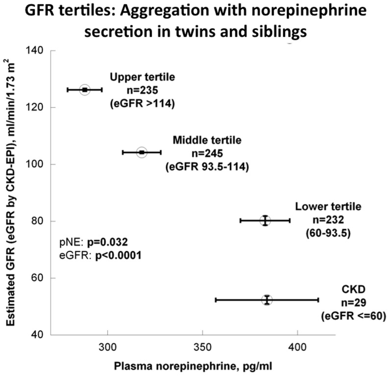
Norepinephrine secretion rose across eGFR tertiles while eGFR fell (p<0.0001). eGFR was heritable, at h2 = 67.3±4.7% (p = 3.0E-18), as were secretion of norepinephrine (h2 = 66.5±5.0%, p = 3.2E-16) and dopamine (h2 = 56.5±5.6%, p = 1.8E-13), and eGFR displayed genetic co-determination (covariance) with norepinephrine (ρG = −0.557±0.088, p = 1.11E-08) as well as dopamine (ρG = −0.223±0.101, p = 2.3E-02). Since dopamine β-hydroxylase (DBH) catalyzes conversion of dopamine to norepinephrine, we studied functional variation at DBH; DBH promoter haplotypes predicted transcriptional activity (p<0.001), plasma DBH (p<0.0001) and norepinephrine (p = 0.0297) secretion; transcriptional activity was inversely (p<0.0001) associated with basal eGFR. Meta-analysis validated DBH haplotype effects on eGFR across 3 samples. In NIDDK-AASK, we established a role for DBH promoter variation in long-term renal decline rate (GFR slope, p = 0.003).
Conclusions
The heritable GFR trait shares genetic determination with catecholamines, suggesting new pathophysiologic, diagnostic and therapeutic approaches towards disorders of GFR as well as CKD. Adrenergic activity may play a role in progressive renal decline, and genetic variation at DBH may assist in profiling subjects for rational preventive treatment.
Introduction
The autonomic nervous system, and in particular its sympathetic branch, plays a role in physiological control of GFR as well as the development of CKD (chronic kidney disease) and eventually end-stage renal disease (ESRD) [1], [2]. Activation of sympathetic activity in CKD is characterized by increased muscle sympathetic nerve traffic [1] [3] and circulating levels of plasma norepinephrine [4]. Renal afferent sensory and efferent sympathetic innervation [5] may mediate the effect of chemoreceptors and baroreceptors in damaged kidneys [1] [3], [6], resulting in juxta-glomerular cell renin release, BP elevation, and acceleration of progressive renal dysfunction [7]. Indeed, agents that decrease sympathetic outflow have selective beneficial effects in progression of CKD, even at sub-antihypertensive doses [8], and renal sympathetic denervation is also an emerging therapy for intractable hypertension with progressive renal disease [9]. Sympathetic activation may also influence renal function by other means, including augmented renal vascular resistance (efferent or afferent arteriole), or increased tubular sodium reabsorption.
CKD (often defined as a chronic loss of GFR to <60 ml/min/1.73 m2) is an increasingly recognized syndrome, with substantial elevations in cardiovascular morbidity and mortality [10], [11]. CKD was responsible for the death of nearly 45,000 people in 2006, ranking as the ninth leading cause of death in the United States [12].
An improved understanding of the role of heredity in adrenergic control of GFR as well as progressive CKD may reveal novel pathways that could be exploited for preventive or even therapeutic strategies in CKD. Here we employed a twin pair design to explore the role of heredity in coupling of adrenergic and renal function, in healthy individuals as well as patients with progressive CKD.
Results
eGFR tertiles
Demographic traits
Table S1 in file S1 stratifies the twin/sibling study population by eGFR tertiles (using the CKD-EPI method) in those without CKD (eGFR>60 ml/min), as compared to those with CKD (eGFR≤60 ml/min). Demographic parameters differing by eGFR stratum included age (subsequently adjusted for), ethnicity and family history of hypertension.
Physical/physiological traits
SBP/DBP decreased in the higher eGFR tertiles (P<0.001), though the association disappeared once adjusted for age. BMI was slightly higher in the middle eGFR tertile.
Renal traits
Each estimator of GFR was significantly different by tertile in the age-adjusted model, as was eGFR between individuals with and without CKD (P<0.0001). Urine albumin excretion was elevated in subjects with CKD (p = 0.0164).
Adrenergic traits
Individuals in the lower eGFR strata displayed higher plasma norepinephrine concentrations (Table S1 in file S1; Figure 1; p = 0.032), though other catecholamines were not different. To probe the relationship in individual detail, we found a significant inverse correlation between eGFR and plasma norepinephrine, whether tested in all subjects (ρ = −0.263, p<0.0001) or only in those without CKD (ρ = −0.266, p<0.0001) (Figure S1 in file S2).
Figure 1. Adrenergic function and GFR: eGFR tertiles.
Aggregation of eGFR tertiles with plasma norepinephrine in the entire study cohort, including those with CKD (eGFR≤60).
eGFR trait-on-trait correlations and h2 in twins
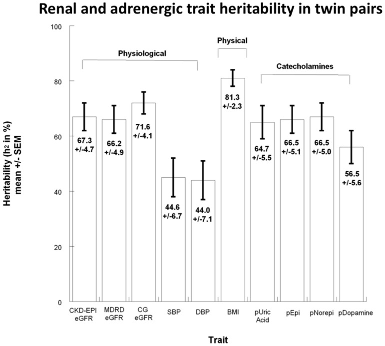
Inter-individual correlations between eGFR and physiological or adrenergic traits are presented in Table S2 in file S1. As shown there and in Figure 2, heritability for eGFR was h2 = 67.0±5.0% (p<0.0001); heritability values for other physiological, physical and adrenergic traits are also displayed graphically in Figure 2. Heritability was highly significant for all adrenergic and renal traits: plasma norepinephrine (h2 = 66.5±5.0%, p<0.0001), plasma epinephrine (h2 = 66.5±5.1%, p<0.0001) and plasma dopamine (h2 = 56.5±5.6%, p<0.0001). Other physical traits such as BMI displayed an even higher rate of heritability, at h2 = 81.3±2.3%, p<0.0001.
Figure 2. Heritability h2 and genetic covariance: Heritability (h2) estimates.
of CKD-EPI estimated glomerular filtration rate (eGFR) and renal, physiological, physical and catecholamine risk factors in the twin study cohort. The h2 estimates, expressed as % (± standard error of mean) of trait variance (h2 = VG/VP), obtained using SOLAR, suggest general agreement of the present cohort with other previously published research. h2 estimates arose from n = 340 (renal traits) to n = 357 (adrenergic traits) individuals. BMI indicates body mass index; SBP, systolic blood pressure; DBP, diastolic blood pressure; pEpi, plasma norepinephrine; pNorepi, plasma norepinephrine; pDopamine, plasma dopamine; CKD-EPI, Chronic Kidney Disease Epidemiology Collaboration formula; CG, Cockroft-Gault formula; MDRD, Modification of Diet in Renal Disease study formula.
Shared heritability in twins: Genetic covariance between eGFR and catecholamines
Several phenotypic traits that correlated with eGFR were analyzed for shared heredity (genetic covariance or pleiotropy) in twin pairs (Table S2 in file S1). As expected, CKD-EPI eGFR displayed substantial genetic covariance with MDRD eGFR (ρG = 0.98±0.008, p<0.0001) and Cockroft/Gault eGFR (ρG = 0.65±0.01, p<0.0001). Two catecholamine traits showed significant genetic covariance with eGFR: plasma norepinephrine (ρG = −0.56±0.09, p<0.0001) and plasma dopamine (ρG = −0.22±0.010, p<0.05). Figure S2 in file S2 depicts shared genetic (ρG) versus environmental (ρE) co-determination of eGFR with associated variables of interest, illustrating significant pleiotropy for eGFR with these two adrenergic traits.
DBH functional genetic variation: Promoter haplotypes
DBH promoter transcription
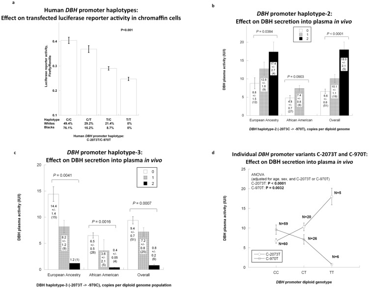
In reference to our previous work in the DBH promoter region [13], [14], there are two common functional variants that alter transcription of the gene: C-2073T (rs1989787) and C-970T (rs1611115), separated by only 1103 bp. We thus evaluated functional consequences of haplotypic variation at C-2073T→C-970T, using transfected promoter haplotype/luciferase reporter plasmids (Figure 3a), and found haplotype C→C to exhibit the highest gene expression, followed by C→T, then T→C, and finally T→T (note that T→T does not occur in human samples; Table S3 in file S1). Thus, haplotypes naturally occurring in humans (C→C, C→T, T→C) have pronounced effects on DBH transcription (p<0.001).
Figure 3. DBH promoter haplotypes (C-2073T→C-970T): Results for haplotype/luciferase reporter enzymatic activity in transfected chromaffin cells, as well as DBH secretion in humans.
a. DBH promoter haplotype expression in the nucleus: Transcription in luciferase reporter plasmids transfected into chromaffin (PC12) cells. Each promoter transfection was done in 8 replicates. b. DBH promoter haplotypes in vivo : Effects on plasma DBH activity. Two functional promoter SNPs constituting a haplotype are shown in subjects of European ancestry, African American and the overall population. Haplotype-2 (C→T) is significantly associated with DBH activity in subjects from European ancestry as well as the overall study population. c. Haplotype-3 (T→C) is significantly associated with DBH activity in all groups. d. Common promoter variants C-2073T and C-970T analyzed individually for effects on DBH secretion in vivo. Plasma DBH activity shows significant association with each of the common variants, both C-2073T and C-970T. To attain specificity, C-2073T or C-970T (as appropriate) were included as covariates, along with age and sex.
DBH secretion into plasma
Across biogeographic ancestries (white versus black), we found that DBH plasma activity substantially increased (p<0.0001) with increasing copy number (0,1,2 per genome) for haplotype-2 (C→T), with a directionally opposite effect (p = 0.0007) for haplotype-3 (T→C) (Figures 3b, 3c). We also found that the C→C haplotype-1 increased (p = 0.0124) plasma DBH activity (Figure S3 in file S1), but most prominently in African Americans, perhaps because of the relatively high frequency of haplotype C→C in that population (76.6% of chromosomes; Tables S3c and S4b in file S1). The theoretical T→T haplotype was not observed in any subject (0/458 chromosomes, Table S3c in file S1), reflecting the minor allele frequencies of T-alleles at both loci.
We explored directional effects for individual promoter SNP genotypes on DBH activity (Figure 3d): the minor (T) allele increased (p<0.0001) DBH activity at SNP C-2073T, while decreasing (p = 0.032) DBH activity at SNP C-970T.
Frequencies, LD, and biogeographic ancestry
Across biogeographic ancestry groups (Tables S3a and S3b in File S1), the two nearby promoter variants differed in allele and diploid genotype frequencies as well as extent of LD (linkage disequilibrium, as D′, r2, or chi2). Within the overall study population and specifically in the white ethnic subpopulation, we found minimal historical recombination as indicated by high LD values (p<0.0001; Table S3b in file S1). Based on this degree of LD, the experiment-wide significance threshold as determined by Nyholt's SNPSpD to maintain the type I (false positive) error rate at 5% or less is P≤0.026. However, greater historical recombination effects were reflected as reduced LD in the African-American (NIDDK-AASK) study population (Table S3b in file S1).
Of the 4 possible haplotypes across these two tightly linked DBH promoter variants in twin/siblings and AASK subjects, we imputed the existence of three common haplotypes: C→C, C→T, and T→C (Table S3c in file S1); theoretical haplotype T→T was not observed in at least 2n = 458 chromosomes (Tables S3c and S4a in file S1), consistent with the T-allele as the minor allele at both C-2073T and C-970T (Tables S3a and S3c in file S1).
DBH promoter functional genetic variation: Effects on renal and adrenergic traits
Since secretion of both norepinephrine and dopamine displayed pleiotropy with eGFR (Table S2 in File S1; Figure S2 in file S1), and DBH catalyzes the conversion of dopamine to norepinephrine, we also explored the effects of DBH genetic variation on eGFR and catecholamines.
Norepinephrine secretion
Individuals homozygous for the most transcriptionally active (Figure 3a) promoter haplotype, C→C/C→C, displayed higher plasma norepinephrine concentration than all others (by ∼16%, p = 0.029; Table S4b in file S1; Figure 4a). In bivariate though not univariate models (Table S4a in file S1), promoter haplotypes also jointly influenced plasma norepinephrine and eGFR (C→C, p = 0.0075; T→C, p = 0.0022).
Figure 4. DBH promoter haplotype with adrenergic or GFR traits in twins and siblings.
a. DBH promoter diploid haplotype-1 (C→C) association with norepinephrine secretion. b. DBH promoter haplotypes: Inverse association between transcriptional activity (transfected promoter/reporter plasmids in PC12 chromaffin cells) and eGFR. Each promoter transfection was done in 8 replicates. Haplotype numbers for this population were: C→C (n = 226; 29.4%), C→T (n = 134; %), T→C (n = 98; 21.4%), and T→T (n = 0). c. DBH promoter haplotype-3 (T→C): Significant association (P<0.0001) with eGFR in an age-adjusted model.
Basal eGFR
Overall, DBH promoter transcriptional activity displayed a clearly inverse relationship with basal eGFR (p<0.0001, Figure 4b). DBH promoter common haplotype-3 (T→C, across C-2073T→C-970T) displayed the most significant (P<0.0001; Table S4a in file S1) effect on eGFR, with the T→C haplotype diminishing the trait (Figure 4c). When results were tested in the largest biogeographic ancestry group (white, n = 229) alone, the effects persisted (p = 0.0377; Table S4c in file S1). DBH promoter diploid haplotypes also influenced eGFR (C→C/C→T, p = 0.0072; C→T/T→C, p = 0.0019; Table S4b in file S1). Individual SNP C-2073T predicted eGFR, though not norepinephrine (Table S4c in file S1). DBH promoter genetic variation did not influence urine albumin excretion in these subjects.
Extension of DBH promoter haplotype effects into additional population samples: Meta-analysis
Meta-analysis combining the twin/sibling pairs and two additional independent population samples (Kaiser-1 and Kaiser-2), for a total of n = 3063 subjects, indicated allelic effects consistent in magnitude (beta, or effect size per allele) and direction (sign on slope) across groups; the overall slope of the meta-analysis regression on haplotype-2 (C-2073→-970T, C→T) was significant, at beta (slope) = −1.321, with SE (of beta) = 0.560, and p = 0.018 (Table S5 in file S1). In a similar meta-analysis, haplotype T→C maintained marginal statistical significance on eGFR (p = 0.081).
DBH genetic variation and longitudinal progression of CKD (GFR slope) in the NIDDK-AASK trial
Very different DBH haplotype frequencies were observed in the African-American subjects as compared to other ethnicities in the twins/siblings (Table S3c in file S1; p = 0.0041), though not between blacks in the twin/sibling versus AASK (p = 0.99); especially prominent was the increased frequency of haplotype C→C in blacks (from 49.5% of chromosomes in whites, to 76.1% in AASK).
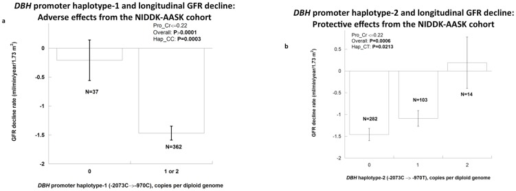
DBH promoter haplotypes C→C and C→T displayed significant associations with GFR slope (Table S6a in file S1, each P<0.01). While the presence of haplotype C→C seemed to accelerate renal decline (p = 0.003, Figure 5a), haplotype C→T was protective, as a function of its copy number (0,1,2 copies/diploid genome; p = 0.0006, Figure 5b). At individual SNPs, DBH promoter variant C-970T retained significant association with GFR slope (Table S6b in file S1; p = 0.029). In the same individuals, DBH promoter genetic variation did not influence baseline (pre-study) urinary protein excretion (protein/creatinine ratio, mg/gm).
Figure 5. DBH promoter haplotype activity: Longitudinal effects on GFR decline in the NIDDK-AASK study population.
GFRs were determined longitudinally by [125I]-iothalamate clearance. Subjects without excessive proteinuria at entry (urine protein/creatinine ratio ≤0.22 gm/gm at entry, N = 428) were analyzed. a. DBH promoter haplotype-1 (C→C) displaying a significant association (p = 0.0006) with GFR slope in an age-adjusted model. b. DBH promoter haplotype-2 (C→T) displaying a significant association (p = 0.0006) with GFR slope in an age-adjusted model.
Discussion
Overview
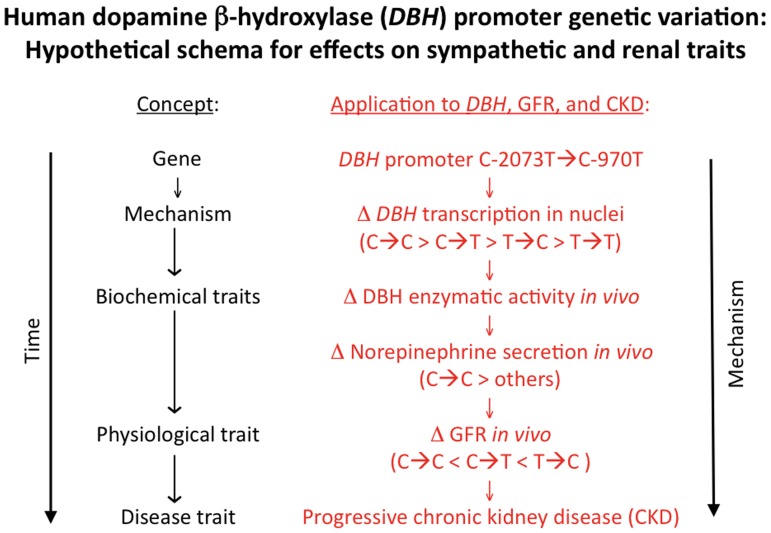
Here we probed the aggregation of renal function with physical, physiological, and adrenergic traits, focusing on the role of heredity in control of GFR (as estimated with the CKD-EPI algorithm). We found plasma norepinephrine to be inversely associated with eGFR (Figure 1; Figure S1 in file S2) with an R2 (explanatory coefficient) of ∼6.5%, indicating that sympathetic over-activity is not restricted to End-Stage Renal Disease (ESRD) [15], but occurs in earlier stages of progressive renal failure [16]. As schematically depicted in Figure 6, sympathetic activation seems to play a fundamental role in control of renal function, which may contribute to progression of CKD. Indeed, other evidence suggests that decreased renal function mediated through sympathetic over-activation may contribute to arterial hypertension in humans [2] and experimental animals [8]. Other studies suggest a role for genetic variation in adrenergic receptor loci on progressive renal disease [17], [18].
Figure 6. Human DBH genetic variation and its effects on sympathetic and renal traits: a schematic hypothesis.
Framework for application of experimental results with DBH genetic variation at the promoter region: Alteration of DBH transcription and enzymatic activity affects norepinephrine, ultimately influencing GFR (physiological and disease traits).
DBH functional genetic variation and renal function
Since norepinephrine is formed from dopamine in the catecholamine biosynthetic pathway through an enzymatic reaction catalyzed by DBH, and GFR displays genetic covariance with both norepinephrine and dopamine (Table S2 in File S1; Figure S2 in file S2), we targeted variation at the DBH gene for heritable effects on GFR. We began by looking at these two functional DBH promoter SNPs [13], [14] across four biogeographic ancestry groups (Tables S3b, S3c in file S1) and found that both SNPs are in close LD, especially in individuals of European ancestry.
Transcriptional activity of the DBH promoter variants was evaluated by transfection of promoter→luciferase reporter plasmids. Previously we identified two functional variants in the proximal human DBH promoter that alter transcription: C-970T [13] and C-2073T [14]. Transcriptional activity of the 4 haplotypes themselves ranked (Figure 3a): C→C>C→T>T→C>T→T. At C-970T, promoter activity ranked T>C allele [13], while at C-2073T the activity ranked C>T allele [14]. However, the individual allelic effects on transcription were background (i.e., haplotype) –specific [14]. A similar rank order of haplotype effects was established on DBH secretion in vivo (Figure 4a): C→C>C→T>T/C (note that the dual minor allele haplotype T→T was not observed in human samples).
In previous studies, we linked individual DBH promoter variants to DBH secretion as well as elevated blood pressure [13], [14]; since hypertension is coupled to progression of kidney dysfunction, we searched for DBH promoter haplotype effects on renal function, beginning with normal variation of eGFR in the healthy twin/sibling sample [17], [19]. In studies of basal (resting) eGFR, we found that each of the 3 DBH haplotypes – C→C (p = 0.0017), C→T (p = 0.0013), and T→C (p<0.0001) – displayed significant effects on the eGFR trait (Table S4a in file S1), and the overall effect was such that higher transcriptional activity paralleled basal eGFR (Figure 4b). Despite this overall picture, individual results on occasion deviated from expectation. For example, haplotype-3 (T→C) was associated with a decrease in not only plasma DBH (Figure 3c) but also eGFR (Figure 4c); here we cannot exclude reciprocal effects of central versus peripheral noradrenergic systems on sympathetic and renal traits [13] [20].
We extended the approach into progressive renal disease with the NIDDK-AASK longitudinal GFR decline (Table S5 in file S1; Figure 5); here the C→C haplotype, which is far more common in African Americans (at >76%, Table S3c in file S1), exerted a hazardous (steeper) effect on downward GFR slope (Figure 5a), while haplotype C→T seemed to exert a protective (flattening) effect on GFR slope (Figure 5b).
Role of biogeographic ancestry and disease phenotype (GFR, basal versus change)
In order to minimize artificial marker on trait associations due to population admixture, we limited our genetic analyses on basal eGFR to people of European ancestry in the twin/sibling study (Figure 4), and to sub-Saharan ancestry in the NIDDK-AASK longitudinal cohort (Figure 5). Since the C-alleles at each SNP had the higher frequencies, it was perhaps not surprising to find effects of haplotype C→C on both basal eGFR (Figure 4b) and iothalamate GFR slope (Figure 5).
Why was the C→C haplotype associated with higher basal eGFR in healthy individuals (Figure 4b), but instead a risk haplotype for longitudinal renal decline in the NIDDK-AASK cohort (Figure 5a)? In accord with the “remnant nephron” hypothesis, initially higher GFR or “hyperfiltration”, while benign in healthy individuals (Figure 4b) may actually become a risk factor for more rapid decline of GFR in patients with of already damaged kidneys in CKD (Figure 5a) [21].
Advantages and limitations of this study
Particular strengths of this study include: a) Well-characterized cohorts with multiple phenotypes profiled; b) Twin study design to allow genetic analyses such as heritability of GFR, as well as and genetic and environmental covariance for associated traits; c) Pleiotropic effects of DBH genetic variation on GFR and adrenergic traits; d) Novel effects of two functional DBH promoter variants on the GFR, shedding light on the adrenergic influence on renal function; and e) Extension of the findings into an African American cohort with progressive renal disease.
Caveats to interpretation of these results include the primarily white twin/sibling study sample, which may limit generalization of findings to other populations, particularly other ethnic groups; however, the relatively homogeneous biogeographic ancestry does limit the potential artifactual influence of admixture. In addition, our healthy study subjects (twins/siblings) were predominantly female, and the majority was recruited from southern California. Thus, the twin/sibling results might not be demographically generalizable to the population as a whole. Nonetheless, the NIDDK-AASK results do extend our findings into a minority (and disease) population. Heritability and genetic covariance results emerge from correlations, and it may not be possible to impute causality or directionality from such findings; such findings do however suggest hereditary control of physiological or disease pathways. ∼9% of our twin/sibling subjects reported antihypertensive drug treatment; vasoactive drugs may influence GFR, and this potential confounder is of uncertain significance here.
Finally, the phenotype of plasma norepinephrine steady-state concentration is not solely dependent on rate of norepinephrine biosynthesis or secretion; inter-individual variability in the steady-state concentration of plasma catecholamines can also be determined by removal processes including reuptake (by NET1 or OCT3) or metabolism/degradation (by COMT or MAOA/MAOB); indeed, when plasma norepinephrine is elevated in uremia, one causative factor seems to be diminished plasma clearance of the amine [22]. Why did we more frequently observe DBH haplotype predictions of eGFR than of plasma norepinephrine (Table S4a in file S1)? As noted above, plasma norepinephrine is only an imperfect index of sympathetic tone, and in addition its assay CV of ∼10% exceeds the 1.0–1.5% assay CV for creatinine, the basis of eGFR calculation.
After initial positive findings, we focused thereafter on the role of DBH genetic variation. During genome wide association studies (GWAS), other loci, such as UMOD, have been documented to have small but reproducible effects on eGFR [23]; here we took a different approach, focusing on a particular candidate pathway (Figure 6) suggested by the aggregation of adrenergic and renal traits in twin pairs (Table S2 in File S1; Figure S2 in file S2).
Conclusions and perspectives
We conclude that GFR is a highly heritable trait that shares genetic determination with adrenergic phenotypes, in particular norepinephrine and dopamine secretion. Linking these observations (Figure 6), GFR is influenced by functional variation in the promoter region of the catecholamine biosynthetic enzyme DBH, and variants at the DBH locus seem to confer risk in progressive renal disease, and may assist in profiling appropriate subjects for rational preventive treatment. With this knowledge in hand, subjects with an initial decline in renal function could be profiled for risk of future accelerated decline. Furthermore, understanding the linkage between adrenergic (e.g., DBH) polymorphism and renal decline would suggest rational intervention with agents that interfere with the formation, release, or renal actions of the sympathetic neurotransmitter norepinephrine.
Methods
Twin and sibling study population and design (TSP)
Recruitment and characterization of the twin/sibling cohort has been previously described in detail [13], [24], [25]. With ongoing recruitment from a southern California twin registry and newspaper advertisement, the overall study consists of 208 male and 533 female subjects; ∼0.93% had a diagnosis of diabetes mellitus, while ∼4.8% had urine albumin excretion greater than the upper limit of normal (>30 mg albumin/gm creatinine). 237 twin pairs (from 237 nuclear families) were recruited, of which more than 75% (n = 178 pairs, 2n = 356 individuals; 238 monozygotic, 118 dizygotic) were self-identified as ethnically. Zygosity was both self-reported and further verified through extensive single nucleotide polymorphism (SNP) genotype analysis. Ethnicity was self-identified, where white ethnic groups were of European ancestry and African Americans of sub-Saharan ancestry. The study population had an average recorded age at the time of initial visit at 41.7±0.5 years. ∼9% reported drug treatment for hypertension. None of the participants had a history of renal failure. More information on the study cohort health status can be seen from previous work published from our laboratory, as well as Table S1 in file S1. Plasma DBH enzymatic activity (see below) was evaluated in an additional sample of n = 25 white and n = 35 African American individuals, none of whom displayed renal insufficiency.
Approval
Human studies were approved by the UCSD HRPP (Human Research Protection Program, the name for our Institutional Review Board <http://irb.ucsd.edu/>), and subjects gave written informed consent.
Phenotyping: Catecholamines, DBH enzymatic activity, and creatinine
Blood and urine samples were obtained after at least 3 hours of fasting. Seated individuals, with a heparin-lock intravenous cannula previously in place for 5 minutes, had blood drawn into EDTA (for catecholamines or creatinine) or heparin (for DBH activity) tubes. Anticoagulated blood was promptly chilled on ice (at 0°C) prior to centrifugation within 30 min for preparation of plasma, which was then frozen at −70°C prior to assays in batch. In untimed urine specimens, analytes were normalized to endogenous creatinine concentration in the same sample. Urine albumin excretion was measured as previously described [26] and normalized to creatinine in the same sample. A sensitive radioenzymatic assay was used to obtain plasma or urine catecholamine through the catechol-O-methylation process [4]. The catecholamine assay involved transfer of a 3H label to catecholamines (dopamine, norepinphrine, or epinephrine) from S-adenosylmethionine during O-methylation, mediated by the enzyme catechol-O-methyltransferase (COMT). Prior to O-methylation, plasma catecholamines were extracted into dilute acetic acid to remove COMT inhibitors in plasma. Assay sensitivities (lower limits of detection) were ∼10 pg for dopamine and norepinephrine, and ∼6 pg for epinephrine. Inter-assay coefficients of variation were ∼10% for urine dopamine, ∼20% for plasma dopamine, ∼10% for norepinephrine and ∼13% for epinephrine.
DBH enzymatic activity was measured in heparinized plasma by a modification of the spectrophotometric procedure [27], [28]. The synthetic DBH substrate tyramine is converted (in the presence of Cu2+, N-ethylmaleimide, and fumarate) to octopamine, which is then oxidized to parahydroxybenzaldehyde by sodium periodate; the oxidation is terminated by sodium metabisulfite, and the parahydroxybenzaldehyde product is then quantified by its absorbance at 330 nm. Results are expressed as international units per liter (IU/L) of plasma. The DBH activity inter-assay coefficient of variation was 4.5%.
An autoanalyzer (Beckman-Coulter; Brea, CA) measured levels of plasma and urine creatinine, by the Kinetic Jaffe Reaction with traceable isotope dilution-mass spectrometry [29], with a CV of 1.0–1.5%.
Calculations
Algorithms to estimate glomerular filtration rate (GFR) included the Chronic Kidney Disease Epidemiology Collaboration (CKD-EPI) method, with a simplified equation [30]: eGFR = 141×min(sCr/k, 1)a×max(sCr/k, 1)−1.209×0.993Age×1.018 [if female]×1.159 [if black], where pCr is plasma creatinine (mg/dL), “k” is 0.7 for females and 0.9 for males, “a” is −0.329 for females and −0.411 for males, “min” indicates the minimum of sCr/k or 1, and “max” indicates the maximum of sCr/k or 1. Although the CKD-EPI formula is proposed to be superior to the Modification of Diet in Renal Disease Study (MDRD) and Cockcroft-Gault (C–G) equations in study populations including normal renal function, we estimated GFR by all three algorithms. Aside from the C–G formula, both CKD-EPI and MDRD formulas require plasma creatinine, age, sex and ethnicity. Creatinine was measured in plasma in the twin/sibling study, while serum creatinine was used in the Kaiser/replication study, and the NIDDK AASK study.
Body surface area (BSA, m2) was calculated from weight and height as measure of body size [31]. Body mass index (BMI, kg/m2) was obtained as a function of weight and height to measure body fat [32]. We normalized biochemical (urine epinephrine, norepinephrine, and dopamine) traits in urine to creatinine concentration in the same sample, to control for variation in urine flow rate (water excretion).
Longitudinal significance of DBH promoter polymorphism in progressive renal disease: NIDDK-AASK trial
Subjects were recruited through a prospective trial involving 21 centers that has been previously described [33], [34]. Briefly, African Americans (by self-identification) with hypertension and hypertensive renal disease (entry GFR range 20–65 ml/min/1.73 m2 BSA, by [125I]-iothalamate clearance) were randomized to one of two blood pressure goal groups (“usual” mean arterial pressure goal of 102–107 mmHg, or to a lowered mean arterial pressure goal of ≤92 mmHg), and one of three blinded medication classes: ACE inhibition with ramipril; beta-adrenergic blockade with metoprolol; or calcium channel blockade with amlodipine. Since drugs such as ACE inhibitors influence GFR slope most prominently in patients without substantial proteinuria [35], analyses were limited to the 428 individuals, aged 21 to 71, with urine protein/creatinine ratio of ≤0.22 gm/gm at entry, and analyses were further adjusted for blood pressure goal and medication group. In the NIDDK-AASK trial, GFR was assessed by renal clearance of [125I]-iothalamate twice at baseline, then at 3 and 6 months from baseline, and then every 6 months to end of follow-up. Serum and urine creatinine and protein were measured by a central laboratory (Cleveland Clinic) at 6-month intervals. The primary AASK trial was completed before the inception of ClinicalTrials.gov. Availability of data and samples from the AASK trial is described at <https://www.niddkrepository.org/home/>.
Human DBH promoter genomics and molecular biology
The human DBH promoter was re-sequenced as described for variant discovery, on an ABI-3100 capillary device, using Sanger-dideoxy chemistry and specific fluorescent base incorporation [13]. SNP polymorphisms (DBH promoter C-970T, rs1611115; and C-2073T, rs1989787) were scored in a two-stage assay [36]. In stage one, PCR primers flanking the polymorphism were used to amplify the target region from 5 ng of genomic DNA. In stage two, an oligonucleotide primer flanking the variant was annealed to the amplified template, and extended across the variant base. The mass of the extension product (wild-type versus variant) was scored by MALDI-TOF mass spectrometry (low mass allele versus high mass allele). The human DBH promoter was excised as a 3050-bp fragment by PCR from a large-insert (BAC) genomic clone spanning the entire DBH locus (clone RP11-317B10; from CHORI, at <http://bacpac.chori.org)>), and the 3050 bp promoter was subcloned into the polylinker (promoter insertion site) of a firefly luciferase reporter plasmid as described [13], followed by site-directed mutagenesis to create common haplotypes (verified by re-sequencing), and then transfected into PC12 chromaffin cells for assessment of promoter strength, normalizing firefly luciferase activity to Renilla luciferase from a co-transfected transfection efficiency control plasmid [13]. Each promoter transfection experiment was done in 8 replicates.
Statistical analyses
Descriptive and inferential statistics were obtained at baseline from the entire study population using generalized estimating equations (GEE) implemented in SAS PROC GENMOD in order to account for intra-family correlations between/among twins, siblings and parents [37]. Tertiles of eGFR by CKD-EPI were classified by 33rd and 67th percentiles, used to compare traits of interest while adjusting for age as a covariate. GEE analysis tested for associations between DBH promoter variants or haplotypes with renal and adrenergic traits. Further inferential statistics through chi2 (or Fisher's Exact test) and logistic regression analysis provided information on distribution of haplotypes between/among ethnic groups and in determining the effect of DBH promoter genotype on eGFR. Univariate analyses through general linear models assessed the effect of DBH promoter haplotypes on eGFR and plasma norepinephrine.
In order to maintain the false positive (Type I error) rate at α<0.05 during genetic associations at a single locus, we used the Nyholt's single nucleotide polymorphism spectral decomposition (SNPSpD) correction to determined an experiment-wide significance threshold p-value [38], available at <http://gump.qimr.edu.au/general/daleN/SNPSpD/>. Haplotypes were imputed from unphased diploid genotypes on the DBH promoter region using an expectation-maximization algorithm in PLINK (version 1.07, Harvard University; Cambridge, MA), [39], available at <http://pngu.mgh.harvard.edu/~purcell/plink/>.
Heritability (h2) estimates of physiological and adrenergic traits were obtained through a variance component method in Sequential Oligogenic Linkage Analysis Routines (SOLAR) [40] available at <http://solar.txbiomedgenetics.org/download.html>. Heritability is estimated by the following equation: h2 = VG/VP, where VG is additive genetic variance and VP is total phenotypic variance. Through this method, a normal distribution in twin (monozygotic and dizygotic) pairs is assumed in order to maximize the likelihood with a mean dependent on traits of interest. Genetic covariance (or pleiotropy) of eGFR with other correlated heritable traits was estimated as ρG in SOLAR, while environmental covariance was estimated as parameter ρE.
Analyses were conducted in SAS version 9.1 (Statistical Analysis System; Cary, NC), SPSS (IBM, Chicago, IL), Nyholt's SNPSpD <http://gump.qimr.edu.au/general/daleN/SNPSpD/>, Purcell's PLINK <http://pngu.mgh.harvard.edu/~purcell/plink/>, or SOLAR <http://solar.txbiomedgenetics.org/download.html>. Interested parties should contact the authors for further details on data and statistics in these studies.
Replication and meta-analyses
Three observational study cohorts were examined, including not only the twins/siblings (previously described) but also two population samples from the Kaiser primary care Caucasian population whose medical information was obtained through routine health visits to Kaiser Permanente Medical Group, a primary healthcare organization in San Diego (CA, USA). Although previously described [41], the Kaiser study population from the first cohort included 1616 individuals and 735 individuals from the second cohort, providing us with a total sample size of 3063 subjects. Male and female subjects from this primary care organization were studied. Similar to the twin/sibling cohort, only individuals with eGFR by CKD-EPI>60 ml/min/1.73 m2 are included for analyses. Serum creatinine was determined in the Kaiser-Permanente clinical laboratory by spectrophotometric autoanalyzer (Jaffe reaction).
The genotype effect size (beta, or slope per allele), its SE (standard error) and p value were obtained by regression analysis in SPSS-17. Meta-analyses were carried out with the command META, testing fixed effect (i.e., genotype) models in STATA-12 (College Station, TX), after individual study regression analysis in SPSS, to derive significance as well as pooled genotype effect size (beta, or slope per allele) and its SE (standard error).
Supporting Information
Supporting tables. Table S1. eGFR (by CKD-EPI): Stratification into tertiles. Descriptive and inferential statistics for the twin and sibling study population. Values are Mean ± (SEM), or (n%). Inferential statistics were derived from GEEs (PROC GENMOD in SAS) to account for correlations between twins. CKD-EPi eGFR is grouped by tertiles (formed through SAS using 33th and 67th percentile cut off points). Numbers (N) describe the number of individuals analyzed. As a control, we observed CKD-EPi eGFR trait based on CKD-EPI eGFR tertiles to be significant (P<0.0001). BMI indicates body mass index; BSA, body surface area; SBP, systolic blood pressure; DBP, diastolic blood pressure; CKD-EPI, Chronic Kidney Disease Epidemiology Collaboration formula; CG, Cockroft-Gault formula; MDRD, Modification of Diet in Renal Disease study formula. Significant differences (P<0.05) are bold. Table S2. Heritability (h2) as well as genetic and environmental covariance of eGFR. Heritability, shared genetic determination (genetic covariance, ρG also known as pleiotropy) and environmental determination (environmental covariance, ρE) for traits correlated with CKD-EPI eGFR. ρG and ρE are fractions, scaled from −1 to +1, and determined in SOLAR. Pearson parametric trait on trait correlations is also reported. BMI indicates body mass index; eGFR, estimated glomerular filtration rate; SBP, systolic blood pressure; DBP, diastolic blood pressure; CKD-EPI, Chronic Kidney Disease Epidemiology Collaboration formula; CG, Cockroft-Gault formula; MDRD, Modification of Diet in Renal Disease study formula. Significant differences (P<0.05) are bold. Analysis was undertaken in MZ and DZ twin pairs of European ancestry. Table S3a. Frequencies of DBH promoter variant diploid genotypes across biogeographic ancestry groups. *Fisher's Exact Test p-value. Significant differences (P<0.05) are bold. Table S3b. LD (Linkage Disequilibrium) between DBH promoter variants C-2073T and C-970T in twin/sibling and NIDDK-AASK study populations. DBH, dopamine β-hydroxylase. * Fisher's Exact Test p-values. Significant differences (P<0.05) are bold. Table S3c. Frequency of DBH promoter alleles and haplotypes by ethnicity in the twin/sibling and NIDDK-AASK study populations. Allele and haplotype frequencies of DBH promoter variants in twin/sibling (N = 229, 2N = 458) and the NIDDK-AASK populations (N = 428, 2N = 856). Analysis is restricted to CKD-EPI eGFR>60 in twin/sibling population and with entry urine protein/creatinine ratio ≤0.22 gm/gm in NIDDK-AASK. Significant differences (P<0.05) are bold. Table S4a. DBH promoter haplotypes (C-2073T→C-970T): Renal and adrenergic trait associations in the twin/sibling population. Haplotypes are imputed from PLINK using the twin/sibling population (N = 229). Inferential and descriptive statistics between haplotypes and on trait were obtained through GEE. Significant differences (P<0.05) are bold. Haplotype T→T was not observed. Table S4b. DBH promoter diploid haplotype (C-2073T→C-970T) effects on renal and adrenergic traits in twins/siblings. One-Way ANOVA between DBH diploid haplotype and CKD-EPI eGFR and plasma norepinephrine in an age-adjusted model. P-values are age adjusted. Diploid genotype; No, 0 copies; Yes, 1 copy or more. Significant differences (P<0.05) are bold. Table S4c. Associations of renal and adrenergic traits with DBH promoter variant C-2073T in twins/siblings. DBH, dopamine β-hydroxylase. P-values are obtained through GEEs (PROC GENMOD). Significant differences (P<0.05) are bold. Table S5. Effect of DBH promoter haplotypes on eGFR: Extension to multiple independent groups, by meta-analysis. eGFR: Estimated glomerular filtration rate.CKD-EPI: Chronic Kidney Disease Epidemiology algorithm for GFR. KWE: Kaiser primary-care Caucasian population samples (cohorts 1&2). Table S6a. DBH promoter haplotype effects on progressive renal disease: Longitudinal GFR slope in the NIDDK-AASK cohort. One-Way ANOVA analysis of change in iothalamate GFR overtime with haplotypes adjusted for influential variables. Model I indicates adjustment for Pro/Cr, blood pressure (BP) goal, drug group, and mean baseline glomerular filtration rate (MB GFR). Model II indicates adjustment for Pro/Cr, BP goal, drug group, MB GFR, age at randomization and sex. DBH indicates dopamine β-hydroxylase. In this analysis, we focused on AASK individuals with entry urine protein/creatinine ratio ≤0.22 gm/gm (N = 428; see Methods). Significant differences (P<0.05) are bold. 2N = number of chromosomes. Table S6b. DBH SNP genotype effects on longitudinal GFR slope in the NIDDK-AASK population of African Americans with progressive renal disease. ANOVA analysis of change in GFR (by iothalamate clearance) over time adjusted for influential variables. Model-I indicates adjustment for Pro/Cr, blood pressure (BP) goal, drug group, and mean baseline glomerular filtration rate (MB GFR). Model-II indicates adjustment for Pro/Cr, BP goal, drug group, MB GFR, Age at randomization and sex. DBH indicates dopamine β-hydroxylase. In this analysis, we focused on AASK individuals with entry urine protein/creatinine ratio ≤0.22 gm/gm (N = 428; see Methods). Significant differences (P<0.05) are bold.
(DOC)
Supporting figures. Figure S1. Adrenergic function and GFR. Trait-on-trait correlation. Association of CKD-EPI eGFR with plasma norepinephrine in the segment of the cohort without CKD (i.e., eGFR>60). An inverse relationship is seen between plasma norepinephrine and estimated GFR by CKD-EPI. The inverse correlation was maintained when subjects with CKD were included. Figure S2. Heritability h2 and genetic covariance: Shared genetic and environmental co-determination for traits correlated with CKD-EPI eGFR. Covariance estimates (± SEM) are from analyses in SOLAR, and plotted with ρE as a function of ρG, with a diagonal line of identity (Y = X). h2 estimates arose from n = 340 (renal traits) to n = 357 (adrenergic traits) individuals. P-values are given in Table S2 in File S1. Figure S3. DBH promoter haplotypes (C-2073T→C-970T): Results for DBH secretion in humans. Haplotype-1 (C→C) association with plasma DBH activity in African Americans, where C→C has a higher haplotype frequency (at ∼76% of chromosomes).
(PPTX)
Funding Statement
Support provided by National Institutes of Health (NIH) [DK094894; HL58120; UL1 TR000100 (University of California San Diego (UCSD) Clinical and Translational Research Institute); MD000220 (UCSD Comprehensive Research Center in Health Disparities]; Department of Veterans Affairs and NIH DK079337 (O'Brien AKI center). The funders had no role in study design, data collection and analysis, decision to publish, or preparation of the manuscript.
References
- 1. Hausberg M, Kosch M, Harmelink P, Barenbrock M, Hohage H, et al. (2002) Sympathetic nerve activity in end-stage renal disease. Circulation 106: 1974–1979. [DOI] [PubMed] [Google Scholar]
- 2. Grisk O, Rettig R (2004) Interactions between the sympathetic nervous system and the kidneys in arterial hypertension. Cardiovasc Res 61: 238–246. [DOI] [PubMed] [Google Scholar]
- 3. Grassi G, Bertoli S, Seravalle G (2012) Sympathetic nervous system: role in hypertension and in chronic kidney disease. Curr Opin Nephrol Hypertens 21: 46–51. [DOI] [PubMed] [Google Scholar]
- 4. Kennedy B, Ziegler MG (1990) A more sensitive and specific radioenzymatic assay for catecholamines. Life Sci 47: 2143–2153. [DOI] [PubMed] [Google Scholar]
- 5. Campese VM, Kogosov E (1995) Renal afferent denervation prevents hypertension in rats with chronic renal failure. Hypertension 25: 878–882. [DOI] [PubMed] [Google Scholar]
- 6. Grassi G, Quarti-Trevano F, Seravalle G, Arenare F, Volpe M, et al. (2011) Early sympathetic activation in the initial clinical stages of chronic renal failure. Hypertension 57: 846–851. [DOI] [PubMed] [Google Scholar]
- 7. Schlaich MP, Socratous F, Hennebry S, Eikelis N, Lambert EA, et al. (2009) Sympathetic activation in chronic renal failure. J Am Soc Nephrol 20: 933–939. [DOI] [PubMed] [Google Scholar]
- 8. Amann K, Rump LC, Simonaviciene A, Oberhauser V, Wessels S, et al. (2000) Effects of low dose sympathetic inhibition on glomerulosclerosis and albuminuria in subtotally nephrectomized rats. J Am Soc Nephrol 11: 1469–1478. [DOI] [PubMed] [Google Scholar]
- 9. Krum H, Schlaich M, Whitbourn R, Sobotka PA, Sadowski J, et al. (2009) Catheter-based renal sympathetic denervation for resistant hypertension: a multicentre safety and proof-of-principle cohort study. Lancet 373: 1275–1281. [DOI] [PubMed] [Google Scholar]
- 10. Coresh J, Selvin E, Stevens LA, Manzi J, Kusek JW, et al. (2007) Prevalence of chronic kidney disease in the United States. JAMA 298: 2038–2047. [DOI] [PubMed] [Google Scholar]
- 11. Vanholder R, Massy Z, Argiles A, Spasovski G, Verbeke F, et al. (2005) Chronic kidney disease as cause of cardiovascular morbidity and mortality. Nephrol Dial Transplant 20: 1048–1056. [DOI] [PubMed] [Google Scholar]
- 12.Heron M, Hoyert D, Xu J, Scott C, Tejada-Vera B (2008) Deaths: Preliminary data for 2006. Hyattsville, MD: National Center for Health Statistics. [Google Scholar]
- 13. Chen Y, Wen G, Rao F, Zhang K, Wang L, et al. (2010) Human dopamine beta-hydroxylase (DBH) regulatory polymorphism that influences enzymatic activity, autonomic function, and blood pressure. J Hypertens 28: 76–86. [DOI] [PMC free article] [PubMed] [Google Scholar]
- 14. Chen Y, Zhang K, Wen G, Rao F, Sanchez AP, et al. (2011) Human dopamine beta-hydroxylase promoter variant alters transcription in chromaffin cells, enzyme secretion, and blood pressure. Am J Hypertens 24: 24–32. [DOI] [PMC free article] [PubMed] [Google Scholar]
- 15. Masuo K, Lambert GW, Esler MD, Rakugi H, Ogihara T, et al. (2010) The role of sympathetic nervous activity in renal injury and end-stage renal disease. Hypertens Res 33: 521–528. [DOI] [PubMed] [Google Scholar]
- 16. Grassi G, Seravalle G, Arenare F, Buccianti G, Furiani S, et al. (2009) Behaviour of regional adrenergic outflow in mild-to-moderate renal failure. J Hypertens 27: 562–566. [DOI] [PubMed] [Google Scholar]
- 17. Chen Y, Lipkowitz MS, Salem RM, Fung MM, Bhatnagar V, et al. (2010) Progression of chronic kidney disease: Adrenergic genetic influence on glomerular filtration rate decline in hypertensive nephrosclerosis. Am J Nephrol 32: 23–30. [DOI] [PMC free article] [PubMed] [Google Scholar]
- 18. Fung MM, Chen Y, Lipkowitz MS, Salem RM, Bhatnagar V, et al. (2009) Adrenergic beta-1 receptor genetic variation predicts longitudinal rate of GFR decline in hypertensive nephrosclerosis. Nephrol Dial Transplant 24: 3677–3686. [DOI] [PMC free article] [PubMed] [Google Scholar]
- 19. Hsu CY, McCulloch CE, Darbinian J, Go AS, Iribarren C (2005) Elevated blood pressure and risk of end-stage renal disease in subjects without baseline kidney disease. Arch Intern Med 165: 923–928. [DOI] [PubMed] [Google Scholar]
- 20. Chen Y, Zhang K, Wen G, Rao F, Sanchez AP, et al. (2011) Human dopamine beta-hydroxylase promoter variant alters transcription in chromaffin cells, enzyme secretion, and blood pressure. American journal of hypertension 24: 24–32. [DOI] [PMC free article] [PubMed] [Google Scholar]
- 21. Neuringer JR, Brenner BM (1993) Hemodynamic theory of progressive renal disease: a 10-year update in brief review. Am J Kidney Dis 22: 98–104. [DOI] [PubMed] [Google Scholar]
- 22. Ziegler MG, Kennedy B, Morrissey E, O'Connor DT (1990) Norepinephrine clearance, chromogranin A and dopamine beta hydroxylase in renal failure. Kidney Int 37: 1357–1362. [DOI] [PubMed] [Google Scholar]
- 23. Kottgen A, Glazer NL, Dehghan A, Hwang SJ, Katz R, et al. (2009) Multiple loci associated with indices of renal function and chronic kidney disease. Nat Genet 41: 712–717. [DOI] [PMC free article] [PubMed] [Google Scholar]
- 24. Rao F, Zhang L, Wessel J, Zhang K, Wen G, et al. (2007) Tyrosine hydroxylase, the rate-limiting enzyme in catecholamine biosynthesis: discovery of common human genetic variants governing transcription, autonomic activity, and blood pressure in vivo. Circulation 116: 993–1006. [DOI] [PubMed] [Google Scholar]
- 25. Zhang L, Rao F, Wessel J, Kennedy BP, Rana BK, et al. (2004) Functional allelic heterogeneity and pleiotropy of a repeat polymorphism in tyrosine hydroxylase: prediction of catecholamines and response to stress in twins. Physiol Genomics 19: 277–291. [DOI] [PubMed] [Google Scholar]
- 26. Rao F, Wessel J, Wen G, Zhang L, Rana BK, et al. (2007) Renal albumin excretion: twin studies identify influences of heredity, environment, and adrenergic pathway polymorphism. Hypertension 49: 1015–1031. [DOI] [PubMed] [Google Scholar]
- 27. Nagatsu T, Udenfriend S (1972) Photometric assay of dopamine- -hydroxylase activity in human blood. Clin Chem 18: 980–983. [PubMed] [Google Scholar]
- 28. O'Connor DT, Levine GL, Frigon RP (1983) Homologous radio-immunoassay of human plasma dopamine-beta-hydroxylase: analysis of homospecific activity, circulating plasma pool and intergroup differences based on race, blood pressure and cardiac function. J Hypertens 1: 227–233. [DOI] [PubMed] [Google Scholar]
- 29. Peake M, Whiting M (2006) Measurement of serum creatinine–current status and future goals. Clin Biochem Rev 27: 173–184. [PMC free article] [PubMed] [Google Scholar]
- 30. Levey AS, Stevens LA, Schmid CH, Zhang YL, Castro AF 3rd, et al. (2009) A new equation to estimate glomerular filtration rate. Ann Intern Med 150: 604–612. [DOI] [PMC free article] [PubMed] [Google Scholar]
- 31. Mosteller RD (1987) Simplified calculation of body-surface area. N Engl J Med 317: 1098. [DOI] [PubMed] [Google Scholar]
- 32. Eknoyan G (2008) Adolphe Quetelet (1796–1874)–the average man and indices of obesity. Nephrol Dial Transplant 23: 47–51. [DOI] [PubMed] [Google Scholar]
- 33. Wright JT Jr, Kusek JW, Toto RD, Lee JY, Agodoa LY, et al. (1996) Design and baseline characteristics of participants in the African American Study of Kidney Disease and Hypertension (AASK) Pilot Study. Control Clin Trials 17: 3S–16S. [DOI] [PubMed] [Google Scholar]
- 34. Agodoa LY, Appel L, Bakris GL, Beck G, Bourgoignie J, et al. (2001) Effect of ramipril vs amlodipine on renal outcomes in hypertensive nephrosclerosis: a randomized controlled trial. Jama 285: 2719–2728. [DOI] [PubMed] [Google Scholar]
- 35. Wright JT Jr, Bakris G, Greene T, Agodoa LY, Appel LJ, et al. (2002) Effect of blood pressure lowering and antihypertensive drug class on progression of hypertensive kidney disease: results from the AASK trial. JAMA 288: 2421–2431. [DOI] [PubMed] [Google Scholar]
- 36. Buetow KH, Edmonson M, MacDonald R, Clifford R, Yip P, et al. (2001) High-throughput development and characterization of a genomewide collection of gene-based single nucleotide polymorphism markers by chip-based matrix-assisted laser desorption/ionization time-of-flight mass spectrometry. Proc Natl Acad Sci U S A 98: 581–584. [DOI] [PMC free article] [PubMed] [Google Scholar]
- 37. Do KA, Broom BM, Kuhnert P, Duffy DL, Todorov AA, et al. (2000) Genetic analysis of the age at menopause by using estimating equations and Bayesian random effects models. Stat Med 19: 1217–1235. [DOI] [PubMed] [Google Scholar]
- 38. Nyholt DR (2004) A simple correction for multiple testing for single-nucleotide polymorphisms in linkage disequilibrium with each other. Am J Hum Genet 74: 765–769. [DOI] [PMC free article] [PubMed] [Google Scholar]
- 39.Purcell S (2009) Whole genome association analysis toolset.
- 40. Almasy L, Blangero J (1998) Multipoint quantitative-trait linkage analysis in general pedigrees. Am J Hum Genet 62: 1198–1211. [DOI] [PMC free article] [PubMed] [Google Scholar]
- 41. Rana BK, Insel PA, Payne SH, Abel K, Beutler E, et al. (2007) Population-based sample reveals gene-gender interactions in blood pressure in White Americans. Hypertension 49: 96–106. [DOI] [PubMed] [Google Scholar]
Associated Data
This section collects any data citations, data availability statements, or supplementary materials included in this article.
Supplementary Materials
Supporting tables. Table S1. eGFR (by CKD-EPI): Stratification into tertiles. Descriptive and inferential statistics for the twin and sibling study population. Values are Mean ± (SEM), or (n%). Inferential statistics were derived from GEEs (PROC GENMOD in SAS) to account for correlations between twins. CKD-EPi eGFR is grouped by tertiles (formed through SAS using 33th and 67th percentile cut off points). Numbers (N) describe the number of individuals analyzed. As a control, we observed CKD-EPi eGFR trait based on CKD-EPI eGFR tertiles to be significant (P<0.0001). BMI indicates body mass index; BSA, body surface area; SBP, systolic blood pressure; DBP, diastolic blood pressure; CKD-EPI, Chronic Kidney Disease Epidemiology Collaboration formula; CG, Cockroft-Gault formula; MDRD, Modification of Diet in Renal Disease study formula. Significant differences (P<0.05) are bold. Table S2. Heritability (h2) as well as genetic and environmental covariance of eGFR. Heritability, shared genetic determination (genetic covariance, ρG also known as pleiotropy) and environmental determination (environmental covariance, ρE) for traits correlated with CKD-EPI eGFR. ρG and ρE are fractions, scaled from −1 to +1, and determined in SOLAR. Pearson parametric trait on trait correlations is also reported. BMI indicates body mass index; eGFR, estimated glomerular filtration rate; SBP, systolic blood pressure; DBP, diastolic blood pressure; CKD-EPI, Chronic Kidney Disease Epidemiology Collaboration formula; CG, Cockroft-Gault formula; MDRD, Modification of Diet in Renal Disease study formula. Significant differences (P<0.05) are bold. Analysis was undertaken in MZ and DZ twin pairs of European ancestry. Table S3a. Frequencies of DBH promoter variant diploid genotypes across biogeographic ancestry groups. *Fisher's Exact Test p-value. Significant differences (P<0.05) are bold. Table S3b. LD (Linkage Disequilibrium) between DBH promoter variants C-2073T and C-970T in twin/sibling and NIDDK-AASK study populations. DBH, dopamine β-hydroxylase. * Fisher's Exact Test p-values. Significant differences (P<0.05) are bold. Table S3c. Frequency of DBH promoter alleles and haplotypes by ethnicity in the twin/sibling and NIDDK-AASK study populations. Allele and haplotype frequencies of DBH promoter variants in twin/sibling (N = 229, 2N = 458) and the NIDDK-AASK populations (N = 428, 2N = 856). Analysis is restricted to CKD-EPI eGFR>60 in twin/sibling population and with entry urine protein/creatinine ratio ≤0.22 gm/gm in NIDDK-AASK. Significant differences (P<0.05) are bold. Table S4a. DBH promoter haplotypes (C-2073T→C-970T): Renal and adrenergic trait associations in the twin/sibling population. Haplotypes are imputed from PLINK using the twin/sibling population (N = 229). Inferential and descriptive statistics between haplotypes and on trait were obtained through GEE. Significant differences (P<0.05) are bold. Haplotype T→T was not observed. Table S4b. DBH promoter diploid haplotype (C-2073T→C-970T) effects on renal and adrenergic traits in twins/siblings. One-Way ANOVA between DBH diploid haplotype and CKD-EPI eGFR and plasma norepinephrine in an age-adjusted model. P-values are age adjusted. Diploid genotype; No, 0 copies; Yes, 1 copy or more. Significant differences (P<0.05) are bold. Table S4c. Associations of renal and adrenergic traits with DBH promoter variant C-2073T in twins/siblings. DBH, dopamine β-hydroxylase. P-values are obtained through GEEs (PROC GENMOD). Significant differences (P<0.05) are bold. Table S5. Effect of DBH promoter haplotypes on eGFR: Extension to multiple independent groups, by meta-analysis. eGFR: Estimated glomerular filtration rate.CKD-EPI: Chronic Kidney Disease Epidemiology algorithm for GFR. KWE: Kaiser primary-care Caucasian population samples (cohorts 1&2). Table S6a. DBH promoter haplotype effects on progressive renal disease: Longitudinal GFR slope in the NIDDK-AASK cohort. One-Way ANOVA analysis of change in iothalamate GFR overtime with haplotypes adjusted for influential variables. Model I indicates adjustment for Pro/Cr, blood pressure (BP) goal, drug group, and mean baseline glomerular filtration rate (MB GFR). Model II indicates adjustment for Pro/Cr, BP goal, drug group, MB GFR, age at randomization and sex. DBH indicates dopamine β-hydroxylase. In this analysis, we focused on AASK individuals with entry urine protein/creatinine ratio ≤0.22 gm/gm (N = 428; see Methods). Significant differences (P<0.05) are bold. 2N = number of chromosomes. Table S6b. DBH SNP genotype effects on longitudinal GFR slope in the NIDDK-AASK population of African Americans with progressive renal disease. ANOVA analysis of change in GFR (by iothalamate clearance) over time adjusted for influential variables. Model-I indicates adjustment for Pro/Cr, blood pressure (BP) goal, drug group, and mean baseline glomerular filtration rate (MB GFR). Model-II indicates adjustment for Pro/Cr, BP goal, drug group, MB GFR, Age at randomization and sex. DBH indicates dopamine β-hydroxylase. In this analysis, we focused on AASK individuals with entry urine protein/creatinine ratio ≤0.22 gm/gm (N = 428; see Methods). Significant differences (P<0.05) are bold.
(DOC)
Supporting figures. Figure S1. Adrenergic function and GFR. Trait-on-trait correlation. Association of CKD-EPI eGFR with plasma norepinephrine in the segment of the cohort without CKD (i.e., eGFR>60). An inverse relationship is seen between plasma norepinephrine and estimated GFR by CKD-EPI. The inverse correlation was maintained when subjects with CKD were included. Figure S2. Heritability h2 and genetic covariance: Shared genetic and environmental co-determination for traits correlated with CKD-EPI eGFR. Covariance estimates (± SEM) are from analyses in SOLAR, and plotted with ρE as a function of ρG, with a diagonal line of identity (Y = X). h2 estimates arose from n = 340 (renal traits) to n = 357 (adrenergic traits) individuals. P-values are given in Table S2 in File S1. Figure S3. DBH promoter haplotypes (C-2073T→C-970T): Results for DBH secretion in humans. Haplotype-1 (C→C) association with plasma DBH activity in African Americans, where C→C has a higher haplotype frequency (at ∼76% of chromosomes).
(PPTX)


