Abstract
Entamoeba histolytica is a protozoan parasite responsible for invasive intestinal and extraintestinal amebiasis. The pathology of amebiasis is still poorly understood, which can be largely attributed to lack of molecular tools. Here we present the optimization of SNAP-tag technology via codon optimization specific for E. histolytica. The resultant SNAP protein is highly expressed in amebic trophozoites, and shows proper localization when tagged with an endoplasmic reticulum retention signal. We further demonstrate the capabilities of this system using super resolution microscopy, done for the first time in E. histolytica.
Introduction
The protozoan parasite Entamoeba histolytica is a major cause of dysenteric disease throughout the developing world. Although most cases are asymptomatic, approximately 10% of cases develop into invasive amebiasis [1]. During invasive amebiasis trophozoites degrade the mucosal and epithelial layers of the host’s colon and migrate into the underlying tissue [2]–[4]. In rare cases there is formation of liver and even brain abscesses [1].
The underlying basis for an invasive E. histolytica infection is only beginning to become clear. It has recently been shown that a Q223R mutation in the human leptin receptor is associated with an increased infection rate [5]. This mutation is believed to decrease leptin-dependent STAT3-mediated activation, which renders host cells more susceptible to amebic cytotoxicity [6]. However, the mechanism of amebic cytotoxicity is still largely a mystery. It is known that E. histolytica secretes cysteine proteases and pore-forming peptides known as amoebapores [4], [7]–[11]. Yet host cell cytotoxicity appears to be largely apoptotic and contact dependent [12]–[14]. Understanding the basis of this observed pathology has been slow, mainly due to a lack of genetic and molecular tools.
Live cell imaging in E. histolytica is particularly challenging because the parasite is an obligate fermenter that can only withstand small amounts of molecular oxygen [15]. The most widely used tracking technique in the field is the expression of a green fluorescent protein (GFP) hybrid with the protein of interest. However, since GFP and its derivative protein tags rely on oxygen activation to achieve maximum fluorescence, it is necessary to express GFP-fusion proteins at high levels for visualization in E. histolytica, which can cause aberrant protein localization [16]. Here we present an alternative to GFP, a SNAP protein tag that has been optimized for the codon usage bias of E. histolytica. The SNAP protein is derived from the human DNA repair protein O6-alkylguanine-DNA alkyltransferase (hAGT), and acts irreversibly on O6-benzylguanine (BG) derivatives [17]. BG derivatives are amenable to a wide variety of labeling, from small molecules to fluorophores [18]. This allows for a greater versatility in label color and chemistry. The SNAP protein is not innately fluorescent, but rather becomes fluorescent when it binds irreversibly to fluorophore labeled O6-benzylguanine (BG) derivatives [17], [18]. This important aspect allows for the researcher to control the labeling process in both time and space.
Materials and Methods
Cell Culture
Entamoeba histolytica trophozoites, HM-1:IMSS strain grown in TYI-S-33 growth media, were used for all experiments [19]. During normal cell culture and antibiotic selection, amebas were grown in 15 mL glass tubes.
SNAP Vector Construction
The backbone of the SNAP plasmid incorporates a previously described hygromycin selected tetracycline-inducible expression vector [20]. The codon optimized SNAP gene insert sequence is available as a supplementary data (Text S1). The oligos used to construct the upstream signal peptide and FLAG-tag were as follows: (forward) 5′-CATGAAATTATTATTATTAAATATCTTATTATTATGTTGTCTTGCAGATAAGCTAGATTATAAGGATGATGATGATAAGG-3′, (reverse) 5′- CTAGCCTTATCATCATCATCCTTATAATCTAGCTTATCTGCAAGACAACATAATAATAAGATATTTAATAATAATAATTTCATGGTAC-3′. The oligos used to construct the downstream KDEL amino acid endoplasmic retention signal were as follows: (forward) 5′-GATCTAAAGATGAGCTTTAACGATCGC-3′, (reverse) 5′-TCGAGCGATCGTTAAAGCTCATCTTTA-3′. The SNAP gene insert and all primers were produced by Integrated DNA Technologies, and all restriction enzymes used were produced by New England BioLabs. Trophozoites were transfected with 20 µg of plasmid DNA using the reagent Attractene (Qiagen) according to a previously described protocol [21]. Transfected amebas were selected using hygromycin, beginning with 1.5 µg/mL 24 hours following transfection. The concentration of hygromycin was slowly increased over the next 4 weeks to reach a final concentration of 15 µg/mL.
Flow Cytometry and Western Blotting
Trophozoites were grown in 15 mL glass tubes, and then SNAP protein expression was induced for indicated times with 1 µg/mL tetracycline prior to prepping for flow cytometry. Tubes were placed on ice for 15 minutes to dislodge amebas, and then amebas were washed with phosphate buffered saline (PBS) and fixed with 4% paraformaldehyde for 20 minutes at room temperature. Amebas were permeabilized with 0.2% Triton-X, and then blocked for 30 minutes in 10% goat serum/5% bovine serum albumin in PBS. SNAP substrate (505-STAR (New England BioLabs)) was added to a final concentration of 5 µM, and amebas were incubated for 1 hr at room temperature. Trophozoites were washed 4 times with PBS, and then fluorescence was measured using a Beckman Coulter EPICS XL-MCL flow cytometer. Results represent two biological replicates from each time point, with 10,000 cells measured in each.
Amebas were grown in 15 mL glass tubes for immunoblotting, and received 1 µg/mL tetracycline 24 hours prior to harvest. Trophozoites were resuspended in cold lysis buffer (50 mM Tris-Cl, 300 mM NaCl, 1.0% Triton X-100) containing protease inhibitors (400 µM AEBSF, 200 µM EDTA, 60 nM aprotinin, 200 µM leupeptin, 2.8 µM E64, and 26 µM bestatin). 40 micrograms of protein from each sample was loaded into a 12% SDS-PAGE gel in reducing conditions. The gel was transferred to a PVDF membrane and blocked for 1 hour in Odyssey Blocking Buffer with 0.1% (v/v) Tween. Antibody to the SNAP protein (New England BioLabs) was used at 1∶1000, and labeled using IRDye 680 CW at 1∶5000. Infrared fluorescence was measured using an Odyssey LiCor CLx.
Fluorescence Microscopy
For colocalization of the SNAP protein with native calreticulin, ameba were grown to mid-log phase in 15 mL glass tubes, and then induced with tetracycline for 24 hrs at a concentration of 1 µg/mL. Amebas were dislodged from these glass tubes and allowed to adhere to sterile glass coverslips for 30 minutes at 37°C. Cells were washed with PBS, and then fixed using 4% paraformaldehyde. Following fixation, amebas were again washed with PBS, and then blocked using 10% goat serum/5% bovine serum albumin in PBS. SNAP tagged substrate was used at 5 µM concentration for 1 hr. Calreticulin was visualized using a previously described scFv monoclonal antibody at 1 µg/mL [22]. Trophozoites were imaged using a Nikon Eclipse Ti2000 microscope with a 60× (1.4 NA) oil immersion objective. Images were taken using a Z-spacing of 0.267 µm, and then deconvolved using Autoquant X software (MediaCybernetics).
For live cell microscopy, trophozoites were grown to mid-log phase in 2 mL glass tubes, and then induced with tetracycline for 24 hrs at a concentration of 1 µg/mL. Following induction, SNAP substrate (TMR-STAR (New England BioLabs)) was added to a final concentration of 3 µM, and amebas were incubated at 37°C for 6 hrs. Media was then changed with new TYI-S-33 and labeled trophozoites were dislodged and moved to a 35 mm MatTek plate. Ameba were allowed to adhere for 30 minutes at 37°C, and then growth media was removed and replaced with PBS containing 1% low melting point agarose. Amebas were imaged using a Nikon Eclipse Ti2000 microscope with a 60×(1.4 NA) oil immersion objective and a heated stage (37°C). Images were taken using a Z-spacing of 0.500 µm, and then deconvolved using Autoquant X software (MediaCybernetics).
STORM Microscopy
Entamoeba histolytica trophozoites were grown to mid-log phase in 2 mL glass tubes, and then induced with tetracycline for 24 hrs at a concentration of 1 µg/mL. Following induction, SNAP substrate (SNAP-Surface 647 New England BioLabs) was added to a final concentration of 5 µM, and ameba were incubated at 37°C for 6 hrs. Cells were washed with PBS, fixed using 4% paraformaldehyde, and again washed with PBS. Stochastic optical reconstruction microscopy (STORM) imaging was done as described previously, using a Nikon Eclipse Ti microscope base operating Nikon N-STORM software within NIS Elements (version AR 4.13.04) [23]. Image acquisition was performed using a 150 mW 647 nm laser in TIRF mode on continuous illumination. The STORM imaging buffer was composed of 50 mM Tris-HCl, 10 mM NaCl, 10% glucose, and 0.1 M cysteamine (Sigma). Buffer was supplemented with an enzymatic oxygen scavenging system using glucose oxidase (Sigma) and catalase (Sigma). 30,000 frames per image were collected at a rate of 50 Hz using a 100×PlanApo 1.45NA Nikon objective projected on an Andor iXon DU897 EMCCD camera. Single molecule fitting and image rendering was performed with N-STORM software within NIS Elements (version AR 4.13.04).
Results
Codon Optimized SNAP-tag Expression in E. histolytica
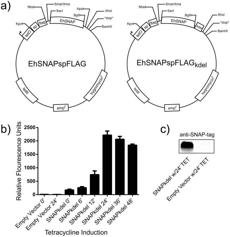
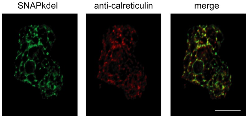
Our preliminary experiments of SNAP-tag protein expression in E. histolytica using the commercially available mammalian codon based DNA template proved unsuccessful (data not shown). Because the parasite has a highly AT rich genome, we hypothesized that altering the codons for the bias of E. histolytica might allow for increased SNAP-tag protein production. A new DNA sequence for the SNAP protein was constructed using synonymous codons reported to be overrepresented in E. histolytica genes with high expression [24]. Following codon optimization, the nucleotide composition of the SNAP-tag consisted of ∼55% AT, which more closely resembles that of the parasite genome, which is ∼75% AT (full sequence in Text S1) [15]. In order to test the expression and localization of the codon optimized SNAP-tag, we attached a KDEL localization signal specific for the endoplasmic reticulum, and used a tetracycline inducible vector for expression (Figure 1a). Tetracycline induction of SNAP protein expression, measured via flow cytometry, showed a peak in fluorescence at 24 hours that slowly tapered off (Figure 1b). The expression profile over time is in concordance with previously published data for other proteins expressed using this vector [20]. At 24 hours of tetracycline induction, SNAP protein expression was measured by quantitative infrared Western blot at approximately 76 times over background expression compared to lysate from trophozoites transfected with an empty vector control (Figure 1c). Visualization of the SNAP-tag in fixed trophozoites using a fluorescent O6-benzylguanine derivative together with antibody tagged calreticulin showed proper colocalization within the endoplasmic reticulum (Figure 2).
Figure 1. Tetracycline-regulated SNAP protein expression in E. histolytica.
(A) Vector maps of EhSNAPspFLAG with and without the KDEL endoplasmic reticulum retention signal. (B) Time course of SNAP-tag protein expression induction using tetracycline, measured by flow cytometry. Times indicated in hours post addition of tetracycline. Values are mean and standard deviation for two experiments. (C) Western blot of trophozoite lysate from transfected cells induced with tetracycline for 24 hours. SNAP protein expression was measured to be approximately 76 times greater in trophozoites expressing SNAP-kdel compared to an empty vector control.
Figure 2. Colocalization of the SNAP-kdel protein with E. histolytica calreticulin.
This ameba was fixed and labeled using a SNAP-Cell 505 Star reagent and anti-calreticulin (scale bar is 10 µm).
Live Cell Imaging
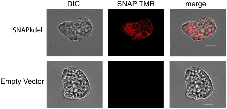
The SNAP substrate was able to effectively cross the Entamoeba cell membrane in live trophozoites, allowing for real-time visualization of protein dynamics (Figure 3, and see Movie S1). Images here are taken using the cell permeable SNAP-Cell TMR-STAR reagent in TYI-S-33 amebic growth media, which can be visualized using a rhodamine filter set. The SNAP-Cell 505-Star reagent, which can be visualized using a fluorescein filter set, proved unsuccessful for live cell imaging due to the high amount of background fluorescence in the growth media (data not shown).
Figure 3. Live cell microscopy using the SNAP-tag.
Trophozoites were labeled using a SNAP-Cell TMR Star reagent for 6 hours prior to microscopy (scale bar is 10 µm).
STORM
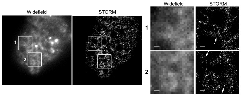
One feature of the SNAP-tag system that particularly intrigued us was the ability to use the super resolution microscopy technique, stochastic optical reconstruction microscopy (STORM). STORM allows for the visualization of cellular structures at a spatial resolution below the diffraction limit of light [25]. This is accomplished through rapid imaging as fluorophores switch between on and off states. For each image, only small subsets of fluorophore molecules are visible, and thus the positions of individual molecules do not overlap, allowing for the precise determination of their location. Processing these images together allows for accurate mapping of many individual florescent molecules. However, the method typically requires direct labeling of a primary antibody with an appropriate fluorophore, which is not always practical in the E. histolytica system given the limited availability of quality antibodies. Because of the many fluorophores available, SNAP-tag fusion proteins are well suited to STORM imaging in either live or fixed cells with little optimization required. Here we present the first super resolution light microscopy imaging of E. histolytica (Figure 4). The enhanced spatial resolution allowed us to see the three-way bifurcation typical of the endoplasmic reticulum with a localization precision of ∼50 nm. The same image taken without STORM optic settings (widefield) showed the power of this method.
Figure 4. STORM imaging of an E. histolytica trophozoite.
Super resolution microscopy of the amebic endoplasmic reticulum (ER) using a SNAP-tag protein with a KDEL ER retention signal. Inlayed boxes and arrows show the three way bifurcations that are characteristic of the ER, and the improved resolution that the STORM method provided (scale bar is 500 nm).
Discussion
Here, we have presented a new method for protein localization in the parasitic protist E. histolytica. By optimizing the codon usage in the mammalian gene encoding a SNAP protein tag, E. histolytica trophozoites can now express the SNAP-tag, and expression can be controlled using available tetracycline-regulated expression plasmids. SNAP-tag fusion proteins can be detected in fixed or living trophozoites using a variety of commercially available reagents, and as one demonstration of new possibilities for E. histolytica researchers, we used the SNAP-tag to enable STORM for super resolution light microscopy in E. histolytica for the first time. However, we have only begun to scratch the surface of the SNAP-tag system capabilities.
SNAP substrates can be transiently applied, yet become covalently bound to their tagged protein of interest. These attributes make the SNAP-tag system ideal for pulse-chase experiments, whereby the trafficking dynamics of labeled proteins can be explored in vitro or in vivo [26]. Protein-protein interactions can be quantified using SNAP technology coupled with fluorescence resonance energy transfer (FRET) [27]. SNAP labeled proteins can be locally inactivated in living cells when coupled with chromophore-assisted laser inactivation (CALI) [28]. SNAP substrates can also be targeted to specific organelles, conjugated to biotin for pull-down experiments, or even used to immobilize tagged proteins onto printed structures [29], [30].
The SNAP-tag system represents a substantial improvement in the ability to track and image proteins in E. histolytica. The large variety of molecules that can be conjugated onto BG derivatives enables the adaptation and creation of new methods to study this protozoan parasite. We hope that these methods will enable the E. histolytica research community to better understand the pathology of amebiasis.
Supporting Information
E. histolytica codon optimized SNAP protein sequence.
(TXT)
E. histolytica trophozoites expressing the SNAP protein with an endoplasmic reticulum localization signal. SNAP protein was visualized using a SNAP-Cell TMR Star reagent (New England Biolabs). Shown at 3× normal speed.
(AVI)
Acknowledgments
The authors wish to thank Kovi Bessoff, Jose Teixeira, and Peter Miller for their helpful conversations and advice.
Funding Statement
This work was funded by NIAID R01 A1072021 to Christopher Huston. STORM microscopy was funded by National Institutes of Health grant R01 AI080302 to Markus Thali. Nathan Roy was supported by T32 AI055402. The funders had no role in study design, data collection and analysis, decision to publish, or preparation of the manuscript.
References
- 1. Haque R, Huston CD, Hughes M, Houpt E, Petri WA (2003) Amebiasis. New England Journal of Medicine 348: 1565–1573. [DOI] [PubMed] [Google Scholar]
- 2. Ravdin JI, Guerrant RL (1981) Role of adherence in cytopathogenic mechanisms of Entamoeba histolytica. Study with mammalian tissue culture cells and human erythrocytes. Journal of Clinical Investigation 68: 1305–1313. [DOI] [PMC free article] [PubMed] [Google Scholar]
- 3. Petri WA, Haque R, Mann BJ (2002) The bittersweet interface of parasite and host: lectin-carbohydrate interactions during human invasion by the parasite Entamoeba histolytica . Annual Reviews of Microbiology 56: 39–64. [DOI] [PubMed] [Google Scholar]
- 4. Moncada D, Keller K, Chadee K (2003) Entamoeba histolytica cysteine proteinases disrupt the polymeric structure of colonic mucin and alter its protective function. Infection and Immunity 71: 838–844. [DOI] [PMC free article] [PubMed] [Google Scholar]
- 5. Duggal P, Guo X, Haque R, Peterson KM, Ricklefs S, et al. (2011) A mutation in the leptin receptor is associated with Entamoeba histolytica infection in children. J Clin Invest 121: 1191–1198. [DOI] [PMC free article] [PubMed] [Google Scholar]
- 6. Marie CS, Verkerke HP, Paul SN, Mackey AJ, Petri Jr WA (2012) Leptin protects host cells from Entamoeba histolytica cytotoxicity by a STAT3-dependent mechanism. Infect Immun 80: 1934–1943. [DOI] [PMC free article] [PubMed] [Google Scholar]
- 7. Leippe M, Ebel S, Schoenberger OL, Horstmann RD, Muller-Eberhard HJ (1991) Pore-forming peptide of pathogenic Entamoeba histolytica . Proceedings of the National Academy of Sciences of the United States of America 88: 7659–7663. [DOI] [PMC free article] [PubMed] [Google Scholar]
- 8. Leippe M, Andra J, Nickel R, Tannich E, Muller-Eberhard HJ (1994) Amoebapores, a family of membranolytic peptides from cytoplasmic granules of Entamoeba histolytica: isolation, primary structure, and pore formation in bacterial cytoplasmic membranes. Mol Microbiol 14: 895–904. [DOI] [PubMed] [Google Scholar]
- 9. Ankri S, Stolarsky T, Bracha R, Padilla-Vaca F, Mirelman D (1999) Antisense inhibition of expression of cysteine proteinases affects Entamoeba histolytica-induced formation of liver abscess in hamsters. Infection and Immunity 67: 421–422. [DOI] [PMC free article] [PubMed] [Google Scholar]
- 10. Que X, Reed SL (2000) Cysteine proteinases and the pathogenesis of amebiasis. Clinical Microbiology Reviews 13: 196–206. [DOI] [PMC free article] [PubMed] [Google Scholar]
- 11. Bruchhaus I, Loftus BJ, Hall N, Tannich E (2003) The intestinal protozoan parasite Entamoeba histolytica contains 20 cysteine protease genes, of which only a small subset is expressed during in vitro cultivation. Eukaryotic Cell 2: 501–509. [DOI] [PMC free article] [PubMed] [Google Scholar]
- 12. Huston CD, Houpt ER, Mann BJ, Hahn CS, Petri WA (2000) Caspase 3-dependent killing of host cells by the parasite Entamoeba histolytica . Cellular Microbiology 2: 617–625. [DOI] [PubMed] [Google Scholar]
- 13. Huston CD, Boettner DR, Miller-Sims V, Petri WA (2003) Apoptotic killing and phagocytosis of host cells by the parasite Entamoeba histolytica . Infection and Immunity 71: 964–972. [DOI] [PMC free article] [PubMed] [Google Scholar]
- 14. McCoy JJ, Mann BJ, Petri WAJ (1994) Adherence and cytotoxicity of Entamoeba histolytica or how lectins let parasites stick around. Infect Immun 62: 3045–3050. [DOI] [PMC free article] [PubMed] [Google Scholar]
- 15. Loftus B, Anderson I, Davies R, Alsmark UC, Samuelson J, et al. (2005) The genome of the protist parasite Entamoeba histolytica . Nature 433: 865–868. [DOI] [PubMed] [Google Scholar]
- 16. Heim R, Prasher DC, Tsien RY (1994) Wavelength mutations and posttranslational autoxidation of green fluorescent protein. Proc Natl Acad Sci U S A 91: 12501–12504. [DOI] [PMC free article] [PubMed] [Google Scholar]
- 17. Juillerat A, Gronemeyer T, Keppler A, Gendreizig S, Pick H, et al. (2003) Directed evolution of O6-alkylguanine-DNA alkyltransferase for efficient labeling of fusion proteins with small molecules in vivo. Chem Biol 10: 313–317. [DOI] [PubMed] [Google Scholar]
- 18. Keppler A, Pick H, Arrivoli C, Vogel H, Johnsson K (2004) Labeling of fusion proteins with synthetic fluorophores in live cells. Proc Natl Acad Sci U S A 101: 9955–9959. [DOI] [PMC free article] [PubMed] [Google Scholar]
- 19. Diamond LS, Harlow DR, Cunnick C (1978) A new medium for axenic cultivation of Entamoeba histolytica and other Entamoeba . Transactions of the Royal Society for Tropical Medicine and Hygiene 72: 431–432. [DOI] [PubMed] [Google Scholar]
- 20. Ramakrishnan G, Vines RR, Mann BJ, Petri WAJ (1997) A tetracycline-inducible gene expression system in Entamoeba histolytica . Mol Biochem Parasitol 84: 93–100. [DOI] [PubMed] [Google Scholar]
- 21. Buss SN, Hamano S, Vidrich A, Evans C, Zhang Y, et al. (2010) Members of the Entamoeba histolytica transmembrane kinase family play non-redundant roles in growth and phagocytosis. Int J Parasitol 40: 833–843. [DOI] [PMC free article] [PubMed] [Google Scholar]
- 22. Vaithilingam A, Teixeira JE, Miller PJ, Heron BT, Huston CD (2012) Entamoeba histolytica cell surface calreticulin binds human c1q and functions in amebic phagocytosis of host cells. Infect Immun 80: 2008–2018. [DOI] [PMC free article] [PubMed] [Google Scholar]
- 23. Roy NH, Chan J, Lambele M, Thali M (2013) Clustering and mobility of HIV-1 Env at viral assembly sites predict its propensity to induce cell-cell fusion. J Virol 87: 7516–7525. [DOI] [PMC free article] [PubMed] [Google Scholar]
- 24. Ghosh TC, Gupta SK, Majumdar S (2000) Studies on codon usage in Entamoeba histolytica. International Journal for Parasitology 30: 715–722. [DOI] [PubMed] [Google Scholar]
- 25. Rust MJ, Bates M, Zhuang X (2006) Sub-diffraction-limit imaging by stochastic optical reconstruction microscopy (STORM). Nat Methods 3: 793–795. [DOI] [PMC free article] [PubMed] [Google Scholar]
- 26. Bojkowska K, Santoni de Sio F, Barde I, Offner S, Verp S, et al. (2011) Measuring in vivo protein half-life. Chem Biol 18: 805–815. [DOI] [PubMed] [Google Scholar]
- 27. Maurel D, Comps-Agrar L, Brock C, Rives ML, Bourrier E, et al. (2008) Cell-surface protein-protein interaction analysis with time-resolved FRET and snap-tag technologies: application to GPCR oligomerization. Nat Methods 5: 561–567. [DOI] [PMC free article] [PubMed] [Google Scholar]
- 28. Keppler A, Ellenberg J (2009) Chromophore-assisted laser inactivation of alpha- and gamma-tubulin SNAP-tag fusion proteins inside living cells. ACS Chem Biol 4: 127–138. [DOI] [PubMed] [Google Scholar]
- 29. Srikun D, Albers AE, Nam CI, Iavarone AT, Chang CJ (2010) Organelle-targetable fluorescent probes for imaging hydrogen peroxide in living cells via SNAP-Tag protein labeling. J Am Chem Soc 132: 4455–4465. [DOI] [PMC free article] [PubMed] [Google Scholar]
- 30. Iversen L, Cherouati N, Berthing T, Stamou D, Martinez KL (2008) Templated protein assembly on micro-contact-printed surface patterns. Use of the SNAP-tag protein functionality. Langmuir 24: 6375–6381. [DOI] [PubMed] [Google Scholar]
Associated Data
This section collects any data citations, data availability statements, or supplementary materials included in this article.
Supplementary Materials
E. histolytica codon optimized SNAP protein sequence.
(TXT)
E. histolytica trophozoites expressing the SNAP protein with an endoplasmic reticulum localization signal. SNAP protein was visualized using a SNAP-Cell TMR Star reagent (New England Biolabs). Shown at 3× normal speed.
(AVI)


