



Abstract
The saliva of ticks is critical to their survival as parasites and hematophagous animals. In this study, we have purified an enzyme with trypsin-like activity from the saliva of the tick vector of Lyme Disease, Ixodes scapularis. This enzyme, named as IXOSP (Ixodes scapularis salivary serine protease), is a 29.9 kDa molecule with N-terminus FPxMVxLRIKxR. A BLAST search identified IXOSP as a secreted serine protease (AAY66740) with a conserved catalytic triad His, Asp, and Ser. In vitro studies demonstrated that IXOSP cleaves chromogenic substrates with arginine in the P1 position, by a mechanism inhibited by PMSF or aprotinin. Gene expression studies revealed that IXOSP is expressed at different tick developmental stages, including eggs, and unfed or fed adult tick salivary glands, but not in nymphs or in the midgut. While the physiological substrate for IXOSP remains to be identified, we demonstrated that I. scapularis saliva activate protein C (PC) resulting in the production of activated PC, a potent anticoagulant that also regulates a myriad of inflammatory responses through protease activated receptors. In contrast, the salivary glands of Anopheles gambiae, An. stephensi, An. albimanus, Aedes aegypti, Lutzomyia longipalpis, and Phlebotomus ariasi did not activate protein C. These discoveries are discussed in the context of blood coagulation, inflammation and vector-host interactions.
Keywords: Serine protease, saliva, Ixodes scapularis, protease activated receptors (PAR), activated protein C (APC)
1. Introduction
Ticks are considered to be second to mosquitoes as vectors of human diseases worldwide (Parola and Raoult, 2001), transmitting a variety of microbes that cause significant human disease with serious public health repercussions to rural, suburban, and even peri-urban populations especially in north-temperate regions of the globe. A better understanding of tick biology may lead to improved strategies for disrupting pathogen transmission and tick feeding on vertebrate hosts (Piesman and Eisen, 2008). Both pathogen transmission and successful blood feeding are dependent on tick saliva which is injected at the site of their attachment to a host to inhibit hemostasis, inflammation, as well as innate and adaptive immune responses (Francischetti et al., 2009). As the tick prepares to insert its hypostome through the skin of the host, contraction of the salivary ducts initiates the flow of saliva to the tip of the mouthparts. Saliva is pumped into the feeding site before any blood is consumed, and thereafter the tick goes through alternating cycles of feeding and salivating which are critical for survival (Stone et al., 1983).
Tick salivary components include amine binding proteins, and cytokine inhibitors which act as anti-hemostatic, anti-inflammatory and immunomodulators, antimicrobials, enzymes and enzyme inhibitors (Francischetti et al., 2009, Kazimirova and Stibraniova, 2013, Mans and Neitz, 2004, Maritz-Olivier et al., 2007, Montgomery et al., 2004, Oliveira et al., 2011, Oliveira et al., 2011, Pichu et al., 2011, Steen et al., 2006). Surprisingly, while insects proteolytic enzymes have been studied in detail (Ashida et al., 1990, Jiang and Kanost, 2000, Lee et al., 2007, Lopes and Terra, 2003), in ticks, there has been very few reports on the function of enzymes in saliva, despite an abundancy of genes coding for serine proteases and metalloproteases in this tissue for several species (Francischetti et al., 2009, Mulenga et al., 2001, Valenzuela et al., 2002). In fact, the only two enzymes biologically identified in I. scapularis saliva are metalloproteases, which regulate angiogenesis (Francischetti et al., 2005) and fibrinolysis (Francischetti et al., 2003). Due to the pleiotropic nature of serine proteases in activating anticoagulant, fibrinolysis, or anti-inflammatory processes, these enzymes are likely important for successful blood feeding and digestion and perhaps, pathogen transmission (McNally et al., 2012, Miyoshi et al., 2008, Ribeiro and Francischetti, 2003, Ullmann et al., 2013).
In the course of fractionating I. scapularis saliva, we sought to determine amidolytic activity of saliva. A novel proteolytic enzyme was purified as a serine protease (IXOSP) of 29.9 KDa that displays activity compatible with trypsin-like enzymes. We also tested and discovered that tick saliva activates protein C.
2. Materials and Methods
2.1 Source of ticks and blood sucking insects
I. scapularis ticks were collected from forested sites in southern Rhode Island. For some experiments, adult ticks were allowed to feed on New Zealand white rabbits under controlled conditions (Mather and Mather, 1990). A restraining collar was placed around the neck of each rabbit, and their ears were covered with cotton stockinette prior to tick exposure. For these experiments, different development stages of ticks were collected. All animal studies were approved by the University of Rhode Island Institutional Animal Care and Use Committee (protocol number AN01-12-014). Anopheles gambiae, An. stephensi, An. albimanus, Aedes aegyptii, Lutzomyia longipalpis, and Phebotomus ariasi were reared at the LMVR/NIAID/NIH.
2.2. Tick saliva collection
Adult-stage ticks weighing 200–300 μg were used for tick saliva extraction. The pilocarpine induction method was used to induce ticks to salivate (Ribeiro et al., 2004). Ticks were permitted to engorge for 4–5 days on the ear of a rabbit, after which they were removed by traction using pointed tweezers. Harvested ticks were rinsed in distilled water and immediately fixed to glass slides with double-sided tape, and a sterile glass micropipette was placed around the hypostome to collect saliva. Salivation was induced by applying 2 μl of pilocarpine (50 μg/ml) in 95% ethanol to the scutum of the tick. Additional 1-μl volumes of pilocarpine were applied at 20-min intervals when little salivation was observed. Ticks were incubated at 35°C in a humid chamber until salivation ceased (2 to 3 h). Micropipettes were removed from the ticks and amount of saliva collected was determined. Typically, volumes of 10 μl per tick were collected. The saliva was pooled and stored at −70°C.
2.3. IXOSP purification
Saliva (300 μl) was diluted with equal volume of Milli Q Water and centrifuged for 10 min at 14,000×g. The supernatant was chromatographed in a HiTrap benzamidine column (GE Healthcare, Piscataway, NJ) using fast-performance liquid chromatography (FPLC) equilibrated in 20 mM Tris-HCl, pH 8.0. The unbound protein was removed by washing buffer containing 0.05 M Tris HCl, 0.5 M NaCl, pH7.4, until absorbance at 215nm was zero. Bound proteins were eluted with 0.05 M glycine, pH 3.0 and the fractions were immediately collected and neutralized in 200 μl of 1 M Tris HCl, pH 9.0. The peak obtained was pooled, concentrated in a speed-vac, and checked for amidolytic activity (see below).
Active fractions were applied into a reverse-phase high-performance liquid chromatography (HPLC) C18 column (0.5 mm × 150 mm) (Phenomenex, Torrance, CA) equilibrated with a flow 5 or 10 μl/min using an ABI 140D pump and 785A UV detector from Applied Biosystems (Foster City, CA). Solution A contained water and 0.1% formic acid (FA), and solution B contained 0.1% FA in acetonitrile. After injecting the sample into the column, a gradient from 10% to 80% B was carried out for 40 min at a flow rate of 10 μl/min. Fractions were collected using the Gilson 203B fraction collector (Gilson, Inc., Middleton, WI) at 1-min volume intervals. Protein values for each of the fractions were measured using a Nano Drop UV/Vis Spectrometer, and amidolytic activity was determined as described below. The amidolytic peak fraction was collected, pooled, and concentrated in a speed-vac. All experiments were carried out with the purified fraction.
2.4. Mass spectrometry (MS)
Molecular mass of the protein was determined by matrix-assisted laser desorption/ionization time-of-flight (MALDI/TOF) MS analysis. The experiments were conducted on a Ciphergen ProteinChip® System (Ciphergen Biosystems, Fremont, CA) using the matrix solution of α-cyano-4-hydroxycinnamic acid (Sigma Chemical, St. Louis, MO) suspended in 100% acetonitrile.
2.5. Amino acid sequencing
N-terminal amino acid sequencing of the isolated protein was obtained by automated Edman degradation. Phenylthiohydantoin derivatives were detected using a pulse liquid automatic sequencer (Applied Biosystems, Fullerton, CA). The resulting sequences were analyzed using the expert protein analysis system (ExPASy) server of the Swiss Institute of Bioinformatics (SIB; Lausanne, Switzerland).
2.6. Sequence analysis
Sequence identity analysis of the serine protease gene was performed using the blastx, and protein by blastp program (Altschul et al., 1997) and comparing it with Genbank/protein bank databases. Analysis for putative signal peptide cleavage sites was carried out with SignalP 3.0 (Bendtsen et al., 2004) and calculations of theoretical isoelectric point was conducted with WINPEP and PROTPARAM (Hennig, 2001). ClustalX 1.83 software (Thompson et al., 1997) was used for multiple sequence alignments, and alignments were displayed with GENEDOC and refined manually.
2.7. Enzyme activity
Enzymatic activity was determined by the release of p-nitroanilide (pNA) from the different synthetic substrates (Chromogenix, Milano, Italy). Activity was measured at 405 nm by using an ELISA plate reader (Labsystem Multiskan, USA). The assays were performed in 96-well tissue culture plates in 50 μl of 100 mM Tris-HCl, pH 8.0, and 0.3 mM of different substrates. The samples were incubated at 37°C, and the reaction was stopped by the addition of 25 μl of 30% acetic acid. One enzymatic unit was defined by the release of 1 μmol of pNA per min at 37°C at pH 8.0. In some experiments, protease inhibitors were incubated with the enzymes as described below.
2.8. Characterization of proteolytic activity
All substrates and inhibitors were obtained from Sigma-Aldrich (Taufkirchen, Germany), prepared, and stored according to the manufacturer’s instructions. Influences of solvents were excluded by measuring the appropriate solvent only. Optimal pH of proteolytic activity was determined using three independent samples. Briefly, 0.5 μl purified IXOSP was incubated at 37°C for 150 min in a 400 μl reaction mixture (120 mM Britton Robinson buffer pH 4–10, 0.3 mM BAPNA, w/v). Reactions were stopped by adding 400 μl 10% trichloracetic acid (w/v). After 10 min precipitation on ice, samples were centrifuged for 10 min at 13,000×g at 4°C. Supernatant (700 μl) was mixed with 300 μl 5M NaOH, and the optical density was determined at 405 nm. Effects of inhibitors were determined as described above with 0.3 mM BAPNA as substrate. The following substrates were tested: 95 μM PMSF, 100 μM TLCK, 30 μM antipain, 10 μM aprotinin, 1.8 μM SBTI, and 25 μM E-64. Three independent samples were used in duplicate measurements. The effect of reducing agent was determined with 1 mM DTT.
2.9-Proteolytic activity determined by gel electrophoresis or zymograms
Zymogram were performed with SDS-PAGE (12.5%) prepared with 3.0 mg/ml of gelatin (DIFCO; Becton Dickinson and Company, San José, CA) to detect proteolytic activity in the gel (Hanspal et al., 1983). After electrophoresis, gels were washed twice (30 s) with 2% Triton-X-100 to remove SDS and incubated at 37°C in 100 mM Tris-HCl, pH 8.0. After incubation, gels were stained with Coomassie brilliant blue R250 in ethanol:water:acetic acid (4:4:1) and destained with a solution of ethanol:water: acetic acid (4:4:1).
2.10. Tissue expression profile analysis
Gene-specific primers were designed from the tick-specific expressed sequence tag (EST) sequence for IXOSP (GenBank accession number DQ066103) to amplify a cDNA fragment from different stages of tick tissues [20–30 eggs, fed and unfed larvae, fed and unfed nymphs, and fed and unfed adult salivary glands (SGs) and midguts] dissected from all stages of I. scapularis. Total RNA was isolated from each tick tissue using an RNAqueous® total RNA isolation kit (Ambion, Inc., Austin, TX). Concentration of total RNA was determined spectrophotometrically at 260 and 280 nm, and aliquots were stored at −80°C until use. Total RNA was reverse transcribed using Maloney murine leukemia virus (M-MLV) reverse transcriptase according to an Invitrogen kit protocol. For each set, cDNA was PCR amplified using gene-specific primers for IXOSP (Forward: 5′-ATGGGGTTCCATACACGAAA-3′; Reverse: 5′-GTGTAT CCACCCGGCATATC-3′); β-actin (Forward: 5′-AAACATCCGACATGTGTGACGACGA-3′; Reverse: 5′-TGTGGTGCCAGATCTTCTCCATGT-3′) as a control (Stratagene, La Jolla, CA) using the PCR program of 94°C for 4 min, 35 cycles of 94°C for 1 min, 49°C for 1 min, and 72°C for 1 min. All experiments were carried out in triplicate.
2.11. Tick saliva fractionation
Two hundred μl of saliva was loaded in a Sephadex G-75 10/300 column (GE Healthcare Bio-Sciences) equilibrated in PBS, pH 7.4, at 0.5 ml/min. Fractions of 0.5 ml were collected in polypropylene 0.5-ml volume 96-well plates (Costar 3956) and frozen.
2.12. Protein C activation
0.5 μl of Ixodes saliva, or 1 pair/assay of the salivary gland homogenate (SGH) from other blood-sucking arthropods, or 20 μl of fractionated saliva was added to 100 μl EBM-2-BSA 0.3% medium (used for endothelial cells culture without FBS or growth factors), supplemented with 100 nM of Protein C (Enzyme Research Laboratory). After overnight incubation, 5 μl of S2236 (250 μM, Chromogenix) was added to the wells, and substrate hydrolysis was detected at 405 nm essentially as described (Collin et al., 2012).
3. Results and Discussion
Several genes coding for serine proteases have been sequenced from Ixodes scapularis salivary gland cDNA libraries (Francischetti et al., 2009, Ribeiro et al., 2006, Valenzuela et al., 2002). Following a preliminary assay, saliva was found to hydrolyze small chromogenic substrate for trypsin-like proteases, such as BAPNA. In an attempt to identify the enzyme involved in this activity, saliva was loaded in a benzamidine-affinity column, eluted (not shown), and active fractions were combined and loaded on a reverse-phase HPLC C18 column. Two peaks (1 and 2) were obtained (Fig. 1A). Peak 2 showed maximum amidolytic activity (5.3 Units/mg) and was used for further characterization. Table 1 presents the yield for protein purification steps. Edman degradation identified the N-terminus FPxMVxLRIKxR and MALDI/TOF analysis calculated a molecular weight of 29.9 kDa (Fig. 1B). Purified IXOSP was evaluated by SDS-PAGE revealing that it migrates at a molecular weight of ~29 kDa (Figure 1C).
Fig. 1. Purification of IXOSP.
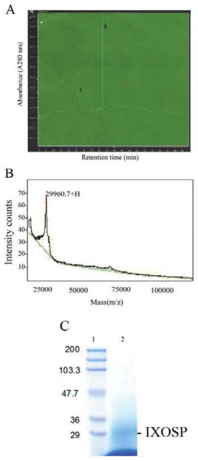
(A) Saliva was applied to benzamidine-affinity column chromatography, eluted, and tested for amidolytic activity. Active fractions were applied to a HPLC-Reverse C18 column using a gradient from 10% to 80% acetonitrile, formic acid 0.1% for 40 min at a flow rate of 10 μl/min. The eluate was monitored at 225 nm. All fractions were concentrated in a speed-vac and checked for amidolytic activity. (B) MALDI/TOF MS analysis of purified IXOSP. Spectra were analyzed to the most intense matrix peak at m/z 29960.7. (C) SDS-PAGE. Purified IXOSP was loaded in a 12.5% SDS-PAGE. Gels were Coomassie brilliant blue stained. Lanes: 1, Prestained molecular weight marker; 2, HPLC purified protein (peak 2).
Table 1.
Purification profile of IXOSP.a
| Sl. No. | Sample | Total protein (mg/ml) | Activity (BAPNA) b | Specific activity of enzyme (Units/mg) |
|---|---|---|---|---|
| 1 | Saliva | 0.97 ± 0.08 | 0.48 ± 0.07 | 0.494 |
| 2 | Protein peak I (HPLC) | 0.01 ± 0.001 | 0.02 ± 0.001 | 2.0 |
| 3 | Protein peak II (HPLC) | 0.03 ± 0.002 | 0.16 ± 0.01 | 5.3 |
Protease activity was assayed by using BAPNA as a substrate. Values are mean ± SD for three experiments. The peak fractions were assayed as described in Materials and Methods. The assay was monitored at 405 nm.
1 unit, 1 μl of pNA released per min at 37°C.
IXOSP is a serine protease
Based on the N-terminus, a BlastP search showed this protein to be a putative 29.9-kDa secreted protein (AAY66740) from I. scapularis salivary gland. Figure 2 shows the open reading frame and translation for IXOSP. The predicted protein exhibits a signal-peptide of 19 aa, followed by 31 amino acid residues which appears to be the result of proteolytic cleavage, and the N-terminus identified by Edman degradation. The mature protein is composed of 223 amino acids (mol mass 24.7 kDa) with serine protease conserved domain motifs, including the catalytic triad His, Asp, and Ser. To confirm that the proteolytic activity of IXOSP exists, zymograms were performed, where gelatinolytic activity was detected at the molecular weight expected for the purified enzyme (Figure 2B). The discrepancy between the predicted mass and the mass calculated by MS is possibly due to glycosylation or other post-translational modifications.
Fig. 2. cDNA of IXOSP.

(A) The full length clone for AY 66740 is depicted. The N-teminus is underlined. The putative activation peptide is boxed, and the N-terminus identified by Edman degradation is italicized. The catalytic triad His (H), Asp (D) and Ser (S) is circled. (B) Zymogram of purified IXOSP. Purified IXOSP was loaded in a 12.5% SDS-PAGE containing 3.0mg/ml of gelatin. Following electrophoresis, the gels were washed with 2% Triton-X-100 to remove SDS and incubated at 37°C in 100 mM Tris-HCl, pH 8.0. Gels were Commassie Blue stained. Lanes: 1, Prestained molecular weight marker; 2, HPLC purified protein (peak 2) from Figure 1A.
IXOSP exhibits serine protease activity
Next, substrate specificity of IXOSP was tested against several substrates used to detect serine protease activity. As shown in Table 2, the best-cleaved substrate was N-alpha-benzoyl-DL-arginine-p-nitroanilide (BAPNA) as well as N-p-Tosyl-Gly-Pro-Lys-p-nitroanilide, both of which are substrates for trypsin; similarly, IXOSP exhibits specificity for arginine- or lysine-containing substrates at the P1 position. The proteolytic activity of the IXOSP against BAPNA was highest at neutral to slightly basic pH (Table 3).
Table 2.
Hydrolysis of chromogenic substrates by IXOSP.a
| Substrate | Substrate specific for | Specific activity(Units/mg) b (Purified protein IXOSP) |
|---|---|---|
| Benzoyl-DL-Arg-p-nitroanilide (BAPNA) | Trypsin | 5.32 ± 0.40 |
| N-p-Tosyl-Gly-Pro-Lys-p-nitroanilide | Trypsin | 5.12 ± 0.50 |
| H-D-Val-Leu-Lys-p-nitroanilide | Plasmin | 0.140 ± 0.02 |
| Suc-Ala-Ala-Pro-Phe-Arg-p-nitroanilide | Chymotrypsin | 2.18 ± 0.20 |
| N-p-Tosyl–Gly-Phe-Arg-p-nitroanilide | Thrombin | 1.83 ± 0.11 |
IXOSP purified by the HPLC column was incubated with the synthetic substrates listed above in a concentration of 0.3 mM in Tris-HCl, 100 mM, pH 8.0, at 37°C. The numbers are mean ± standard deviation of triplicate measurements.
(Units/mg = μmol of pNA released per min per mg of protein).
Table 3.
Hydrolysis of BAPNA by IXOSP at different pH values.
| pH | Average±SD Specific activity(Units/mg)a | IXOSP % activity |
|---|---|---|
| 4 | 0.523 ± 0.02 | 10 |
| 5 | 0.784 ± 0.06 | 15 |
| 6 | 2.615 ± 0.1 | 50 |
| 7 | 4.707 ± 0.3 | 90 |
|
| ||
| 8 | 5.235 ± 0.5 | 100 |
|
| ||
| 9 | 3.664 ± 0.2 | 70 |
|
| ||
| 10 | 1.046 ± 0.1 | 20 |
0.5 μl purified IXOSP was incubated at 37°C for 150 min in a 400 μl reaction mixture. Reactions were performed as discussed in Materials and Methods. The numbers are mean ± standard deviation of triplicate measurements.
(Units/mg = μmol of pNA released per min per mg of protein).
IXOSP activity was inhibited by DTT, PMSF, aprotinin, soya bean trypsin inhibitor (SBTI), and antipain (Table 4). This inhibitory profile confirms that the enzyme is a serine protease, dependent on disulfide bonds. In contrast, no inhibition was found with cysteine protease inhibitor, E-64. Table 5 compares the amidolytic activity of trypsin and IXOSP.
Table 4.
Effects of protease inhibitors on proteolytic activity of IXOSP.a
| Sl.No | Inhibitor b | Concentration (μM) | Inhibition of activity (% ± SD) |
|---|---|---|---|
| 1 | Antipain | 30 | 55 ± 4 * |
| 2 | SBTI | 1.8 | 72 ± 10 * |
| 3 | E-64 | 25 | n.i. |
| 4 | Aprotinin | 10 | 77 ± 10 * |
| 5 | APMSF | 95 | 75 ± 7 * |
| 6 | DTT | 100 | 85 ± 2 |
Effects of different inhibitors on the proteolytic activity of IXOSP. Experiments were carried out according to Materials and Methods Mean values significantly different from the proteolytic activity in the IXOSP are indicated by * (P < 0.001).
TLCK, tosyl-L-lysyl chloromethane hydrochloride; SBTI, soya bean trypsin inhibitor; APMSF, 4-amidinobenzyl methane sulphonyl fluoride hydrochloride, DTT, Dithiothreitol.
n.i., not inhibited.
Table 5.
Comparison of hydrolysis profile.a
| Substrate | Trypsin | IXOSP |
|---|---|---|
| Ac-Ile-Glu-Gly-Arg-pNA | 1.00 | 0.82 |
| H-D-Ile-Pro-Arg-pNA | 0.24 | 1.00 |
| H-D-val-Leu-Arg-pNA | 0.12 | 0.03 |
| H-D-Val-Leu-Lys-pNA | 0.01 | <0.01 |
Data for trypsin were extracted from Chromogenix catalog and normalized as 1 toward the best substrate.
The function of IXOSP is presently unknown. It is unable to cleave angiotensin and bradykinin, which could mediate pain sensibility. Furthermore, IXOSP has no effect on fibrin clots, i.e., it does not display fibrinolytic activity (data not shown), in contrast to saliva (Francischetti et al., 2003). Given that the maximum activity of IXOSP is at pH 8.0, it also is unlikely that it is involved in a digestive role but more likely targets proteins in the blood, where the pH is about 7.4. It also may cleave other salivary proteins. In fact, some salivary molecules are stored in the glands as inactive zymogens. For example, a trypsin-like protease, triapsin, is stored as a proteolytically activatable zymogen in the lumen Triatoma sp. (Amino et al., 2001). In many insects, such activating proteases are often serine proteases, which can be activated either by themselves or by other serine proteases (Kollien et al., 2004).
Gene expression for IXOSP
To estimate the gene expression levels of IXOSP in different tissues of I. scapularis, RT-PCR experiments were performed using a specific primer for the enzyme. Figure 3A shows that IXOSP gene expression was observed in eggs in addition to unfed and fed adult tick SGs. IXOSP gene expression was not detectable in whole larvae, or nymphs, or in the midgut of adult ticks, either fed or unfed. Tick β-actin gene was used as a control (Fig. 3B).
Fig. 3. Gene expression of IXOSP.

(A) RT-PCR was carried out using gene-specific primers for IXOSP. Total RNA was extracted from tissues from different developmental stages of ticks. All experiments were carried out in triplicate. cDNA from different tick tissues were PCR amplified using gene-specific primers fro (A) IXOSP or (B) β-actin. The tissues used for RNA extraction are indicated in the figure.
Tick saliva activates protein C
While the biological activity of purified IXOSP was not identified in this study, it is known that serine proteases modulate hemostasis through conversion of protein C to activated protein C (APC), a potent anticoagulant (Esmon, 2002). So far, the only molecule that affects APC is a thrombin inhibitor (BmGTI) that enhances the amidolytic activity of the enzyme, but does not activate PC (Ricci et al., 2007). To test whether saliva activates protein C, it was incubated with PC overnight in EBM-2 media, which is used for endothelial cell culture, and therefore contains all divalent ions (e.g. Ca2+, Zn2+) necessary for enzymatic activity. APC activity was detected with S2366 hydrolysis. Figure 4A shows that 1 μl saliva per assay produced substrate hydrolysis, while the SGH from Ae aegypti, Lutzomyia longipalpis, and Phlebotomus ariasi do not. In contrast, SGH from An. stephensi, An. gambiae, and An. albimanus inhibited the basal activity of APC, also suggesting the presence of an inhibitor.
Fig. 4. I. scapularis saliva activates protein C.

(A) Saliva (1 μl) of I. scapularis or SGH (1 pair/assay) from different mosquitoes were incubated with PC (100 nM) overnight in EBM-2 media (100 μl, total volume), followed by addition of substrate specific for PC, S2366 (250 μM). Substrate hydrolysis was detected at 405 nm. (B) Saliva (0.5 μl) was incubated with PC in Hepes saline, 0.3 % BSA overnight, in the presence of CaCl2 (10 mM), ZnCl2 (100 μM) and compared to activation carried out in the presence of EBM-2 containing 0.3% BSA. In some experiments EDTA was added to chelate divalent ions. (C) Saliva (200 μl) was loaded in a G-75 chromatography column, and eluted with PBS. Twenty μl of each fraction was incubate with 100 nM PC overnight in EBM-2-BSA, and S2366 hydrolysis was estimated at 405 nm (squares). As a control, 20 μl of saliva was incubated with S2366, in the absence of PC (up triangle).
Next, the divalent ion dependency for the activity was studied. Figure 4B demonstrates that 0.5 μl saliva activated PC in Hepes-saline (HS) buffer, but this activity was enhanced by Ca2+ and particularly by Zn2+, at levels comparable to results obtained in the presence of EBM-2. EDTA completely abrogated the activity. These results indicate that the molecule involved in PC activation is metal dependent and conceivably this activity is mediated by a serine protease, or metalloproteases. Both enzymes are abundantly expressed in I. scapularis salivary gland based on sialome analyses (Francischetti et al., 2009, Ribeiro et al., 2006, Valenzuela et al., 2002). Activation of PC was dose dependent, since 2 μl saliva more than tripled the activation observed with 0.5 μl. To determine the approximate molecular weight of the PC activator, saliva was loaded in a gel-filtration column, and eluted with PBS. Figure 4C demonstrates that activity for PC activation is found in several fractions containing proteins with molecular weights approximating 30–60 kDa. Activity was not present when saliva was incubated with S2366 in the absence of PC.
Concluding remarks
The findings that a serine protease was purified directly from saliva indicate that these enzymes are expressed in the gland. While the function of this protein was not elucidated, saliva was found to activate protein C. This is a notable finding, and it is the first such report in the salivary gland of any blood sucking arthropod tested so far. Activation of the protein C pathway is a remarkable strategy because APC is a potent anticoagulant through cleavage of FVa and FVIIIa, two cofactors for prothrombinase and intrinsic Xnase complexes, which amplify and consolidate the blood coagulation cascade (Roberts, 2010). In addition, APC down-regulates several pro-inflammatory genes through PAR1 activation in different cell types (Joyce et al., 2001). Discovery that saliva activates PC in addition to several other anticoagulants already identified (Koh and Kini, 2009) indicates that I. scapularis exhibits a repertoire of mechanisms which may act in concert to down-modulate the hemostatic process, particularly that of blood coagulation.
A serine protease (IXOSP) was purified from the saliva of the tick Ixodes scapularis
Saliva from tick, but not from mosquitoes, activates Protein C
Salivary enzymes emerge as novel negative modulators of hemostasis
Acknowledgments
This work was supported by National Institute of Health Grant AI 37230 (TNM) and 1U19 AI 082642 (TNM), a subcontract to USDA Special Grant 2006-34438-17306 (TNM), and by the Intramural Research Program of the Division of Intramural Research, National Institute of Allergy and Infectious Diseases, National Institutes of Health. Experiments were supported by the URI Proteomics Core facility, made available by the RI-BRIN and RI-INBRE (NIH award P20 RR16457). We are grateful to Nathan Miller for helpful technical discussions. We also thank NIAID intramural editor Brenda Rae Marshall for assistance. Because IMBF is a government employee and this is a government work, the work is in the public domain of the United States. Notwithstanding any other agreements, the NIH reserves the right to provide the work to PubMedCentral for display and use by the public, and PubMedCentral may tag or modify the work consistent with its customary practices.
Footnotes
Competing Interests: The authors have declared no competing interests exist.
Author contributions
Conceived and designed the experiments: SP, JMCR, TNM, IMBF. Performed the experiments: SP Analyzed the data: SP, JMCR, TNM, IMBF. Contributed reagents/materials/analysis tools: SP, JMCR, TNM, IMBF. Wrote the paper: SP, JMCR, TNM, IMBF.
We certify that there is not conflict of interest for this paper
We certify that this paper was performed following the ethics principles of research adopted by the NIH and University of Rhode Island
Publisher's Disclaimer: This is a PDF file of an unedited manuscript that has been accepted for publication. As a service to our customers we are providing this early version of the manuscript. The manuscript will undergo copyediting, typesetting, and review of the resulting proof before it is published in its final citable form. Please note that during the production process errors may be discovered which could affect the content, and all legal disclaimers that apply to the journal pertain.
References
- 1.Altschul SF, Madden TL, Schaffer AA, Zhang J, Zhang Z, Miller W, Lipman DJ. Gapped BLAST and PSI-BLAST: a new generation of protein database search programs. Nucleic Acids Res. 1997;25:3389–3402. doi: 10.1093/nar/25.17.3389. [DOI] [PMC free article] [PubMed] [Google Scholar]
- 2.Amino R, Tanaka AS, Schenkman S. Triapsin, an unusual activatable serine protease from the saliva of the hematophagous vector of Chagas’ disease Triatoma infestans (Hemiptera: Reduviidae) Insect Biochem Mol Biol. 2001;31:465–472. doi: 10.1016/s0965-1748(00)00151-x. [DOI] [PubMed] [Google Scholar]
- 3.Ashida M, Kinoshita K, Brey PT. Studies on prophenoloxidase activation in the mosquito Aedes aegypti L. Eur J Biochem. 1990;188:507–515. doi: 10.1111/j.1432-1033.1990.tb15429.x. [DOI] [PubMed] [Google Scholar]
- 4.Bendtsen JD, Nielsen H, von Heijne G, Brunak S. Improved prediction of signal peptides: SignalP 3.0. J Mol Biol. 2004;340:783–795. doi: 10.1016/j.jmb.2004.05.028. [DOI] [PubMed] [Google Scholar]
- 5.Collin N, Assumpcao TC, Mizurini DM, Gilmore DC, Dutra-Oliveira A, Kotsyfakis M, Sa-Nunes A, Teixeira C, Ribeiro JM, Monteiro RQ, Valenzuela JG, Francischetti IM. Lufaxin, a Novel Factor Xa Inhibitor From the Salivary Gland of the Sand Fly Lutzomyia longipalpis Blocks Protease-Activated Receptor 2 Activation and Inhibits Inflammation and Thrombosis In Vivo. Arterioscler Thromb Vasc Biol. 2012;32:2185–2198. doi: 10.1161/ATVBAHA.112.253906. [DOI] [PMC free article] [PubMed] [Google Scholar]
- 6.Esmon CT. New mechanisms for vascular control of inflammation mediated by natural anticoagulant proteins. J Exp Med. 2002;196:561–564. doi: 10.1084/jem.20021088. [DOI] [PMC free article] [PubMed] [Google Scholar]
- 7.Francischetti IM, Mather TN, Ribeiro JM. Cloning of a salivary gland metalloprotease and characterization of gelatinase and fibrin(ogen)lytic activities in the saliva of the Lyme disease tick vector Ixodes scapularis. Biochem Biophys Res Commun. 2003;305:869–875. doi: 10.1016/s0006-291x(03)00857-x. [DOI] [PMC free article] [PubMed] [Google Scholar]
- 8.Francischetti IM, Mather TN, Ribeiro JM. Tick saliva is a potent inhibitor of endothelial cell proliferation and angiogenesis. Thromb Haemost. 2005;94:167–174. doi: 10.1267/THRO05010167. [DOI] [PMC free article] [PubMed] [Google Scholar]
- 9.Francischetti IM, Sa-Nunes A, Mans BJ, Santos IM, Ribeiro JM. The role of saliva in tick feeding. Front Biosci. 2009;14:2051–2088. doi: 10.2741/3363. [DOI] [PMC free article] [PubMed] [Google Scholar]
- 10.Hanspal JS, Bushell GR, Ghosh P. Detection of protease inhibitors using substrate-containing sodium dodecyl sulfate-polyacrylamide gel electrophoresis. Anal Biochem. 1983;132:288–293. doi: 10.1016/0003-2697(83)90010-6. [DOI] [PubMed] [Google Scholar]
- 11.Hennig L. WinPep 2.11: novel software for PC-based analyses of amino acid sequences. Prep Biochem Biotechnol. 2001;31:201–207. [PubMed] [Google Scholar]
- 12.Jiang H, Kanost MR. The clip-domain family of serine proteinases in arthropods. Insect Biochem Mol Biol. 2000;30:95–105. doi: 10.1016/s0965-1748(99)00113-7. [DOI] [PubMed] [Google Scholar]
- 13.Joyce DE, Gelbert L, Ciaccia A, DeHoff B, Grinnell BW. Gene expression profile of antithrombotic protein c defines new mechanisms modulating inflammation and apoptosis. J Biol Chem. 2001;276:11199–11203. doi: 10.1074/jbc.C100017200. [DOI] [PubMed] [Google Scholar]
- 14.Kazimirova M, Stibraniova I. Tick salivary compounds: their role in modulation of host defences and pathogen transmission. Front Cell Infect Microbiol. 2013;3:43. doi: 10.3389/fcimb.2013.00043. [DOI] [PMC free article] [PubMed] [Google Scholar]
- 15.Koh CY, Kini RM. Molecular diversity of anticoagulants from haematophagous animals. Thromb Haemost. 2009;102:437–453. doi: 10.1160/TH09-04-0221. [DOI] [PubMed] [Google Scholar]
- 16.Kollien AH, Waniek PJ, Prols F, Habedank B, Schaub GA. Cloning and characterization of a trypsin-encoding cDNA of the human body louse Pediculus humanus. Insect Mol Biol. 2004;13:9–18. doi: 10.1111/j.1365-2583.2004.00453.x. [DOI] [PubMed] [Google Scholar]
- 17.Lee N, Gannavaram S, Selvapandiyan A, Debrabant A. Characterization of metacaspases with trypsin-like activity and their putative role in programmed cell death in the protozoan parasite Leishmania. Eukaryot Cell. 2007;6:1745–1757. doi: 10.1128/EC.00123-07. [DOI] [PMC free article] [PubMed] [Google Scholar]
- 18.Lopes AR, Terra WR. Purification, properties and substrate specificity of a digestive trypsin from Periplaneta americana (Dictyoptera) adults. Insect Biochem Mol Biol. 2003;33:407–415. doi: 10.1016/s0965-1748(03)00003-1. [DOI] [PubMed] [Google Scholar]
- 19.Mans BJ, Neitz AW. Adaptation of ticks to a blood-feeding environment: evolution from a functional perspective. Insect Biochem Mol Biol. 2004;34:1–17. doi: 10.1016/j.ibmb.2003.09.002. [DOI] [PubMed] [Google Scholar]
- 20.Maritz-Olivier C, Stutzer C, Jongejan F, Neitz AW, Gaspar AR. Tick anti-hemostatics: targets for future vaccines and therapeutics. Trends Parasitol. 2007;23:397–407. doi: 10.1016/j.pt.2007.07.005. [DOI] [PubMed] [Google Scholar]
- 21.Mather TN, Mather ME. Intrinsic competence of three ixodid ticks (Acari) as vectors of the Lyme disease spirochete. J Med Entomol. 1990;27:646–650. doi: 10.1093/jmedent/27.4.646. [DOI] [PubMed] [Google Scholar]
- 22.McNally KL, Mitzel DN, Anderson JM, Ribeiro JM, Valenzuela JG, Myers TG, Godinez A, Wolfinbarger JB, Best SM, Bloom ME. Differential salivary gland transcript expression profile in Ixodes scapularis nymphs upon feeding or flavivirus infection. Ticks Tick Borne Dis. 2012;3:18–26. doi: 10.1016/j.ttbdis.2011.09.003. [DOI] [PMC free article] [PubMed] [Google Scholar]
- 23.Miyoshi T, Tsuji N, Islam MK, Alim MA, Hatta T, Huang X, Fujisaki K. A set of serine proteinase paralogs are required for blood-digestion in the ixodid tick Haemaphysalis longicornis. Parasitol Int. 2008;57:499–505. doi: 10.1016/j.parint.2008.08.003. [DOI] [PubMed] [Google Scholar]
- 24.Montgomery RR, Lusitani D, De Boisfleury Chevance A, Malawista SE. Tick saliva reduces adherence and area of human neutrophils. Infect Immun. 2004;72:2989–2994. doi: 10.1128/IAI.72.5.2989-2994.2004. [DOI] [PMC free article] [PubMed] [Google Scholar]
- 25.Mulenga A, Sugimoto C, Ingram G, Ohashi K, Misao O. Characterization of two cDNAs encoding serine proteinases from the hard tick Haemaphysalis longicornis. Insect Biochem Mol Biol. 2001;31:817–825. doi: 10.1016/s0965-1748(00)00187-9. [DOI] [PubMed] [Google Scholar]
- 26.Oliveira CJ, Sa-Nunes A, Francischetti IM, Carregaro V, Anatriello E, Silva JS, de Miranda Santos IK, Ribeiro JM, Ferreira BR. Deconstructing Tick Saliva: NON-PROTEIN MOLECULES WITH POTENT IMMUNOMODULATORY PROPERTIES. J Biol Chem. 2011;286:10960–10969. doi: 10.1074/jbc.M110.205047. [DOI] [PMC free article] [PubMed] [Google Scholar]
- 27.Oliveira CJ, Sa-Nunes A, Francischetti IM, Carregaro V, Anatriello E, Silva JS, Santos IK, Ribeiro JM, Ferreira BR. Deconstructing tick saliva: non-protein molecules with potent immunomodulatory properties. J Biol Chem. 2011;286:10960–10969. doi: 10.1074/jbc.M110.205047. [DOI] [PMC free article] [PubMed] [Google Scholar]
- 28.Parola P, Raoult D. Tick-borne bacterial diseases emerging in Europe. Clin Microbiol Infect. 2001;7:80–83. doi: 10.1046/j.1469-0691.2001.00200.x. [DOI] [PubMed] [Google Scholar]
- 29.Pichu S, Yalcin EB, Ribeiro JM, King RS, Mather TN. Molecular characterization of novel sulfotransferases from the tick, Ixodes scapularis. BMC Biochem. 2011;12:32. doi: 10.1186/1471-2091-12-32. [DOI] [PMC free article] [PubMed] [Google Scholar]
- 30.Piesman J, Eisen L. Prevention of tick-borne diseases. Annu Rev Entomol. 2008;53:323–343. doi: 10.1146/annurev.ento.53.103106.093429. [DOI] [PubMed] [Google Scholar]
- 31.Ribeiro JM, Alarcon-Chaidez F, Francischetti IM, Mans BJ, Mather TN, Valenzuela JG, Wikel SK. An annotated catalog of salivary gland transcripts from Ixodes scapularis ticks. Insect Biochem Mol Biol. 2006;36:111–129. doi: 10.1016/j.ibmb.2005.11.005. [DOI] [PubMed] [Google Scholar]
- 32.Ribeiro JM, Francischetti IM. Role of arthropod saliva in blood feeding: sialome and post-sialome perspectives. Annu Rev Entomol. 2003;48:73–88. doi: 10.1146/annurev.ento.48.060402.102812. [DOI] [PubMed] [Google Scholar]
- 33.Ribeiro JM, Zeidner NS, Ledin K, Dolan MC, Mather TN. How much pilocarpine contaminates pilocarpine-induced tick saliva? Med Vet Entomol. 2004;18:20–24. doi: 10.1111/j.0269-283x.2003.0469.x. [DOI] [PubMed] [Google Scholar]
- 34.Ricci CG, Pinto AF, Berger M, Termignoni C. A thrombin inhibitor from the gut of Boophilus microplus ticks. Exp Appl Acarol. 2007;42:291–300. doi: 10.1007/s10493-007-9097-7. [DOI] [PubMed] [Google Scholar]
- 35.Roberts HR, Monroe DM, III, Hoffman M. Williams Hematology. 8. The McGraw-Hill Co; New York, NY: 2010. Molecular biology and biochemistry of the coagulation factors and pathways of hemostasis; pp. 1815–1844. [Google Scholar]
- 36.Steen NA, Barker SC, Alewood PF. Proteins in the saliva of the Ixodida (ticks): pharmacological features and biological significance. Toxicon. 2006;47:1–20. doi: 10.1016/j.toxicon.2005.09.010. [DOI] [PubMed] [Google Scholar]
- 37.Stone BF, Commins MA, Kemp DH. Artificial feeding of the Australian paralysis tick, Ixodes holocyclus and collection of paralysing toxin. Int J Parasitol. 1983;13:447–454. doi: 10.1016/s0020-7519(83)80007-1. [DOI] [PubMed] [Google Scholar]
- 38.Thompson JD, Gibson TJ, Plewniak F, Jeanmougin F, Higgins DG. The CLUSTAL_X windows interface: flexible strategies for multiple sequence alignment aided by quality analysis tools. Nucleic Acids Res. 1997;25:4876–4882. doi: 10.1093/nar/25.24.4876. [DOI] [PMC free article] [PubMed] [Google Scholar]
- 39.Ullmann AJ, Dolan MC, Sackal CA, Fikrig E, Piesman J, Zeidner NS. Immunization with adenoviral-vectored tick salivary gland proteins (SALPs) in a murine model of Lyme borreliosis. Ticks Tick Borne Dis. 2013;4:160–163. doi: 10.1016/j.ttbdis.2012.08.006. [DOI] [PMC free article] [PubMed] [Google Scholar]
- 40.Valenzuela JG, Francischetti IM, Pham VM, Garfield MK, Mather TN, Ribeiro JM. Exploring the sialome of the tick Ixodes scapularis. J Exp Biol. 2002;205:2843–2864. doi: 10.1242/jeb.205.18.2843. [DOI] [PubMed] [Google Scholar]