

Abstract
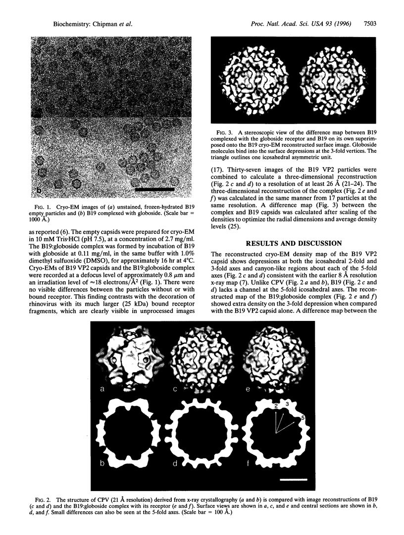
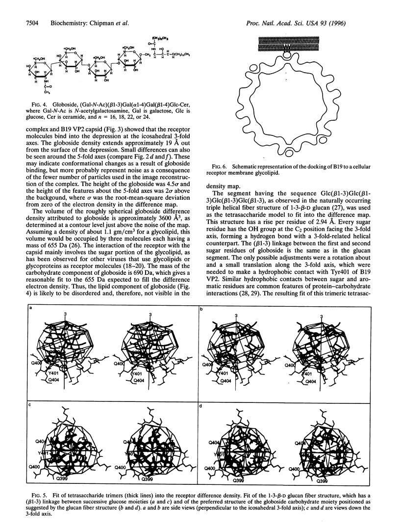
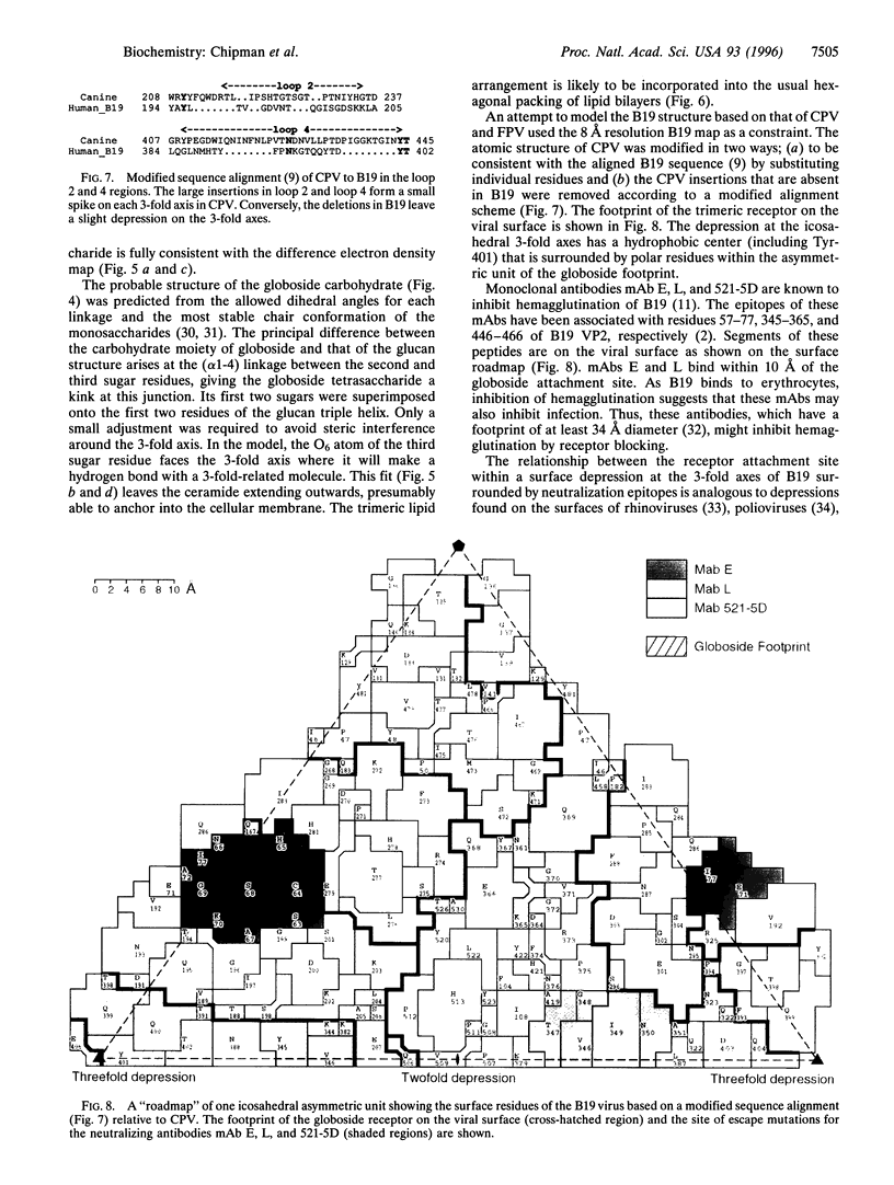
The three-dimensional structures of human parvovirus B19 VP2 capsids, alone and complexed with its cellular receptor, globoside, have been determined to 26 resolution. The B19 capsid structure, reconstructed from cryo-electron micrographs of vitrified specimens, has depressions on the icosahedral 2-fold and 3-fold axes, as well as a canyon-like region around the 5-fold axes. Similar results had previously been found in an 8 angstrom resolution map derived from x-ray diffraction data. Other parvoviral structures have a cylindrical channel along the 5-fold icosahedral axes, whereas density covers the 5-fold axes in B19. The glycolipid receptor molecules bind into the depressions on the 3-fold axes of the B19:globoside complex. A model of the tetrasaccharide component of globoside, organized as a trimeric fiber, fits well into the difference density representing the globoside receptor. Escape mutations to neutralizing antibodies map onto th capsid surface at regions immediately surrounding the globoside attachment sites. The proximity of the antigenic epitopes to the receptor site suggests that neutralization of virus infectivity is caused by preventing attachment of viruses to cells.
Full text
PDF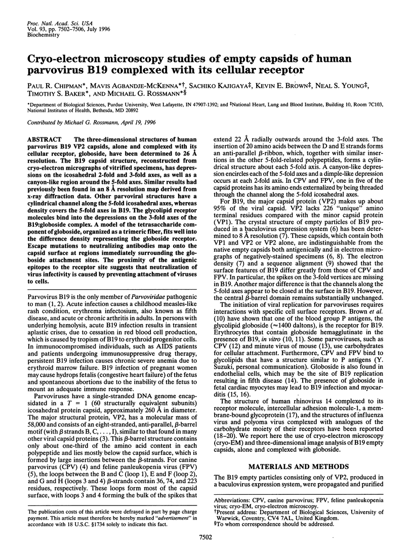
Images in this article
Selected References
These references are in PubMed. This may not be the complete list of references from this article.
- Agbandje M., Kajigaya S., McKenna R., Young N. S., Rossmann M. G. The structure of human parvovirus B19 at 8 A resolution. Virology. 1994 Aug 15;203(1):106–115. doi: 10.1006/viro.1994.1460. [DOI] [PubMed] [Google Scholar]
- Agbandje M., McKenna R., Rossmann M. G., Strassheim M. L., Parrish C. R. Structure determination of feline panleukopenia virus empty particles. Proteins. 1993 Jun;16(2):155–171. doi: 10.1002/prot.340160204. [DOI] [PubMed] [Google Scholar]
- Baker T. S., Cheng R. H. A model-based approach for determining orientations of biological macromolecules imaged by cryoelectron microscopy. J Struct Biol. 1996 Jan-Feb;116(1):120–130. doi: 10.1006/jsbi.1996.0020. [DOI] [PubMed] [Google Scholar]
- Baker T. S., Newcomb W. W., Olson N. H., Cowsert L. M., Olson C., Brown J. C. Structures of bovine and human papillomaviruses. Analysis by cryoelectron microscopy and three-dimensional image reconstruction. Biophys J. 1991 Dec;60(6):1445–1456. doi: 10.1016/S0006-3495(91)82181-6. [DOI] [PMC free article] [PubMed] [Google Scholar]
- Basak S., Turner H., Parr S. Identification of a 40- to 42-kDa attachment polypeptide for canine parvovirus in A72 cells. Virology. 1994 Nov 15;205(1):7–16. doi: 10.1006/viro.1994.1614. [DOI] [PubMed] [Google Scholar]
- Brown C. S., Van Lent J. W., Vlak J. M., Spaan W. J. Assembly of empty capsids by using baculovirus recombinants expressing human parvovirus B19 structural proteins. J Virol. 1991 May;65(5):2702–2706. doi: 10.1128/jvi.65.5.2702-2706.1991. [DOI] [PMC free article] [PubMed] [Google Scholar]
- Brown K. E., Anderson S. M., Young N. S. Erythrocyte P antigen: cellular receptor for B19 parvovirus. Science. 1993 Oct 1;262(5130):114–117. doi: 10.1126/science.8211117. [DOI] [PubMed] [Google Scholar]
- Brown K. E., Cohen B. J. Haemagglutination by parvovirus B19. J Gen Virol. 1992 Aug;73(Pt 8):2147–2149. doi: 10.1099/0022-1317-73-8-2147. [DOI] [PubMed] [Google Scholar]
- Brown K. E., Young N. S., Liu J. M. Molecular, cellular and clinical aspects of parvovirus B19 infection. Crit Rev Oncol Hematol. 1994 Feb;16(1):1–31. doi: 10.1016/1040-8428(94)90040-x. [DOI] [PubMed] [Google Scholar]
- Chapman M. S., Rossmann M. G. Structure, sequence, and function correlations among parvoviruses. Virology. 1993 Jun;194(2):491–508. doi: 10.1006/viro.1993.1288. [DOI] [PubMed] [Google Scholar]
- Conway J. F., Duda R. L., Cheng N., Hendrix R. W., Steven A. C. Proteolytic and conformational control of virus capsid maturation: the bacteriophage HK97 system. J Mol Biol. 1995 Oct 13;253(1):86–99. doi: 10.1006/jmbi.1995.0538. [DOI] [PubMed] [Google Scholar]
- Crawford L. V. A minute virus of mice. Virology. 1966 Aug;29(4):605–612. doi: 10.1016/0042-6822(66)90284-4. [DOI] [PubMed] [Google Scholar]
- Dryden K. A., Wang G., Yeager M., Nibert M. L., Coombs K. M., Furlong D. B., Fields B. N., Baker T. S. Early steps in reovirus infection are associated with dramatic changes in supramolecular structure and protein conformation: analysis of virions and subviral particles by cryoelectron microscopy and image reconstruction. J Cell Biol. 1993 Sep;122(5):1023–1041. doi: 10.1083/jcb.122.5.1023. [DOI] [PMC free article] [PubMed] [Google Scholar]
- Fuller S. D., Butcher S. J., Cheng R. H., Baker T. S. Three-dimensional reconstruction of icosahedral particles--the uncommon line. J Struct Biol. 1996 Jan-Feb;116(1):48–55. doi: 10.1006/jsbi.1996.0009. [DOI] [PubMed] [Google Scholar]
- Kajigaya S., Fujii H., Field A., Anderson S., Rosenfeld S., Anderson L. J., Shimada T., Young N. S. Self-assembled B19 parvovirus capsids, produced in a baculovirus system, are antigenically and immunogenically similar to native virions. Proc Natl Acad Sci U S A. 1991 Jun 1;88(11):4646–4650. doi: 10.1073/pnas.88.11.4646. [DOI] [PMC free article] [PubMed] [Google Scholar]
- Kreusch A., Schulz G. E. Refined structure of the porin from Rhodopseudomonas blastica. Comparison with the porin from Rhodobacter capsulatus. J Mol Biol. 1994 Nov 11;243(5):891–905. doi: 10.1006/jmbi.1994.1690. [DOI] [PubMed] [Google Scholar]
- Matthews B. W. Solvent content of protein crystals. J Mol Biol. 1968 Apr 28;33(2):491–497. doi: 10.1016/0022-2836(68)90205-2. [DOI] [PubMed] [Google Scholar]
- Morey A. L., Patou G., Myint S., Fleming K. A. In vitro culture for the detection of infectious human parvovirus B19 and B19-specific antibodies using foetal haematopoietic precursor cells. J Gen Virol. 1992 Dec;73(Pt 12):3313–3317. doi: 10.1099/0022-1317-73-12-3313. [DOI] [PubMed] [Google Scholar]
- Naides S. J., Weiner C. P. Antenatal diagnosis and palliative treatment of non-immune hydrops fetalis secondary to fetal parvovirus B19 infection. Prenat Diagn. 1989 Feb;9(2):105–114. doi: 10.1002/pd.1970090205. [DOI] [PubMed] [Google Scholar]
- Olson N. H., Kolatkar P. R., Oliveira M. A., Cheng R. H., Greve J. M., McClelland A., Baker T. S., Rossmann M. G. Structure of a human rhinovirus complexed with its receptor molecule. Proc Natl Acad Sci U S A. 1993 Jan 15;90(2):507–511. doi: 10.1073/pnas.90.2.507. [DOI] [PMC free article] [PubMed] [Google Scholar]
- Rossmann M. G., Arnold E., Erickson J. W., Frankenberger E. A., Griffith J. P., Hecht H. J., Johnson J. E., Kamer G., Luo M., Mosser A. G. Structure of a human common cold virus and functional relationship to other picornaviruses. Nature. 1985 Sep 12;317(6033):145–153. doi: 10.1038/317145a0. [DOI] [PubMed] [Google Scholar]
- Rossmann M. G., Johnson J. E. Icosahedral RNA virus structure. Annu Rev Biochem. 1989;58:533–573. doi: 10.1146/annurev.bi.58.070189.002533. [DOI] [PubMed] [Google Scholar]
- Rouger P., Gane P., Salmon C. Tissue distribution of H, Lewis and P antigens as shown by a panel of 18 monoclonal antibodies. Rev Fr Transfus Immunohematol. 1987 Dec;30(5):699–708. doi: 10.1016/s0338-4535(87)80138-1. [DOI] [PubMed] [Google Scholar]
- Sathyanarayana B. K., Rao V. S. Conformational studies of -glucans. Biopolymers. 1972;11(7):1379–1394. doi: 10.1002/bip.1972.360110707. [DOI] [PubMed] [Google Scholar]
- Sauter N. K., Glick G. D., Crowther R. L., Park S. J., Eisen M. B., Skehel J. J., Knowles J. R., Wiley D. C. Crystallographic detection of a second ligand binding site in influenza virus hemagglutinin. Proc Natl Acad Sci U S A. 1992 Jan 1;89(1):324–328. doi: 10.1073/pnas.89.1.324. [DOI] [PMC free article] [PubMed] [Google Scholar]
- Schirmer T., Keller T. A., Wang Y. F., Rosenbusch J. P. Structural basis for sugar translocation through maltoporin channels at 3.1 A resolution. Science. 1995 Jan 27;267(5197):512–514. doi: 10.1126/science.7824948. [DOI] [PubMed] [Google Scholar]
- Stehle T., Yan Y., Benjamin T. L., Harrison S. C. Structure of murine polyomavirus complexed with an oligosaccharide receptor fragment. Nature. 1994 May 12;369(6476):160–163. doi: 10.1038/369160a0. [DOI] [PubMed] [Google Scholar]
- Tsao J., Chapman M. S., Agbandje M., Keller W., Smith K., Wu H., Luo M., Smith T. J., Rossmann M. G., Compans R. W. The three-dimensional structure of canine parvovirus and its functional implications. Science. 1991 Mar 22;251(5000):1456–1464. doi: 10.1126/science.2006420. [DOI] [PubMed] [Google Scholar]
- Weis W., Brown J. H., Cusack S., Paulson J. C., Skehel J. J., Wiley D. C. Structure of the influenza virus haemagglutinin complexed with its receptor, sialic acid. Nature. 1988 Jun 2;333(6172):426–431. doi: 10.1038/333426a0. [DOI] [PubMed] [Google Scholar]
- Wikoff W. R., Wang G., Parrish C. R., Cheng R. H., Strassheim M. L., Baker T. S., Rossmann M. G. The structure of a neutralized virus: canine parvovirus complexed with neutralizing antibody fragment. Structure. 1994 Jul 15;2(7):595–607. doi: 10.1016/s0969-2126(00)00062-9. [DOI] [PMC free article] [PubMed] [Google Scholar]