Abstract
A minireplicon of plasmid pXO2 of Bacillus anthracis was isolated by molecular cloning in Escherichia coli and shown to replicate in B. anthracis, Bacillus cereus, and Bacillus subtilis. The pXO2 replicon included (i) an open reading frame encoding the putative RepS replication initiation protein and (ii) the putative origin of replication. The RepS protein was expressed as a fusion with the maltose binding protein (MBP) at its amino-terminal end and purified by affinity chromatography. Electrophoretic mobility shift assays showed that the purified MBP-RepS protein bound specifically to a 60-bp region corresponding to the putative origin of replication of pXO2 located immediately downstream of the RepS open reading frame. Competition DNA binding experiments showed that the 5′ and central regions of the putative origin were important for RepS binding. MBP-RepS also bound nonspecifically to single-stranded DNA with a lower affinity.
Bacillus anthracis is a gram-positive bacterium that is the etiological agent of anthrax (reviewed in references 18, 24, and 31). There is a high degree of similarity between B. anthracis and members of the Bacillus cereus group (B. cereus, Bacillus thuringiensis, and Bacillus mycoides), with the major differences between these organisms being the presence or absence of two large virulence plasmids, pXO1 and pXO2 (18, 19, 23, 24, 31, 33, 36, 39, 40). Plasmid pXO1 (181.6 kb) encodes the anthrax toxin proteins termed protective antigen, lethal factor, and edema factor (16, 20, 23, 24, 32, 33). Plasmid pXO2 (96.2 kb) contains the capA, capB, and capC genes required for capsule biosynthesis and the dep gene involved in the depolymerization of the capsule (14, 24, 28, 32, 34, 41). In addition, both plasmids carry regulatory genes that control expression of the toxin and capsule genes: atxA and pagR on pXO1 (3, 10, 17, 20, 25, 30, 41, 42) and acpA and acpB on pXO2 (11, 43).
Although pXO1 and pXO2 play central roles in the pathogenesis of anthrax (24, 31, 44), little is known about the mechanism(s) of replication and copy number control of these plasmids. In culture, the pXO1 plasmid is extremely stable and is rarely cured spontaneously, while the pXO2 plasmid is not as stable and much more likely to be cured (14, 24, 31). A recent report suggested that differences in pXO2 copy number in naturally occurring strains may, at least in part, be related to differences in virulence (9). pXO1 and pXO2 replication and maintenance are not limited to B. anthracis. Although self-transmission of the plasmids has not been demonstrated, pXO1 and pXO2 can be mobilized into the closely related species B. cereus and B. thuringiensis by conjugative plasmids found in the B. cereus group (2, 15, 23, 24). Interspecies transduction of pXO2 into B. cereus has also been reported (14).
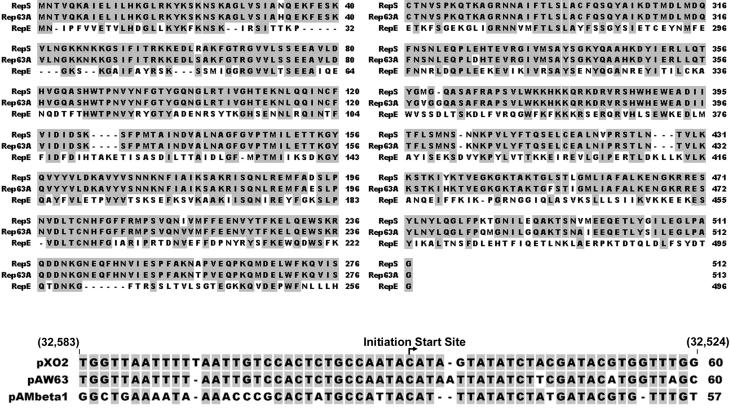
The pXO2 plasmid contains sequences that share homology with the replication regions of plasmids of the pAMβ1 family, such as pAW63, pAMβ1, pIP501, and pSM19035, which are found in gram-positive organisms, suggesting that pXO2 also belongs to this plasmid family (4, 7, 26, 34, 45). These conjugative plasmids are promiscuous and have a broad host range (7). They replicate by a theta-type mechanism, and their replication proceeds unidirectionally from the origin (6, 7). Sequence alignments have shown that the predicted replication initiator protein of pXO2 termed RepS (ORF-38; 512 amino acids; nucleotides [nt] 34115 to 32580 of pXO2, GenBank accession no. NC_002146) has 96% identity with the Rep63A protein of the B. thuringiensis plasmid pAW63 (34, 45). The RepS protein of pXO2 also has approximately 40% identity with the Rep proteins of plasmids pAMβ1 and pRE25 of Enterococcus faecalis, pIP501 and pSM19035 of Streptococcus agalactiae, and pPLI100 of Listeria innocua on the basis of BLAST alignments (1). Similarly, the putative origin of replication (ori) of pXO2 (nt 32583 to 32524) is highly homologous to the postulated ori of pAW63 (34, 45), and the ori of pAMβ1 (4-7, 26, 27).
The replication regions of the pIP501, pSM19035, and pAMβ1 have been identified by the isolation of minimal replicons. The best-studied plasmid of this group is pAMβ1. The RepE protein of pAMβ1 has been isolated and shown to bind specifically to the double-stranded (ds) DNA at the origin and nonspecifically to single-stranded (ss) DNA (27). Binding of the RepE protein to the ds origin results in the formation of an open complex. RepE stays bound to the two melted single strands of the origin region. The pAMβ1 ori and the putative ori of pAW63 are located immediately downstream of the sequence coding for RepE (6, 27, 34, 45). The mRNA of the RepE protein of pAMβ1 also plays a role in providing the RNA primer for the initiation of DNA replication. Transcription of the Rep mRNA terminates approximately 20 nt downstream of the replication start site (5). At the origin, the 3′ end of the RepE mRNA pairs with one strand of the DNA generating an R-loop structure. An RNase H-like activity in the cell or the RepE protein itself (it has been postulated to have an RNase H activity) may then cleave the RNA at the initiation site, and the RNA primer paired to the DNA serves as a primer for leading strand replication by DNA polymerase I (22, 27). After limited synthesis by DNA polymerase I, it is postulated to be replaced by the replisome that carries out coordinated leading and lagging strand synthesis (22, 27). Minimal information is available on the replication properties of pXO2 and the closely related pAW63 plasmid.
We have initiated studies to characterize the replication properties of the pXO2 plasmid. In this report, we describe the isolation and identification of a minireplicon of pXO2. Our results demonstrate that a 2,429-bp region (GenBank accession no. AF188935, pXO2 positions 32423 to 34851) containing the repS gene and the putative origin is sufficient for replication of the miniplasmid pXO2. We also report the overexpression and purification of the RepS initiator protein and demonstrate that RepS interacts specifically with the putative pXO2 origin.
MATERIALS AND METHODS
Cloning of the pXO2 minireplicon in Escherichia coli.
DNA enriched for pXO2 was isolated from B. anthracis strain 9131 containing pXO2 (13, 14). After digestion of the plasmid pXO2 DNA with NsiI, a 4,970-bp DNA fragment (GenBank accession no. AF188935, nt 31241 to 36210) was purified from a 0.7% agarose gel using Zymoclean (Zymo Research, Orange, Calif.). This fragment contains the repS and repB open reading frames (ORFs), the putative origin of replication of pXO2, and additional upstream and downstream sequences. The NsiI fragment was ligated into PstI-cleaved pBSIIKS (Stratagene, La Jolla, Calif.) and transformed into E. coli (38). Finally, the spectinomycin resistance cassette aad9 from pJRS312 (37) was inserted into the BamHI site of the vector to yield pUTE439 (9,811 bp). The sequence of the cloned pXO2 DNA was confirmed using automated DNA sequencing.
We made a subclone of pUTE439 to further reduce the size of miniplasmid pXO2. For this, a 1,463-bp MspI-HindIII fragment containing the chloramphenicol resistance gene from plasmid pC194 of Staphylococcus aureus (nt 973 to 2435; GenBank accession no. NC_002013) was ligated into pBSIIKS digested with AccI and HindIII. The recombinant plasmid pBSCm (4,424 bp) was recovered by electroporating E. coli DH5α with selection for the ampicillin resistance marker. We then amplified a 2,429-bp region of pXO2 (nt 32423 to 34851) containing the repS gene and the putative pXO2 ori. The sequences of the primers used were 5′-CCG GAT CCG TGT TGA AAT GAT TCA GAC CAG TG-3′ for the forward primer (nt 34851 to 34828) and 5′-CCG GATCCC ACA TAC CAT AAT GAG AAT ATA ACC-3′ for the reverse primer (nt 32423 to 32447). The PCR primers contained BamHI linkers at their ends to facilitate cloning. The reaction mixtures contained a 200 μM concentration of each deoxynucleoside triphosphate (dNTP), 50 ng of pUTE439 DNA, a 1 μM concentration of each primer, and 5 U of the Pfu polymerase (Stratagene). The amplification conditions were as follows: (i) 3 min at 94°C; (ii) 25 cycles, with 1 cycle consisting of 1 min at 94°C, 1 min at 60°C, and 6 min at 72°C; and (iii) 10 min at 72°C. The amplified product was gel purified and digested with BamHI. The amplified DNA was then ligated into the BamHI site of the pBSCm plasmid, and the recombinant pBSCmrepS plasmid (6,853 bp) was recovered by transforming E. coli DH5α.
Mutagenesis of the repS gene.
A frameshift mutation in the repS gene was introduced by using the Stratagene QuikChange site-directed mutagenesis kit according to the manufacturer's instructions. Two complementary primers were designed containing pXO2 nt 34011 to 34045 but lacking the A nucleotide at position 34029. This deletion was expected to destroy a BsaBI site and introduce a frameshift at codon 29 of the RepS ORF. The sequences of the primers used were 5′-CAA AAG CTG GAT TAG TTC TAT TGC TAA TCA AGA G-3′ and 5′-CTC TTG ATT AGC AAT AGA ACT AAT CCA GCT TTT G-3′. The reaction mixture (50 μl) contained 20 mM Tris-HCl (pH 8.8), 10 mM KCl, 10 mM (NH4)2SO4, 2 mM MgSO4, 0.1% Triton X-100, 100 μg of nuclease-free bovine serum albumin per ml, a 200 μM concentration of each dNTP, 75 ng of pBSCmrepS plasmid DNA, 125 ng of each primer, and 2.5 U of PfuTurbo (Stratagene). The amplification conditions were as follows: (i) 30 s at 95°C; (ii) 13 cycles, with 1 cycle consisting of 30 s at 95°C, 1 min at 55°C, and 7 min at 68°C; and (iii) 10 min at 68°C. The reaction mixture was treated with 20 U of DpnI for 1 h at 37°C to remove the parental, methylated template DNA, followed by phenol-chloroform-isoamyl alcohol extraction and ethanol precipitation. The mutagenized plasmid was recovered by transforming E. coli DH5α, and miniplasmid preparations were screened by digestion with BsaBI. The deletion of a single nucleotide resulting in a frameshift mutation in the repS gene was confirmed by automated DNA sequencing.
Cloning of the pXO2 repS gene for overexpression.
The RepS ORF of pXO2 (pXO2 positions 34115 to 32580) consists of 1,536 bp and is predicted to encode a protein of 512 amino acids with a molecular weight of 57,000. The RepS ORF was amplified from pUTE439 using PCR to encode amino acids 2 to 512 of RepS. The following primers containing BamHI linkers at their ends (shown in lowercase) were used: 5′-ccggatccaatacagtacaaaaagctatcg-3′ for the forward primer (nt 34112 to 34091 of pXO2) and 5′-ccggatccCACATACCATAATGAGAATATAACC-3′ for the reverse primer (nt 32423 to 32447 of pXO2, 154 bp downstream of the termination codon of RepS). The reaction mixtures contained a 200 μM concentration of each dNTP, 10 ng of pUTE439 plasmid DNA, a 1 μM concentration of each primer, and 5 U of the Pfu polymerase (Stratagene). The amplification conditions were as follows: (i) 3 min at 94°C; (ii) 25 cycles, with 1 cycle consisting of 1 min at 94°C, 1 min at 65°C, and 6 min at 72°C; and 10 min at 72°C. The amplified product was gel purified and digested with BamHI. The repS gene was then ligated in frame to the maltose binding protein (MBP) epitope at the BamHI site of the pMAL-p2X vector from New England Biolabs (Cambridge, Mass.). The ligation mixture was electroporated into E. coli DH5α, and the appropriate clones were isolated. The sequence of the cloned RepS ORF was confirmed by automated DNA sequencing.
Overexpression and purification of the MBP-RepS protein.
To improve the integrity and yield of the MBP-RepS protein, the MBP-RepS expression plasmid was introduced into E. coli BL21. Cells were grown in Luria-Bertani broth supplemented with 10 mM glucose at 37°C to the mid-exponential phase, and the MBP-RepS protein was overexpressed by induction with 1 mM isopropyl-β-d-thiogalactopyranoside (IPTG) at 30°C for 2 h. The cells were lysed by several freeze-thaw cycles in a buffer containing 20 mM Tris-HCl (pH 8), 0.1 mM EDTA, 1 M NaCl, 10% glycerol, and Complete protease inhibitor cocktail tablets (Roche Molecular Biochemicals, Indianapolis, Ind.) as described earlier (8). The MBP-RepS protein was purified by chromatography on an amylase affinity column, and the protein was eluted using the above buffer in the presence of 10 mM maltose (8). The purity of the protein was tested by sodium dodecyl sulfate-polyacrylamide gel electrophoresis and staining with Coomassie brilliant blue. We also purified the native MBP by similar procedures using the pMAL-p2X overexpression plasmid (not shown).
DNA binding assays.
The binding of the RepS protein to various DNA substrates was studied using electrophoretic mobility shift assays (EMSA). ds or ss oligonucleotides were labeled at the 5′ ends with 32P using T4 polynucleotide kinase (38). Approximately 1 ng of various probes was incubated with the indicated amounts of MBP-RepS in a reaction buffer consisting of 10 mM Tris-HCl (pH 7.5), 70 mM NaCl, 2.5 mM MgCl2, 50 ng of poly(dI-dC), 1 mM dithiothreitol, and 10% ethylene glycol (27). The reaction mixtures were incubated at room temperature for 15 min, and the DNA-protein complexes were resolved by electrophoresis on 6% native polyacrylamide gels. The gels were dried and subjected to autoradiography. In competition DNA binding experiments, various amounts of cold competitor oligonucleotides were also included in the above reaction mixtures.
RESULTS AND DISCUSSION
Replication of miniplasmid pXO2 in B. anthracis and other gram-positive bacteria.
Sequence alignment showed that the RepS protein of pXO2 has 96% identity with the Rep63A protein of plasmid pAW63 of B. thuringiensis (Fig. 1). BLAST alignment also showed that RepS has 39% identity and 56% similarity to the better-studied RepE protein of plasmid pAMβ1 of E. faecalis (Fig. 1). Similarly, the putative origin of pXO2 is highly homologous to the postulated ori of pAW63 and the ori of pAMβ1 (Fig. 1).
FIG. 1.
Alignment of the RepS protein of pXO2, Rep63A protein of plasmid pAW63, and RepE protein of pAMβ1 as well as the origins of replication of these three plasmids (1, 7, 27, 34, 45). The alignment was done using the ClustalW program, and the shaded letters indicate identical amino acids or nucleotides. Gaps introduced to maximize alignment are indicated by hyphens. Nucleotide coordinates of the pXO2 origin of replication are indicated in parentheses.
We generated plasmid pBSCmrepS (6,853 bp) containing a 2,429-bp region of pXO2 (nt 32423 to 34851, including the RepS ORF and the putative ori) and a 1,463-bp fragment from plasmid pC194 containing the Cmr gene (Fig. 2). The pBSCmrepS plasmid was isolated from E. coli and introduced into the plasmid-free B. anthracis strain UM23C1-1 using electroporation and selection for chloramphenicol resistance (12, 29, 35). Plasmid DNA was isolated and digested with BamHI and EcoRI. The digestion pattern of the plasmid DNA from the B. anthracis isolates was identical to that of pBSCmrepS from E. coli (Fig. 3). These results indicate that the functional replicon of pXO2 is contained within a 2,429-bp region. This region includes the RepS ORF (nt 34115 to 32580) and the putative ori of pXO2 present immediately downstream of repS (nt 32583 to 32524).
FIG. 2.
Schematic representation of the construction of plasmid pBSCmrepS containing the pXO2 minireplicon. The numbers in parentheses correspond to the nucleotide coordinates of pC194 or pXO2. The direction of transcription of the various genes is indicated by the arrows.
FIG. 3.
Restriction analysis of the pBSCmrepS plasmid isolated from E. coli, B. anthracis, B. cereus, and B. subtilis. Plasmid was digested with BamHI (B) or EcoRI (E). L lanes contain size markers (in kilobases).
The pBSCmrepS plasmid was also introduced into B. cereus and B. subtilis by electroporation (12, 21). The restriction patterns of plasmid DNA isolated from these gram-positive hosts were identical to those from B. anthracis and E. coli (Fig. 3). These results show that pXO2 miniplasmid has a broad host range, since it can be established in at least three different species, B. anthracis, B. cereus, and B. subtilis. To test whether the RepS protein was essential for pXO2 replication, we generated a frameshift mutation at codon 29 of RepS in the context of the pBSCmrepS plasmid to generate pBSCmrepSmut. In three independent experiments, no Cmr colonies were obtained when plasmid pBSCmrepSmut was electroporated into B. anthracis, B. cereus, and B. subtilis. These results showed that, as expected, RepS is essential for pXO2 replication. Previous studies with the pAW63 plasmid have suggested that in addition to the Rep63A protein (homolog of pXO2 RepS), the Rep63B protein (homolog of RepB of pXO2) may also be involved in plasmid replication (45). Our studies demonstrate that the RepB protein is dispensable for replication of the pXO2 miniplasmid.
Overexpression and purification of the MBP-RepS protein.
The MBP-RepS protein was purified by chromatography on an amylase affinity column. The purity of the protein was tested by sodium dodecyl sulfate-polyacrylamide gel electrophoresis and staining with Coomassie brilliant blue. The purified protein contained the full-length MBP-RepS protein of approximately 100 kDa as well as some breakdown products (Fig. 4). Protease inhibitors were used throughout the purification procedures, and various times and temperatures for IPTG induction were attempted. However, the breakdown products were always observed. Presumably, the MBP-RepS protein is subject to partial breakdown in vivo as well as during the purification procedures.
FIG. 4.
Purification of the RepS protein of pXO2. Lanes: U, lysates from uninduced E. coli cells; I, lysates from IPTG-induced cells overexpressing the MBP-RepS protein; P, purified MBP-RepS; M, protein molecular mass standards (in kilodaltons).
ds and ss DNA binding activity of the RepS protein.
On the basis of sequence homology to the pAW63 and pAMβ1 origins, a region immediately downstream of the RepS ORF was postulated to contain the ori of pXO2. We utilized a 60-bp ds oligonucleotide containing the putative pXO2 ori (oligonucleotide a, nt 32583 to 32524) to study the DNA binding activity of the RepS protein. In the case of the pAMβ1 plasmid, the 5′ and central regions of the ori have been shown to be critical for RepE binding (27). We, therefore, also utilized several pXO2 ori derivatives lacking the 5′, 3′, or central region of the 60-bp ori (Table 1) in competition EMSA studies. We also studied the binding of the MBP-RepS protein to ss origin DNA and to nonspecific ss DNA (Table 1).
TABLE 1.
Oligonucleotides used in the EMSA studiesa
| Oligonucleotide | Sequence |
|---|---|
| a | 5′-TGGTTAATTTTTAATTGTCCACTCTGCCAATACATAGTATATCTACGATACGTGGTTTGG-3′ |
| b | 5′-AATTGTCCACTCTGCCAATACATAGTATATCTACGATACGT-3′ |
| c | 5′-TGGTTAATTTTTAATTGTCCATCTACGATACGTGGTTTGG-3′ |
| d | 5′-ATCTACGATACGTGGTTTGGTTAGCCAGTCTTGGAATTACAGGATTCACTAGTTTAGGAT-3′ |
| e | 5′-AACAAGAAACACTATACGGCATATTGGAAGGGCTACCAGCTGGTTAATTTTTAATTGTCC-3′ |
| f | 5′-GGCTACCAGCTGGTTAATTTTTAATTGTCCATCTACGATACGTGGTTTGGTTAGCCAGTC-3′ |
| ss nonspecific oligonucleotide | 5′-GATCCAACCGGCTACTCTAATAGCCGGTTGGACGCACATACTGTGTGCATATG-3′ |
Only the top strands are shown.
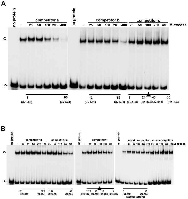
When MBP-RepS was incubated with the 60-bp ori probe (oligonucleotide a, nt 1 to 60), a single DNA-protein complex was observed in a RepS dose-dependent manner (Fig. 5A). This band presumably corresponds to the RepS-ori complex. Purified native MBP did not bind to the ori (Fig. 5A), demonstrating that the DNA binding activity of the MBP-RepS fusion was due to the RepS protein. We also tested whether RepS can bind to ss DNA. For this purpose, two probes were used: one corresponding to the bottom strand of the origin (complementary to oligonucleotide a in Table 1; specific ss probe), while the second consisted of an unrelated 53-nt ss sequence (nonspecific ss probe). EMSA results showed that RepS bound to both specific and nonspecific ss DNA in a dose-dependent manner, generating a single DNA-protein complex (Fig. 5B). These results showed that RepS has both ds and ss DNA binding activities.
FIG. 5.
Binding of the RepS protein to ds origin DNA (A) or to ss DNA (B). The indicated amounts of the MBP-RepS or MBP protein were incubated with 5′-end-labeled probes, and the DNA-protein complexes were resolved by electrophoresis on native 6% polyacrylamide gels. The probes used were as follows: a 60-bp region containing the putative ori of pXO2 (ds-ori; oligonucleotide a in Table 1); ss-ori, 60-nt bottom strand of the ori; ss-ns, a 53-nt nonspecific ss oligonucleotide. The positions of free probe (P) and RepS-DNA complex (C) are shown to the left of the gels.
Binding of RepS to ds DNA is origin specific.
We also tested the specificity of RepS binding to DNA using competition EMSA experiments. The DNA-protein complex formed in the presence of the ds ori probe was disrupted in the presence of excess cold ds ori oligonucleotide (oligonucleotide a) (Fig. 6A). A 100-fold molar excess of cold ds ori disrupted more than 50% of the binding. The central 41-bp sequence of the ori (oligonucleotide b, nt 13 to 53) also disrupted the RepS-ori complex, although at a higher molar excess (Fig. 6A). On the other hand, a 40-bp region of ori that lacks the central 20 nt (oligonucleotide c) did not significantly affect the RepS-ori complex (Fig. 6A).
FIG. 6.
Competition EMSA experiments using the 60-bp ds ori (oligonucleotide a) as the probe. 32P-labeled ds ori DNA was incubated in the presence or absence of molar (M) excesses of ds oligonucleotide competitors a, b, and c (A) or ds oligonucleotides d, e, f, ss ori, and nonspecific ss oligonucleotide (B). The “wild-type” ori consists of 60 bp (nt 1 to 60). Positions with a minus sign refer to sequences upstream of position 1 of the ori. Numbers higher than 60 indicate sequences present downstream of the ori. The positions of free probe (P) and RepS-DNA complex (C) are shown to the left of the gels.
We also used additional 60-bp oligonucleotides in competition EMSA that include 20 nt of the ori and the adjacent 40 nt on either side of the ori (Table 1). The 5′ region of ori is located immediately adjacent to the repS gene, while its 3′ region is distal to repS. The RepS-ori complex was not appreciably affected in the presence of an excess of oligonucleotide d that lacks nt 1 to 40 of the 60-bp ori, whereas oligonucleotides e and f that lack the central 20 nt of the ori but contain the 5′ 20 bp of the ori (nt 1 to 20 in Fig. 1) were more effective as competitors (Fig. 6B). Also, the binding of RepS to ori was not detectably affected in the presence of unrelated 44- and 65-bp ds oligonucleotides (not shown). We conclude from the above experiments that the central 20 nt of the ori (nt 21 to 40 [Fig. 1]) are critical for the recognition of the ori by the RepS protein. Furthermore, nt 1 to 20 at the 5′ end of the ori also contribute to RepS binding. On the other hand, the 3′ region of ori (nt 41 to 60 in Fig. 1) does not appear to be critical for RepS binding. Finally, neither the ss bottom strand of the origin DNA nor a nonspecific ss DNA disrupted RepS-ori binding (Fig. 6B). The top strand of the ori also did not affect RepS-ori binding (not shown). Taken together, the above results suggest that RepS interacts with the ds ori in a sequence-specific manner and that its affinity for ds ori DNA appears to be much stronger than its affinity for ss DNA.
The results of our studies demonstrating that miniplasmid pXO2 containing the repS gene can replicate in B. anthracis, B. cereus, and B. subtilis suggest that RepS corresponds to the replication initiator protein of pXO2. This conclusion is supported by results of our DNA binding studies demonstrating that RepS binds efficiently to the putative origin of replication of pXO2. The pAMβ1 ori is located immediately downstream of the RepE-coding sequence and shares homology with the corresponding regions of pXO2 and pAW63 (Fig. 1). Our results suggest that the 60-bp region of pXO2 (nt 32583 to 32524) located immediately downstream of the RepS ORF corresponds to the pXO2 origin. Within this region, the central 20-bp region (pXO2 positions 32563 to 32544) is critical for RepS binding, since oligonucleotides lacking this region competed poorly with the 60-bp ori in EMSA (Fig. 6). On the basis of the homology of the pXO2 and pAMβ1 ori, the RNA-DNA transition point during the initiation of pXO2 replication is expected to correspond to the conserved C residue at position 33 of ori (nt 32551 of pXO2). Our observation that nt 21 to 40 of ori are critical for RepS binding are consistent with the possibility that this region may play an important role in the generation of a RepS-dependent RNA primer for pXO2 replication. Our data are also consistent with the results obtained with the pAMβ1 plasmid in which the 5′ and central regions of the ori were found to be important for RepE binding, whereas the 3′ region of the ori did not play a significant role in RepE binding (27).
The RepS protein of pXO2 is likely to be involved in the generation of an RNA primer in a manner similar to RepE during the initiation of plasmid replication. The RepE protein of pAMβ1 has been isolated and shown to bind specifically to the ds DNA at the origin and nonspecifically to ss DNA (27). Interestingly, RepE of pAMβ1 binds to ss DNA (both ori specific and nonspecific) with a higher affinity than to specific, ds ori DNA (27). RepS protein of pXO2, on the other hand, shows a stronger interaction with the ds origin than to ss DNA (Fig. 6). Further biochemical studies should identify the mechanism of initiation of pXO2 replication and the significance of the differences in the relative ds and ss DNA binding affinities of the RepS and RepE initiator proteins.
Since the pXO2 plasmid is important for the virulence of B. anthracis, further studies are necessary for a better understanding of its replication and transfer. Such studies will provide insight regarding the potential for generation of recombinant microorganisms in nature and may reveal new molecular targets for therapeutics that affect plasmid replication and/or maintenance during infection.
Acknowledgments
We thank members of our laboratory for helpful discussions.
This work was supported in part by grants GM31685 (to S.A.K.) and AI33537 (to T.M.K.) from the National Institutes of Health. E. Tinsley was supported by NIH Training Grant 5 T32 AI49820 (Molecular Microbial Persistence and Pathogenesis).
REFERENCES
- 1.Altschul, S. F., W. Gish, W. Miller, E. W. Myers, and D. J. Lipman. 1990. Basic local alignment search tool. J. Mol. Biol. 215:403-410. [DOI] [PubMed] [Google Scholar]
- 2.Battisti, L., B. D. Green, and C. B. Thorne. 1985. Mating system for transfer of plasmids among Bacillus anthracis, Bacillus cereus, and Bacillus thuringiensis. J. Bacteriol. 162:543-550. [DOI] [PMC free article] [PubMed] [Google Scholar]
- 3.Bourgogne, A., M. Drysdale, S. G. Hilsenbeck, S. N. Peterson, and T. M. Koehler. 2003. Global effects of virulence gene regulators in a Bacillus anthracis strain with both virulence plasmids. Infect. Immun. 71:2736-2743. [DOI] [PMC free article] [PubMed] [Google Scholar]
- 4.Brantl, S., D. Benhke, and J. Alonso. 1990. Molecular analysis of the replication region of the conjugative Streptococcus agalactiae plasmid pIP501 in B. subtilis. Comparison with plasmids pAMβ1 and pSM19035. Nucleic Acids Res. 18:4783-4789. [DOI] [PMC free article] [PubMed] [Google Scholar]
- 5.Bruand, C., and S. D. Ehrlich. 1998. Transcription-driven DNA replication of plasmid pAMβ1 in Bacillus subtilis. Mol. Microbiol. 30:135-145. [DOI] [PubMed] [Google Scholar]
- 6.Bruand, C., S. D. Ehrlich, and L. Janniere. 1991. Unidirectional theta replication of the structurally stable Enterococcus faecalis plasmid pAMβ1. EMBO J. 10:2171-2177. [DOI] [PMC free article] [PubMed] [Google Scholar]
- 7.Bruand, C., E. Le Chatelier, S. D. Ehrlich, and L. Jannerie. 1993. A fourth class of theta replicating plasmids: the pAMβ1 family from Gram-positive bacteria. Proc. Natl. Acad. Sci. USA 90:11668-11672. [DOI] [PMC free article] [PubMed] [Google Scholar]
- 8.Chang, T.-L., M. G. Kramer, R. A. Ansari, and S. A. Khan. 2000. Role of individual monomers of a dimeric initiator protein in the initiation and termination of plasmid rolling circle replication. J. Biol. Chem. 275:13529-13534. [DOI] [PubMed] [Google Scholar]
- 9.Coker, P. R., K. L. Smith, P. F. Fellows, G. Rybachuck, K. G. Kousoulas, and M. E. Hugh-Jones. 2003. Bacillus anthracis virulence in guinea pigs vaccinated with anthrax vaccine adsorbed is linked to plasmid quantities and clonality. J. Clin. Microbiol. 41:1212-1218. [DOI] [PMC free article] [PubMed] [Google Scholar]
- 10.Dai, Z., J. C. Sirard, M. Mock, and T. M. Koehler. 1995. The atxA gene product activates transcription of the anthrax toxin genes and is essential for virulence. Mol. Microbiol. 16:1171-1181. [DOI] [PubMed] [Google Scholar]
- 11.Drysdale, M., A. Bourgogne, S. G. Hilsenbeck, and T. M. Koehler. 2004. atxA controls Bacillus anthracis capsule synthesis via acpA and a newly discovered regulator, acpB. J. Bacteriol. 186:306-315. [DOI] [PMC free article] [PubMed] [Google Scholar]
- 12.Dunny, G. M., L. N. Lee, and D. J. LeBlanc. 1991. Improved electroporation and cloning vector system for gram-positive bacteria. Appl. Environ. Microbiol. 57:1194-1201. [DOI] [PMC free article] [PubMed] [Google Scholar]
- 13.Etienne-Toumelin, I., J. C. Sirard, E. Duflot, M. Mock, and A. Fouet. 1995. Characterization of the Bacillus anthracis S-layer: cloning and sequencing of the structural gene. J. Bacteriol. 177:614-620. [DOI] [PMC free article] [PubMed] [Google Scholar]
- 14.Green, B. D., L. Battisti, T. M. Koehler, and C. B. Thorne. 1985. Demonstration of a capsule plasmid in Bacillus anthracis. Infect. Immun. 49:291-297. [DOI] [PMC free article] [PubMed] [Google Scholar]
- 15.Green, B. D., L. Battisti, and C. B. Thorne. 1989. Involvement of Tn4430 in transfer of Bacillus anthracis plasmids mediated by Bacillus thuringiensis plasmid pXO12. J. Bacteriol. 171:104-113. [DOI] [PMC free article] [PubMed] [Google Scholar]
- 16.Guidi-Rontani, C., Y. Periera, S. Ruffie, J.-C. Sirard, M. Weber-Levy, and M. Mock. 1999. Identification and characterization of the germination operon on the virulence plasmid pXO1 of Bacillus anthracis. Mol. Microbiol. 33:407-414. [DOI] [PubMed] [Google Scholar]
- 17.Guignot, J., M. Mock, and A. Fouet. 1997. AtxA activates the transcription of genes harbored by both Bacillus anthracis virulence plasmids. FEMS Microbiol. Lett. 147:203-207. [DOI] [PubMed] [Google Scholar]
- 18.Hanna, P. 1998. Anthrax pathogenesis and host response. Curr. Top. Microbiol. Immunol. 225:13-35. [DOI] [PubMed] [Google Scholar]
- 19.Helgason, E., O. A. Okstad, D. A. Caugant, H. A. Johansen, A. Fouet, M. Mock, I. Hegna, and A. B. Kolsto. 2000. Bacillus anthracis, Bacillus cereus, and Bacillus thuringiensis: one species on the basis of genetic evidence. Appl. Environ. Microbiol. 66:2627-2630. [DOI] [PMC free article] [PubMed] [Google Scholar]
- 20.Hoffmaster, A. R., and T. M. Koehler. 1999. Control of virulence gene expression in Bacillus anthracis. J. Appl. Microbiol. 87:279-281. [DOI] [PubMed] [Google Scholar]
- 21.Ito, M., and M. Nagane. 2001. Improvement of the electro-transformation efficiency of facultatively alkaliphilic Bacillus pseudofirmus OF4 by high osmolarity and glycine treatment. Biosci. Biotechnol. Biochem. 65:2773-2775. [DOI] [PubMed] [Google Scholar]
- 22.Janniere, L., V. Bidnenko, S. McGovern, S. D. Ehrlich, and M. A. Petit. 1997. Replication terminus for DNA polymerase I during initiation of pAMβ1 replication: role of the plasmid-encoded resolution system. Mol. Microbiol. 23:525-535. [DOI] [PubMed] [Google Scholar]
- 23.Koehler, T. M. 2000. Bacillus anthracis, p. 519-528. In V. A. Fischetti, R. P. Novick, J. J. Ferretti, D. A. Portnoy, and J. I. Rood (ed.), Gram-positive pathogens. ASM Press, Washington, D.C.
- 24.Koehler, T. M. 2002. Bacillus anthracis genetics and virulence gene regulation. Curr. Top. Microbiol. Immunol. 271:143-164. [DOI] [PubMed] [Google Scholar]
- 25.Koehler, T. M., Z. Dai, and M. Kaufman-Yarbray. 1994. Regulation of the Bacillus anthracis protective antigen gene: CO2 and a trans-acting element activate transcription from one of two promoters. J. Bacteriol. 176:586-595. [DOI] [PMC free article] [PubMed] [Google Scholar]
- 26.Le Chatelier, E., S. D. Ehrlich, and L. Janniere. 1993. Biochemical and genetic analysis of the unidirectional theta replication of the S. agalactiae plasmid pIP501. Plasmid 29:50-56. [DOI] [PubMed] [Google Scholar]
- 27.Le Chatelier, E., L. Janniere, S. D. Ehrlich, and D. Canceill. 2001. The RepE initiator is a double-stranded and single-stranded DNA binding protein that forms an atypical open complex at the onset of replication of plasmid pAMβ1 from Gram-positive bacteria. J. Biol. Chem. 276:10234-10246. [DOI] [PubMed] [Google Scholar]
- 28.Makino, S., I. Uchida, N. Terakado, C. Sasakawa, and M. Yoshikawa. 1989. Molecular characterization and protein analysis of the cap region, which is essential for encapsulation in Bacillus anthracis. J. Bacteriol. 171:722-730. [DOI] [PMC free article] [PubMed] [Google Scholar]
- 29.Marrero, R., and S. L. Welkos. 1995. The transformation frequency of plasmids into Bacillus anthracis is affected by adenine methylation. Gene 152:75-78. [DOI] [PubMed] [Google Scholar]
- 30.Mignot, T., M. Mock, and A. Fouet. 2003. A plasmid-encoded regulator couples the synthesis of toxins and surface structures in Bacillus anthracis. Mol. Microbiol. 47:917-927. [DOI] [PubMed] [Google Scholar]
- 31.Mock, M., and A. Fouet. 2001. Anthrax. Annu. Rev. Microbiol. 55:647-671. [DOI] [PubMed] [Google Scholar]
- 32.Okinaka, R., K. Cloud, O. Hampton, A. Hoffmaster, K. Hill, P. Keim, T. M. Koehler, G. Lamke, S. Kumano, D. Manter, Y. Martinez, D. Ricke, R. Svensson, and P. Jackson. 1999. Sequence, assembly and analysis of pX01 and pX02. J. Appl. Microbiol. 87:261-262. [DOI] [PubMed] [Google Scholar]
- 33.Okinaka, R. T., K. Cloud, O. Hampton, A. R. Hoffmaster, K. K. Hill, P. Keim, T. M. Koehler, G. Lamke, S. Kumano, J. Mahillon, D. Manter, Y. Martinez, D. Ricke, R. Svensson, and P. J. Jackson. 1999. Sequence and organization of pXO1, the large Bacillus anthracis plasmid harboring the anthrax toxin genes. J. Bacteriol. 18:6509-6515. [DOI] [PMC free article] [PubMed] [Google Scholar]
- 34.Pannucci, J., R. T. Okinaka, E. Williams, R. Sabin, L. O. Ticknor, and C. R. Kuske. 2002. DNA sequence conservation between the Bacillus anthracis pXO2 plasmids and closely related bacteria. BMC Genomics 3:34-41. [DOI] [PMC free article] [PubMed] [Google Scholar]
- 35.Quinn, C. P., and B. N. Dancer. 1990. Transformation of vegetative cells of Bacillus anthracis with plasmid DNA. J. Gen. Microbiol. 136:1211-1215. [DOI] [PubMed] [Google Scholar]
- 36.Read, T. D., S. L. Salzberg, M. Pop, M. Shumway, L. Umayam, L. Jiang, E. Holtzapple, J. D. Busch, K. L. Smith, J. M. Schupp, D. Solomon, P. Keim, and C. M. Fraser. 2002. Comparative genome sequencing for discovery of novel polymorphisms in Bacillus anthracis. Science 296:2028-2033. [DOI] [PubMed] [Google Scholar]
- 37.Saile, E., and T. M. Koehler. 2002. Control of anthrax toxin gene expression by the transition state regulator abrB. J. Bacteriol. 184:370-380. [DOI] [PMC free article] [PubMed] [Google Scholar]
- 38.Sambrook, J., E. F. Fritsch, and T. Maniatis. 1989. Molecular cloning: a laboratory manual, 2nd ed. Cold Spring Harbor Laboratory Press, Cold Spring Harbor, N.Y.
- 39.Thorne, C. B. 1993. Bacillus anthracis, p. 113-124. In A. L. Sonenshein, J. A. Hoch, and R. Losick (ed.), Bacillus subtilis and other gram-positive bacteria: biochemistry, physiology, and molecular genetics. American Society for Microbiology, Washington, D.C.
- 40.Uchida, I., K. Hashimoto, and N. Terakado. 1986. Virulence and immunogenicity in experimental animals of Bacillus anthracis strains harboring or lacking 110 MDa and 60 MDa plasmids. J. Gen. Microbiol. 132:557-559. [DOI] [PubMed] [Google Scholar]
- 41.Uchida, I., S. Makino, C. Sasakawa, M. Yoshikawa, C. Sugimoto, and N. Terakado. 1993. Identification of a novel gene, dep, associated with depolymerization of the capsular polymer in Bacillus anthracis. Mol. Microbiol. 9:487-496. [DOI] [PubMed] [Google Scholar]
- 42.Uchida, I., S. Makino, T. Sekizaki, and N. Terakado. 1997. Cross-talk to the genes for Bacillus anthracis capsule synthesis by atxA, the gene encoding the trans-activator of anthrax toxin synthesis. Mol. Microbiol. 23:1229-1240. [DOI] [PubMed] [Google Scholar]
- 43.Vietri, N. J., R. Marrero, T. A. Hoover, and S. L. Welkos. 1995. Identification and characterization of a trans-activator involved in the regulation of encapsulation by Bacillus anthracis. Gene 152:1-9. [DOI] [PubMed] [Google Scholar]
- 44.Welkos, S. L., N. J. Vietri, and P. H. Gibbs. 1993. Non-toxigenic derivatives of the Ames strain of Bacillus anthracis are fully virulent for mice: role of plasmid pXO2 and chromosome in strain-dependent virulence. Microb. Pathog. 14:381-388. [DOI] [PubMed] [Google Scholar]
- 45.Wilcks, A., L. Smidt, O. A. Okstad, A.-B. Kolsto, J. Mahillon, and L. Andrup. 1999. Replication mechanism and sequence analysis of the replicon of pAW63, a conjugative plasmid from Bacillus thuringiensis. J. Bacteriol. 181:3193-3200. [DOI] [PMC free article] [PubMed] [Google Scholar]


