Abstract
Viral RNA is sensed by TLR 7 and 8 or by the RNA helicases LGP2, MDA5 and RIG-I to trigger antiviral responses. Much less is known about sensors for DNA. Here we identify a novel DNA sensing pathway involving RNA polymerase III and RIG-I. AT-rich dsDNA serve as a template for RNA polymerase III, which is transcribed into dsRNA harboring a 5′ triphosphate moiety which signals via RIG-I to activate type I IFN gene transcription and NF-κB. This pathway is also important in sensing Epstein-Barr virus encoded small RNAs, which are transcribed by RNA polymerase III and then trigger RIG-I activation. Thus, RNA Pol III and RIG-I play a pivotal role in coordinating anti-viral defenses in the innate immune response.
Introduction
To survive infection, the immune system must trigger an arsenal of defense measures to combat invading microbes. The innate immune system is the first line of defense1, 2. Innate immunity functions to control infection directly and relay signals to the adaptive immune system. Several classes of germline-encoded pattern recognition receptors (PRRs) have now been implicated in innate defenses. These include the Toll-like receptors (TLRs)3, the C-type lectin receptors (CLRs)4, the RIG-like helicases (RLRs)5, members of the NOD-like receptor (NLR) family6, 7 and cytosolic DNA sensors8–10. Individual PRRs recognize microbial products (also called pathogen associated molecular patterns (PAMPs)) from bacteria, viruses, fungi and parasites and trigger signaling pathways, which regulate immune response genes. These include pro-inflammatory cytokines such as tumor necrosis factor, interleukin-1β and type I interferons2, 7. Accumulating evidence reveals that in addition to sensing microbial products, many of these same sensors detect danger signals (or danger-associated molecular patterns, DAMPs) that are released from damaged or dying cells [reviewed in 11].
A common theme of PAMP recognition is the sensing of non-self nucleic acids. Viruses for example are almost exclusively sensed via their nucleic acid genomes or as a result of their replicative or transcriptional activity12. In the cytosol, RIG-I and MDA5, discriminate between different classes of RNA viruses13, 14. RIG-I senses the nascent 5′ triphosphate moiety of viral genomes or virus derived transcripts of negative-sense ssRNA viruses, whereas MDA5 is activated by long dsRNA, a typical intermediate of the replication of plus-sense ssRNA viruses15, 16. RIG-I also detects short blunt end dsRNAs17. Both RIG-I and MDA5 engage the mitochondrial adapter protein IPS-1 (also known as MAVS, Cardif or VISA). IPS-1 subsequently triggers downstream signaling and activation of the IKKα/β-NFκB pathway or the TBK1-IRF3 pathway and transcriptional regulation of inflammatory cytokine and type I IFN genes, respectively.
DNA is also a potent trigger of innate immunity. In plasmacytoid dendritic cells, CpG DNA engages TLR9 to turn on IFN-α gene transcription. A second DNA sensing pathway elicits activation of the TBK1-IRF3 signaling pathway and transcription of IFNα/β genes, although the underlying mechanisms responsible for these latter events are unclear. Recently, a candidate sensor, DAI (previously called DLM-1 or ZBP1)9 was shown to bind synthetic dsDNA and engage TBK1 and IRF3 to regulate IFN gene transcription. Knockdown experiments indicated that DAI was involved in sensing cytosolic DNA in some cell lines9. However, DAI-deficient embryonic fibroblasts and macrophages responded normally to cytosolic DNA18, and DAI-deficient mice mounted normal adaptive immune responses, indicating possible redundancy with additional sensors. A second DNA sensor, AIM2 has also been identified recently. Absent in melanoma (AIM2) binds dsDNA and ASC to form a caspase-1 activating inflammasome. AIM2 does not regulate type I IFN gene transcription however10, 19–21.
It is likely that in most cell types DNA viruses trigger type I IFN gene transcription via TLR-independent DNA sensing mechanisms. Although type I IFNs are best studied in anti-viral immunity, a role for these cytokines in bacterial, fungal and parasitic infections has also emerged. Francisella tularensis22, Streptococcus agalactiae23, Listeria monocytogenes24, 25, Mycobacterium tuberculosis26, Legionella pneumophila27, Brucella abortus28 and Trypanasomai cruzi29 all trigger type I IFNs by what appears to be a TBK1 and IRF3-dependent pathway. Neither the host sensor nor the microbial ligands that trigger these responses have been clearly identified, although DNA has been suggested as the likely ligand. In addition to microbial triggers of this pathway, endogenous DNAs arising during autoimmunity are also likely to be sensed by this pathway. For example, macrophages from DNase II-deficient mice which fail to digest DNA from engulfed apoptotic cells trigger robust type I IFN and inflammatory cytokine production via IRF3 activation, leading to lethal anemia and chronic arthritis30. Defining the molecular mechanisms responsible for sensing of DNA and type I IFN and cytokine induction therefore holds significant therapeutic promise for infectious as well as autoimmune diseases.
Here we identify a novel DNA sensing pathway involving RIG-I. AT rich dsDNA serves as a template for RNA polymerase III (RNA Pol III), which normally functions to transcribe 5S rRNA, tRNAs and other small RNAs via specific promoter regions. AT rich dsDNA is transcribed by RNA Pol III into dsRNA harboring a 5′ triphosphate moiety, which converts AT rich DNA into a RIG-I ligand. Moreover, we show that Epstein-Barr virus encoded EBER RNAs are also transcribed by RNA Pol III, which then trigger activation of RIG-I and type I IFN gene transcription. The RNA Pol III-RIG-I pathway appears to be functional in both human and mouse cells, but in mouse cells seems to be redundant with additional DNA sensing mechanisms.
Results
RIG-I is required for poly(dA-dT)-mediated type IFN induction in human, but not in murine immune cells
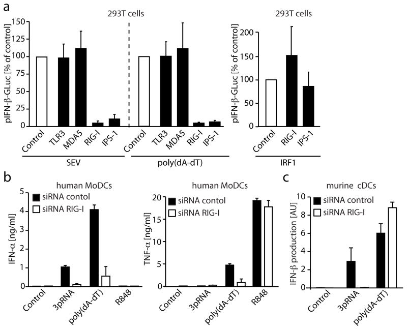
To study the role of known RNA sensors in the detection of the negative-strand ssRNA paramyxovirus virus, Sendai virus (SEV), we transfected human 293T cells with siRNAs targeting TLR3, RIG-I, MDA5 and the downstream adapter IPS-1. The double-stranded DNA mimetic poly(dA-dT) was used as a control stimulus, since it was previously shown to signal independently of these sensors in the murine system31, 32. As expected, SEV-mediated type I IFN induction was completely abolished in 293T cells that had been treated with siRNAs targeting RIG-I and IPS-1. Surprisingly, poly(dA-dT)-triggered type I IFN production was also abolished in 293T cells that had been silenced for RIG-I or IPS-1 (Fig. 1a). Type I IFN induction triggered by overexpression of IRF1 was unaffected by knockdown of RIG-I or IPS-1 (Fig. 1a). Using dominant negative expression constructs for RIG-I or targeting of IPS-1 using the Hepatitis C Virus-derived protease NS3/4A in 293T cells also completely abolished poly(dA-dT)-triggered type I IFN induction (Supplementary Fig. 1a). Similar findings were reported by Chisari and colleagues33. In addition to inducing type I IFN gene transcription, poly(dA-dT) is also a potent inducer of NF-κB dependent gene transcription. Activation of NF-κB by poly(dA-dT) was also impaired in 293T cells in which expression of RIG-I was silenced by siRNA (Supplementary Fig. 1b).
Figure 1. Poly(dA-dT) triggers type I IFN induction via RIG-I in human cells.
a, 293T cells were reverse transfected with three individual siRNAs targeting the indicated genes. 48h after transfection cells were transfected with an pIFN-β reporter plasmid in conjunction with poly(dA-dT) or SEV. After an additional period of 24h transactivation of the IFN-β promoter was measured. Three individual siRNAs were used per gene and each was tested in triplicate. Data are represented as mean values normalized to the set of control siRNAs. b, MoDCs were electroporated with siRNA directed against RIG-I or control siRNA. 48h after electroporation, cells were stimulated with the indicated stimuli and IFN-α or TNF-α production was assessed after 24h by ELISA. c, Murine bone marrow-derived dendritic cells (conventional dendritic cells, cDCs) were electroporated with siRNA targeting RIG-I or a control siRNA. 48h after electroporation cells were stimulated and IFN-β induction was assessed 5h after stimulation by real-time PCR. Mean values ± SEM of one representative experiment out of two (a) or three (b, c) experiments are depicted.
Targeting RIG-I in primary human monocyte-derived dendritic cells (MoDCs) also resulted in a markedly reduced type I IFN and TNF-α response following poly(dA-dT) stimulation (Fig. 1b). TNF-α production in response to the TLR7/8 ligand R848 was unaffected, however. In agreement with previous reports, murine dendritic cells or macrophages genetically deficient for IPS-1 (data not shown) or electroporated with siRNA targeting RIG-I were still responsive to poly(dA-dT) in terms of type I IFN production (Fig. 1c). Altogether these results indicate that RIG-I and IPS-1 were critical for poly(dA-dT)-triggered type IFN induction in human cells, but were dispensable for these responses in murine immune cells.
Poly(dA-dT) triggers RIG-I activation by inducing a stimulatory RNA intermediate
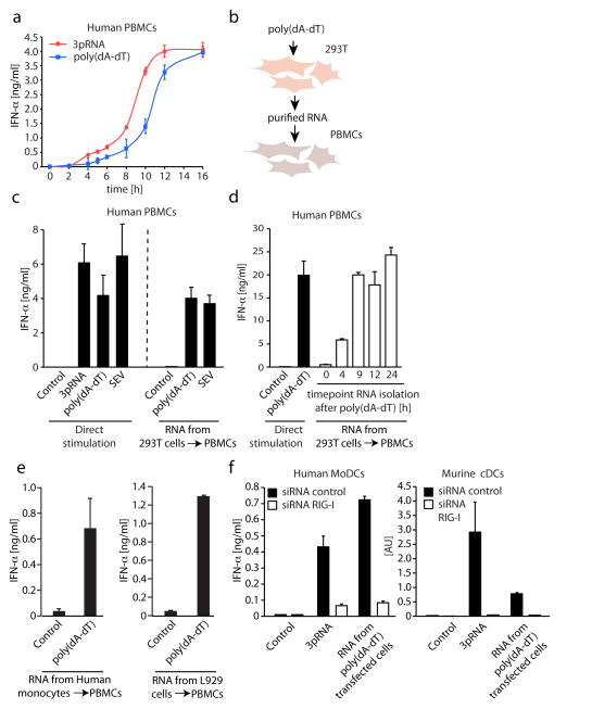
We next sought to determine the underlying mechanisms that could explain how RIG-I, an RNA sensor, could respond to dsDNA. We first compared the kinetics of the IFN response following treatment of human PBMCs with the bona fide RIG-I stimulus 5′ triphosphate RNA (3pRNA) and transfected poly(dA-dT). PBMCs responded to poly(dA-dT) with slower kinetics than that seen with 5′-triphosphate RNA (~ 2 hours slower) (Fig. 2a). These results could indicate that poly(dA-dT) activates RIG-I indirectly possibly by triggering formation of an endogenous, secondary RNA ligand that, would subsequently activate RIG-I. To address this possibility we isolated RNA from poly(dA-dT)-transfected 293T cells (Fig. 2b) and tested this RNA fraction for its ability to stimulate human PBMCs. PBMCs were pretreated with chloroquine to inhibit TLR-dependent type I IFN induction (Supplementary Fig. 2a) and then transfected with RNA isolated from poly(dA-dT) treated or control cells. Indeed, RNA derived from poly(dA-dT)-transfected 293T cells, but not that from untransfected cells induced type I IFN production in PBMCs (Fig. 2c). The magnitude of the IFN response was similar to that observed with SEV infection (Fig. 2c). This response was unaffected by DNase I treatment indicating the response was DNA-independent (Supplementary Fig. 2b). The induction of a stimulatory RNA species by poly(dA-dT) occurred within 4h following transfection and peaked 9h after poly(dA-dT) delivery (Fig. 2d). The ability of poly(dA-dT) to generate a stimulatory RNA ligand was not restricted to 293T cells, as RNA derived from primary cells stimulated with poly(dA-dT), e.g., human monocytes, induced type I IFN induction when transfected into human PBMCs (Fig. 2e). Interestingly, RNA-derived from murine L929 (Fig. 2e) cells or mouse embryonic fibroblasts transfected with poly(dA-dT) also potently induced type I IFN production, indicating that this mechanism was conserved across these two species. As observed in the human system for poly(dA-dT) itself, the signaling pathway induced by the poly(dA-dT)-induced RNA species also required RIG-I in murine as well as in human cells. Knockdown of RIG-I in human MoDCs or murine dendritic cells greatly reduced the type I IFN response triggered by RNA derived from poly(dA-dT) transfected cells (Fig. 2f). Similar findings were made when dominant negative expression constructs of RIG-I or the HCV protease NS3/4A were used in 293T cells (Supplementary Fig. 2c). Altogether these results indicate that poly(dA-dT) transfection led to the generation of RNA species in both human and murine cells, which activated RIG-I. This pathway seems to be solely responsible for poly(dA-dT) triggered type I IFN induction in human cells, however, in mouse immune cells at least one additional poly(dA-dT) sensing mechanism contributes to type I IFN production.
Figure 2. Poly(dA-dT) triggers the formation of an endogenous RIG-I stimulatory RNA.
a, Human PBMCs were transfected with 3pRNA or poly(dA-dT) and IFN-α production was assessed at the time points indicated. b and c, RNA isolated from 293T cells stimulated with SEV or poly(dA-dT) was transfected into chloroquine-treated PBMCs. In addition, PBMCs were stimulated directly with 3pRNA, poly(dA-dT) or SEV. 24h after stimulation IFN-α production was assessed. d, RNA was isolated from 293T cells 0, 4, 9, 12 and 24h after poly(dA-dT) transfection. The obtained RNA was then used to stimulate chloroquine-blocked PBMCs, whereas directly transfected poly(dA-dT) served as a control. e, RNA from poly(dA-dT) transfected monocytes and L929 cells was purified and used to stimulate PBMCs as described above. f, Human MoDCs or murine cDCs were electroporated with siRNA targeting RIG-I or a control siRNA and after 48h cells were stimulated as indicated. IFN-α production or IFN-β induction was assessed 24h or 5h later as described. Mean values ± SEM of one representative experiment out of two (a, d, e), three (f) or four (c) experiments are depicted.
Double stranded DNA molecules of random sequence trigger a distinct type I IFN induction pathway than that of poly(dA-dT)
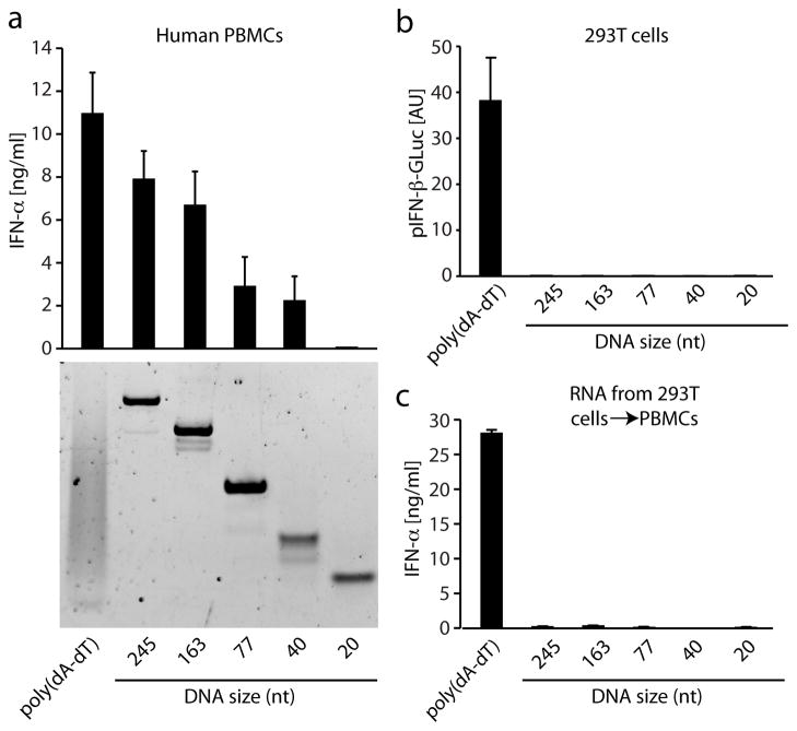
Having established that poly(dA-dT) transfection led to the generation of an endogenous RNA ligand for the RIG-I pathway, we next examined whether double stranded DNA from other sources also triggered the RIG-I pathway by generating an RNA intermediate. We generated dsDNAs of various lengths and with random sequences using PCR and tested the ability of these dsDNA molecules to trigger type I IFN production in 293T cells and in primary human PBMCs. Double stranded DNAs of the size range of the poly(dA-dT) that was used for our studies (~ 20–250 nt in size) were selected (Fig. 3a). Notably, while poly(dA-dT) triggered type I IFN responses in both PBMCs and 293T cells, PCR-generated dsDNAs failed to trigger this response in 293T cells (Fig. 3a, b and Supplementary Fig. 3). In contrast, a size dependent IFN response to dsDNA was observed in PBMCs. Contrary to what we observed for poly(dA-dT), the IFN response to PCR-generated dsDNAs was not abolished in RIG-I silenced human MoDCs (data not shown). In agreement with these findings, RNA derived from 293T cells transfected with poly(dA-dT) induced an IFN response, while RNA generated from PCR-generated dsDNAs failed to do so (Fig. 3c). Collectively, these results indicate that in human cells at least two independent receptor systems/pathways exist to sense dsDNA; an indirect RIG-I pathway for poly(dA-dT) and a second pathway for random long dsDNAs. Both of these pathways are independent of TLR9.
Figure 3. Non poly(dA-dT) double stranded DNA triggers type I IFN induction via an RNA independent pathway.
a and b, double stranded DNAs of different length derived from pcDNA3 by PCR were transfected into chloroquine-blocked PBMCs (200ng/transfection in 96 well) or 293T cells and type I IFN induction was measured 24h after stimulation by ELISA or via reporter gene activity. c, RNA from 293T cells that had been transfected with the above dsDNAs was isolated and transfected into chloroquine-blocked PBMCs. IFN-α production was measured 24h after transfection. Mean values ± SEM of one representative experiment out of four (a, c) or three (b) experiments are depicted.
Poly(dA-dT)-triggered RNA is generated independently of the oligoadenylate synthetase (OAS)/RNAse L pathway
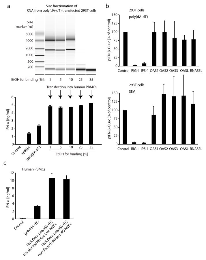
To further characterize the poly(dA-dT)-dependent RNA species, we isolated and fractionated RNA from 293T cells that had been transfected with poly(dA-dT). By using different ethanol concentrations during the binding reaction of RNA to a silica column, we were able to crudely fractionate RNA into different sizes. We then tested the ability of these RNA fractions to trigger IFN and found that the small RNA fraction which was less than 200 nt was sufficient to induce type I IFN (Fig. 4a). It has recently been shown that the antiviral endoribonuclease, RNase L, which is activated by 2′,5′-linked oligoadenylate produces small RNA cleavage products from self-RNA that initiate IFN production via the RIG-I/IPS-1 pathway. In this study, it was shown that small RNAs < 200 nt isolated from RNAse L activated cells contained the stimulatory RNA species. We therefore examined the role of the OAS/RNaseL system in the IFN response to poly(dA-dT). Transient overexpression of OAS1, 2 and 3 or RNase L did not alter poly(dA-dT) induced type I IFN induction (data not shown). Furthermore, overexpression of dominant negative RNAse L mutants (data not shown) or targeting RNAse L, OAS 1, 2, 3 or OASL using siRNA (Fig. 4b) did not inhibit the poly(dA-dT) triggered type I IFN response. Finally, when we isolated RNA from poly(dA-dT) transfected wildtype or RNAse L deficient cells, type I IFN was induced normally (Fig. 4c). Altogether these data indicate that poly(dA-dT) transfection induces the formation of stimulatory small RNAs in an OAS/RNAse L-independent manner.
Figure 4. Poly(dA-dT) triggers the formation of a small stimulatory RNA species independent of the OAS/RNase L pathway.
a, RNA was isolated from poly(dA-dT) transfected 293T cells and fractionated using different concentrations of ethanol during the initial binding step of the RNA in a silica matrix based spin column purification system. The obtained RNA was analyzed on an agilent 6000 pico chip and tested for IFN-α production in chloroquine-treated PBMCs. Direct stimulation with 3pRNA or poly(dA-dT) served as a control. b, 293T cells were reverse transfected with three individual siRNAs targeting the indicated genes. 48h after transfection cells were transfected with a pIFN-β reporter plasmid in conjunction with poly(dA-dT) or SEV. After an additional period of 24h transactivation of the IFN-β promoter was measured. Three individual siRNAs were used per gene and were each tested in triplicate. Data are represented as mean values normalized to the set of control siRNAs. c, RNA was isolated from murine embryonic fibroblasts transfected with poly(dA-dT) and tested in chloroquine-treated PBMCs for IFN-α production. Direct transfection of poly(dA-dT) served as a control. Mean values ± SEM of one representative experiment out of four (a) or two (b, c) experiments are depicted.
Poly(dA-dT)-triggered RNA is a double stranded, 5′-triphosphate RNA that is resistant to RNAse T1 under denaturing conditions
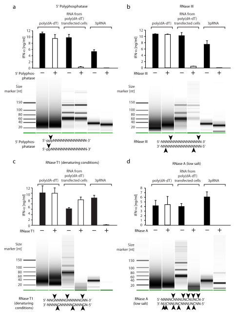
To define the features of the poly(dA-dT)-triggered RNA species important for the IFN response, we took advantage of several RNA modifying enzymes. To determine if phosphate groups were important features of the RNA, we treated poly(dA-dT), poly(dA-dT)-triggered RNA and in vitro transcribed 5′ triphosphate RNA (a 30mer) with alkaline phosphatase (AP) to remove any 5′ or 3′ phosphates that might be present. AP-treatment of 5′ triphosphate RNA and poly(dA-dT)-triggered RNA strongly reduced their stimulatory activity, whereas poly(dA-dT) was unaffected by AP treatment (Supplementary Fig. 4). In addition, employing 5′ polyphosphatase, an enzyme that specifically removes the phosphate group in the γ and β position of 5′ triphosphate or 5′ diphosphate RNA, completely abolished type I IFN induction by both in vitro transcribed 5′ triphosphate RNA and poly(dA-dT)-triggered RNA (Fig. 5a). These results clearly indicate that the poly(dA-dT) induced RNA harbors a 5′ triphosphate or 5′ diphosphate moiety that is critical for its ability to engage the RIG-I pathway. Additionally, we found that treating the stimulatory RNA with RNase III, an enzyme that degrades long dsRNA into short dsRNAs, abolished the stimulatory capacity. These studies indicate that double stranded RNA conformation was essential for the stimulatory activity. Of note, also the in vitro transcribed RNA species were completely sensitive to RNase III treatment, which is in agreement with the finding that in vitro transcribed RNA critically requires dsRNA conformation for its RIG-I stimulatory activity (Schlee et al. in press). Consistent with these observations, RNase T1, an endoribonuclease that specifically degrades ssRNA at guanosine residues did not inhibit the activity of the poly(dA-dT)-triggered RNA species or the in vitro transcribed RNAs (data not shown). However, when we used RNase T1 under conditions that denature dsRNA, the in vitro transcribed RNAs were rendered completely inactive, whereas the poly(dA-dT)-triggered RNA species was not affected (Fig. 5c). This was remarkable, since under denaturing conditions RNase T1 treatment almost completely degraded the RNA from poly(dA-dT) transfected cells (Fig. 5c, lower panel). In contrast, the endoribonuclease RNase A, which cleaves both ssRNA and dsRNA at uridine and cytidine residues under low salt conditions, led to a complete degradation and loss of activity for both in vitro transcribed RNAs and poly(dA-dT)-triggered RNAs (Fig. 5d). Since RNase T1 cleaves single stranded RNA specifically after guanosine residues, we hypothesized that the stimulatory RNA of interest was devoid of guanosine, at least at critical residues required for the stimulatory activity. Indeed, an in vitro transcribed RNA molecule that consisted of alternating uridine and adenosine bases only was not affected by RNase T1 treatment under denaturing conditions and was still active in terms of type I IFN induction (Supplementary Fig. 5). From these results we conclude that poly(dA-dT) transfection triggers the formation of an endogenous double-stranded, 5′ triphosphate RNA molecule, which is devoid of guanosine.
Figure 5. Poly(dA-dT)-induced RNA is a 5′ triphosphate, double stranded RNA species that is devoid of guanosine.
RNA from poly(dA-dT) transfected 293T cells was isolated and treated with RNA 5′ polyphosphatase (a), RNase III (b), RNase T1 under denaturing conditions (c) or RNase A at low salt concentration (d). Poly(dA-dT) or 3pRNA were treated the same way. Chloroquine-treated PBMCs were then transfected with the respective nucleic acids and IFN-α production was assessed 24h later. In addition, the above nucleic acids were analyzed on an agilent small RNA chip. Mean values ± SEM of one representative experiment out of two (a, d) or four (c, b) experiments are depicted.
RNA polymerase III transcribes poly(dA-dT) and is required for poly(dA-dT) triggered type I IFN induction
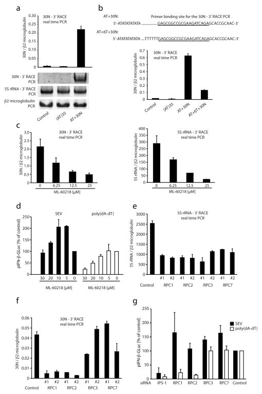
These results led us to hypothesize that poly(dA-dT) might itself serve as a template for the transcription of a 5′ triphosphate poly(rA-rU) molecule by a DNA-dependent RNA polymerase. Indeed, poly(dA-dT) has, for example, been used to study promoter independent transcription by RNA Pol III34, 35. Additionally, it has been shown previously that poly(dA-dT) is transcribed to poly(rA-rU) by RNA Pol III36. We therefore tested the possibility that poly(dA-dT) was transcribed in cells following transfection. Since in vitro transcribed poly(rA-rU) turned out to be an unsuitable template for specific RT-PCR amplification due to its homopolymeric nature (data not shown), we constructed a synthetic 35(dA-dT) template with an additional tail of 30 nucleotides containing a specific primer binding site (AT+30N). Transfection of this synthetic template triggered a type I IFN response in PBMCs and also led to the generation of a stimulatory RNA species, albeit to a lower extent than that observed with poly(dA-dT) (Supplementary Fig. 6a and b). Thus we anticipated that this synthetic poly(dA-dT) construct would be transcribed together with the downstream primer binding site enabling us to utilize it as a specific tag for a 3′ RACE reaction (Supplementary Fig. 7). Indeed, after transfection of this template into 293T cells we were able to detect an RNA transcript containing the specific 3′ RACE tag, indicating that the AT+30N dsDNA had been transcribed through its 30N 3′ end. 5S rRNA, as an established RNA Pol III transcript, was also assessed using the 3′ RACE PCR method. In addition, we analyzed β2 microglobulin transcription as an RNA Pol II dependent control (Fig. 6a). Similar results were obtained when we transfected primary murine dendritic cells, indicating that this phenomenon was not restricted to cells that responded to poly(dA-dT) in a RIG-I dependent fashion (Supplementary Fig. 6c). Using the same template now with a poly T stretch separating the (dA-dT) part from the 30N tag (AT+6T+30N) resulted in a markedly lower transcription of the 30N tag, indicating that the polymerase activity was terminated prior to this part of the template (Fig. 6b). The fact that RNA Pol III terminates transcription at poly T stretches further implicates the involvement of RNA Pol III in the transcription of the AT+30N template. To address this possibility further, we took advantage of a specific inhibitor for RNA Pol III, ML-60218. 293T cells were treated with ML-60218 and subsequently transfected with AT+30N. Consistent with a role for RNA Pol III in mediating the transcription of the AT+30N template, ML-60218 dose-dependently inhibited transcription of 5S rRNA and also the AT+30N DNA when compared to β2 microglobulin transcription (Fig. 6c).
Figure 6. RNA polymerase III transcribes AT rich DNA and is required for poly(dA-dT)-mediated type I IFN induction.
a, The synthetic dsDNA template AT+30N or AT70 was transfected into 293T cells, whereas non transfected cells were used as a control. 8 h after transfection total RNA was isolated, polyadenylated and reverse transcribed as depicted in supplementary Fig. 6. The obtained cDNA was then used for PCR using a specific upstream primer for either 5S rRNA or the 30N transcript and a downstream primer binding within the RT primer. A standard PCR was done with primer pairs for β2 microglobulin. A representative PAGE analysis is shown for a conventional PCR amplification for all three primer combinations. In addition a real-time PCR analysis is shown for the AT+30N transcript normalized to β2 microglobulin expression. b, 293T cells were transfected with (AT)35, AT+30N, AT+6T+30N dsDNA or left untreated. RNA was isolated and treated as above and 3′ RACE real time PCR was performed for the 30N tag. The 30N tag expression data were normalized to β2 microglobulin expression, c, 293T cells were treated with ML-60218 for 2h at the indicated concentrations and transfected with AT+30N dsDNA. 16h later RNA was isolated and the expression of the 30N tag normalized to β2 microglobulin expression was assessed. d, 293T cells were treated with ML-60218 for 2h at the indicated doses. Subsequently cells were transfected with an IFN-β reporter plasmid in conjunction with poly(dA-dT) or SEV. After an additional period of 24h transactivation of the IFN-β promoter was measured. In addition, 293T cells were reverse transfected with two individual siRNAs targeting the indicated genes. After 2 days cells were transfected with AT+30N and after an additional period of 16h transcription of 5S rRNA (e) and AT+30N (f) was determined via 3′ RACE PCR. g, 293T cells were reverse transfected with four individual siRNAs targeting the indicated genes. 48h after transfection cells were transfected with an IFN-β reporter plasmid in conjunction with poly(dA-dT) or SEV. After an additional period of 24h transactivation of the IFN-β promoter was measured. Four individual siRNAs were used per gene and were each tested in triplicates. Data are represented as mean values normalized to the set of control siRNAs. Mean values ± SEM of one representative experiment out of two (c, e, f), three (a, b, g) or four (d) experiments are depicted.
To address the functional consequences of RNA Pol III inhibition on type I IFN induction, we treated 293T cells with ML-60218 and then challenged these cells with poly(dA-dT) or SEV. SEV-triggered type I IFN induction was enhanced at low concentrations of ML-60218 and returned to base line levels at higher concentrations (Fig. 6d). In contrast, poly(dA-dT)-mediated type I IFN induction was dose dependently blocked by ML-60218 at concentrations that did not affect cell viability (Fig. 6d). We also utilized RNAi to knock down essential subunits of the RNA Pol III transcription apparatus to further study the role of RNA Pol III in this response. The two core subunits RPC1 and RPC2 that form the polymerase active center of RNA Pol III were targeted (Supplementary Fig. 8). In addition, we also targeted RPC3 and RPC7, which are not essential for RNA Pol III transcription elongation and termination, but are required for promoter-directed transcription initiation37. As expected, transcription of 5S rRNA was affected by knocking down either RPC1, RPC2, RPC3 or RPC7, all of which are essential subunits in promoter-directed transcription by RNA Pol III (Fig. 6e). However, while the core subunits RPC1 and RPC2 were critically required for transcription of AT+30N, the RPC3 and RPC7 subunits were dispensable (Fig 6f). In line with these data, type I IFN induction triggered by poly(dA-dT) was critically dependent on RPC1 and RPC2, whereas targeting RPC3 or RPC7 had no effect (Fig. 6g). SEV-mediated type I IFN induction was unaffected by knock down of any of these components. These results indicate that the transcriptionally active core units RPC1 and RPC2 of RNA Pol III were required for poly(dA-dT)-transcription and subsequent type I IFN induction, whereas RNA Pol III subunits required for promoter-specific transcription were not essential. We therefore conclude that promoter-independent transcription of poly(dA-dT) by RNA Pol III leads to the formation of 5′ triphosphate RNA that in turn activates RIG-I.
Epstein-Barr virus encoded EBER RNAs are transcribed by RNA Pol III and trigger activation of RIG-I
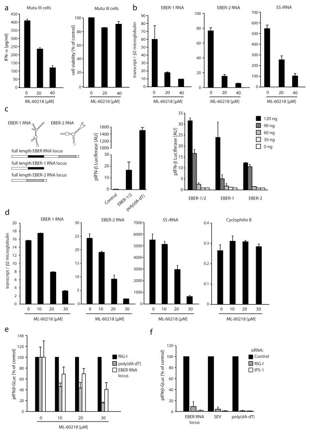
To address the physiological relevance of the RNA Pol III pathway in anti-viral host defenses, we examined the role of RNA Pol III in the regulation of type I IFN responses to a DNA virus. We chose Epstein-Barr virus since it is known that EBV encodes small RNAs that are transcribed by RNA Pol III at very high levels. EBV encoded EBERs are the most abundant viral transcripts in cells with latent EBV infection 38. EBERs are nonpolyadenylated, untranslated RNAs of 167 (EBER-1) or 172 nucleotides (EBER-2) in length 39. Interestingly, EBV-immortalized lymphoblastoid cell lines and EBV-positive burkitt lymphoma (BL) cell lines produce type I IFNs under resting conditions 40, 41, raising the intriguing possibility that PRRs are being activated constitutively in this setting. To test this possibility and define the involvement or RNA Pol III in controlling constitutive production of type I IFNs, we treated the EBV-positive BL Mutu III cell line with the RNA Pol III inhibitor ML-60218. Indeed, inhibition of RNA Pol III dose dependently blocked IFN-α production in Mutu III cells (Fig. 7a). The defect in IFN production correlated with the repression of EBER-1, EBER-2 and 5S rRNA expression in these cells (Fig. 7b). To directly examine the ability of EBER RNAs to activate the RIG-I pathway, we cloned the EBER-1 and 2 gene locus from EBV and transiently overexpressed this construct in IFN-primed 293T cells. Overexpression of EBERs led to a marked induction of type I IFN production, albeit to an lower extent than poly(dA-dT) (Fig. 7c, left panel). Expressing the locus for EBER-1 and EBER-2 RNA separately showed that both EBER RNAs could trigger type I IFN responses independently, whereas EBER-1 RNA was slightly more active at high concentrations (Fig. 7c, right panel). Interestingly, when we expressed a dsDNA template encoding a blunt end version of EBER-1 RNA, higher type I IFN induction was observed (Supplementary Fig. 9). This is in line with the finding that RIG-I favors fully blunt end 5′ triphosphate dsRNA (Schlee et al. in press). As expected, EBER RNA expression in 293T cells was suppressed by inhibiting RNA Pol III activity, whereas RNA Pol II transcription was not affected (Fig. 7d). In addition, inhibition of RNA Pol III led to a reduction in type I IFN induction, indicating that EBER RNAs triggered IFN production in an RNA Pol III dependent fashion (Fig. 7e). Moreover, the induction of type I IFNs was also blocked when RIG-I or IPS-1 was silenced using siRNA (Fig. 7f). Collectively, these results indicated that EBV encoded EBER RNAs are transcribed by RNA Pol III and trigger type I IFN induction via RIG-I.
Figure 7. EBV encoded EBER RNAs are transcribed by RNA Pol III and trigger activation of RIG-I.
a and b, Mutu III cells were treated with ascending doses of ML-60218 for 24h and IFN-α production was assessed by ELISA (a, left panel) and cell viability was assessed using Calcein AM staining (a, right panel), while EBER RNA and 5S rRNA transcription was measured by real-time PCR (b). c, The genomic locus that encodes for EBER-1 and 2 RNA, EBER-1 RNA, EBER-2 RNA was PCR-amplified and transfected into IFN-primed 293T cells (all 120ng or as indicated). PCR-amplified EGFP served as a negative control, whereas poly(dA-dT) was used as a positive control. Subsequently transactivation of the IFN-β promoter was measured after 24h. d, IFN-primed 293T cells transfected with the full length EBER RNA gene locus were treated with ascending doses of ML-60218 and after 24h EBER-1, EBER-2, 5S rRNA and Cyclophilin B transcription was determined. e, In addition, transactivation of the IFN-β promoter was assessed in IFN-primed 293T cells that had been transfected with either poly(dA-dT), the EBER RNA gene locus or RIG-I (all 120ng). Data were normalized to the positive control RIG-I. f, 293T cells were reverse transfected with two individual siRNAs targeting the indicated genes. 48h after siRNA transfection cells were transfected with an IFN-β reporter plasmid in conjunction with the EBER RNA gene locus, poly(dA-dT) (all 120ng) or SEV. After an additional period of 24h transactivation of the IFN-β promoter was measured. Two individual siRNAs were used per gene and were each tested in triplicates. Mean values ± SEM of one representative experiment out of two (b, d, f), three (a, e) or four (c) experiments are depicted.
Discussion
Here we reveal a novel mechanism for sensing of AT-rich double stranded DNA. Our data show that the synthetic dsDNA mimetic, poly(dA-dT) is converted by RNA Pol III of the host into a 5′ triphosphate RNA intermediate, which is in turn is recognized by RIG-I. While the RNA Pol III mediated conversion of poly(dA-dT) is functional both in the murine and in the human system, at least one additional sensing mechanism remains to be defined in murine immune cells that responds to poly(dA-dT) in an RNA-independent manner, presumably as a result of sensing DNA directly. These observations are consistent with previous reports showing that murine cells lacking RIG-I or IPS-1 still induce type I IFNs in response to poly(dA-dT)31, 32. This matter is further complicated by the fact that in the human system at least one additional DNA sensing mechanism exists to recognize double stranded DNA independent of a RIG-I stimulatory RNA intermediate. Only poly(dA-dT) induced the formation of the RIG-I activating RNA species, whereas other double stranded DNAs that were not (dA-dT) homopolymeric in nature failed to do so, despite being active, when directly transfected into PBMC. Nevertheless, our data clearly demonstrate that AT rich DNA transcribed by RNA Pol III is sensed by RIG-I in the human system in a non-redundant manner.
Poly(dA-dT) has been used to study RNA Pol III mediated promoter-independent transcription34–36. This non-specific transcriptional activity requires the core polymerase complex of RNA Pol III, which includes RPC1 and RPC2, yet is independent of subunits that are employed by RNA Pol III for recruitment to specific promoter sites (namely RPC3, RPC6 and RPC7)37. It is currently unclear if RNA Pol III has transcriptional activity independent of its characterized promoter sites in vivo, since no studies have been performed that have addressed the transcriptional activity of RNA Pol III in an unbiased fashion at a global cellular level. It is tempting to speculate that RNA Pol III also transcribes DNA templates from endogenous sources in a promoter-independent manner. Such a mechanism could function as an innate defense strategy tagging DNA as an RNA intermediate to be detected by RIG-I. Poly(dA-dT) or AT rich DNA is an unconventional template for RNA Pol III and thereby indirectly for RIG-I for two main reasons: Firstly, AT rich sequences might be particularly suited for promoter-independent transcription by RNA Pol III due to their propensity to form double stranded DNA regions with low helix stability that are then more accessible as initiation sites for polymerases. Thus AT rich sequences might be especially favored for transcription due to their accessibility for RNA Pol III. However, other DNA templates have also been used to study promoter independent transcription by RNA Pol III and thus it is possible that non-AT DNA is also transcribed by RNA Pol III in a promoter-independent fashion. Secondly and probably most importantly, homopolymeric AT rich DNA is also transcribed into a homopolymeric RNA molecule that has the propensity to form a complete RNA duplex. RIG-I strongly favors blunt end dsRNA over ssRNA or incompletely annealed dsRNA for binding and subsequent activity (Schlee et al. in press). While 5′ triphosphate ssRNA that is not self-complementary is equally active in terms of RIG-I activation when annealed to a complementary ssRNA strand, it shows little or no activity as a single stranded molecule. Poly(A-U) RNA on the other hand is completely self complementary and thus has a strong propensity to form dsRNA with complete blunt end formation, thereby making it ideally suited to be recognized by RIG-I.
Study the role of host RNA Pol III dependent transcription of pathogen-derived DNA will be important, yet technically challenging. During acute infection with DNA viruses it is likely that several DNA sensing pathways will be triggered in the nucleus or in the cytosol simultaneously, making it difficult to study the contribution of a single sensor. In our studies, we focused on cells that are latently infected with EBV and thus harbor viral DNA in the nucleus under steady state conditions. EBV EBERs are transcribed at high levels by RNA Pol III in latently infected EBV-positive cells 42, 43. In these cells we have observed an RNA Pol III dependent activation of type I IFN induction that coincided with EBER RNA expression. Ectopic expression of the EBER RNA gene locus showed a dramatic induction of type I IFN, which was dependent on RIG-I and IPS-1. Similar findings have been published by another group that have implemented EBER RNAs as endogenous triggers for RIG-I 44, 45. While EBER RNAs are already established as RNA Pol III transcripts, we would speculate that other Herpes viruses also encode RNA Pol III transcripts that can trigger RIG-I activation. For example, the genomes of other known gammaherpesviruses contain variable numbers of internal repeats, which are highly conserved across different viruses. In this regard it is noteworthy that a highly repetitive region in the genome of murine Gammaherpesvirus 68 has been shown to trigger type I IFN responses 46.
While some pathogens harbor AT-rich sequence stretches in their genomes it is unlikely that these regions are solely responsible for the detection of these pathogens by the host. Indeed it seems more likely that while AT-rich sequences within the DNA trigger RIG-I activation via the RNA Pol III pathway, non AT-rich DNA sequences are presumably sensed by an additional sensor, which probably directly binds the DNA. It remains to be determined what this alternative receptor is. Previous work has implicated the RIG-I pathway in the sensing of certain DNA viruses, for example Herpes Simplex virus. It is unclear whether the activation of RIG-I in these cases is due to pathogen-dependent RNA polymerase activity or host derived RNA that has been generated by an RNA Pol III mechanism47, 48. Future studies that will allow discrimination between host-derived and pathogen-derived RIG-I ligands could clarify these issues.
Our studies also reveal that the sensing of poly(dA-dT) by the RNA Pol III/RIG-I pathway is not restricted to human cells, but also occurs in murine immune cells. However, in the murine system a second sensor exists in parallel. Although poly(dA-dT) is clearly converted into a RIG-I stimulatory RNA species in murine cells, knockdown of RIG-I does not attenuate the type I IFN response. It is unclear at present what this second sensor is, although it is likely to bind DNA directly. When RIG-I expression is attenuated or when IPS-1 is absent, we have consistently observed a slight upregulation of type I IFN production. We would speculate therefore that in the absence of the alternative poly(dA-dT) sensing mechanism, the RIG-I pathway could compensate, making sensing of AT rich DNA a fully redundant system in the mouse. It seems very unlikely that a single loss of function approach could be successful in indentifying alternative DNA sensors in the murine system. In light of these findings, a reevaluation of the role of DAI and a search for additional sensors is a worthy endeavor.
METHODS
Reagents
Poly(dA:dT) and chloroquine were from Sigma-Aldrich. ML-60218 was from Calbiochem. Human IL-4 and GM-CSF were from ImmunoTools. RNase T1, alkaline phosphatase and DNase I were from Fermentas. RNase III, RNase A, RNA 5′ polyphosphatase and poly(A) polymerase were purchased from Epicentre. R848 and CpG-2216 were from Invivogen.
Cell isolation and culture
Human PBMCs were isolated from whole blood of healthy volunteers by density gradient centrifugation (Biochrom). Red blood cells were lysed with red blood cell lysis buffer (Sigma). Human monocytes were isolated from PBMCs using anti CD14 paramagnetic beads (Miltenyi Biotec) and differentiated into monocyte-derived dendritic cells in the presence of IL-4 (800U/ml) and GM-CSF (800U/ml) for 6 days. Murine bone-marrow was cultured for 6 days with 5% GM-CSF-containing supernatant from J558L cells to generate bone marrow-derived dendritic cells. Primary cells were cultured in RPMI medium supplemented with L-glutamine, sodium pyruvate, 10% (v/v) fetal calf serum (all Invitrogen) and ciprofloxacin (Bayer Schering Pharma). HEK293T cells were cultured in DMEM (Invitrogen) with the same additives as above. RNAse L-deficient MEFs and respective controls were kindly provided by Dr. R. Silverman. Experiments involving PBMCs were approved by the institutional review board of the University Hospital of the University of Bonn.
RNA preparation
RNA isolation was performed using the Mini RNA Isolation II Kit (Zymo Research) with the following modifications: Cells were lysed using TRIZOL (Invitrogen) followed by one round of chloroform extraction. For total RNA extraction, the obtained solution was mixed with 0.7 volumes of ethanol, applied to the separation column and after washing the column bound RNA was eluted. For enrichment of small RNAs, 0.2 volumes of ethanol were added to the chloroform extracted solution, applied to the separation column and now the flow through was collected. This fraction was then mixed with 1.0 volumes of ethanol and passed to a second column. After washing, the column bound RNA was eluted. For some experiments the amount of ethanol used in the first binding step was varied to obtain different sized flow-through fractions.
Enzymatic reactions
2 μg of RNA or poly(dA-dT) were treated with alkaline phosphatase (100 U/ml), RNA 5′ polyphosphatase (1000 U/ml), RNase A (50 μg/ml), RNase III (100 U/ml) or DNase I (1000 – 100 U/ml) in the corresponding buffer solution at 37 °C for 30min (RNA 5′ polyphosphatase) or 1h (alkaline phosphatase, RNase A, RNase III or DNase I) in 10 μl. RNase T1 digestion was performed under denaturing conditions for 30 min at 55 °C in 6 M urea, 17.8 mM Na-citrate at pH 3.5 and 0.9 mM EDTA.
Cell stimulation
293T cells (2×10⁴ cells/96 well) were transfected with 50 ng of an IFN-β promoter reporter plasmid (p125-GLuc) in conjunction with 50 ng poly(dA-dT) or 50 ng of an expression plasmid (pCMV-hsIRF1, pEF-BOS-RIG-I, pEF-BOS-RIG-IΔCARD, pME18-NS3/4A wt, pME18-NS3/4A S139A) using Lipofectamine 2000 (Invitrogen). The total amount of DNA was kept at 200 ng using pcDNA3 as a stuffer plasmid. Transactivation of the IFN-β promoter was measured 24h after stimulation in an Envision 2104 Multilabel Reader (Perkin Elmer). Human PBMCs, human MoDCs and murine bone marrow-derived dendritic cells (2×10⁵ cells/96 well) were pre-incubated with chloroquine (2000 ng/ml) followed by transfection with 200 ng of DNA or RNA using Lipofectamine 2000. Sendai Virus (Cantrell strain, Charles River Laboratories) was used at 300 HAU/ml.
RNAi experiments
siRNA was reverse transfected at a concentration of 25 nM into 293T cells (104 cells/96 well) using 0.5 μl Lipofectamine. 48 h after transfection cells were stimulated as indicated and after an additional period of 24 h luciferase activity was assessed. siRNAs used in Fig. 1 and 4 were obtained from Ambion and siRNAs used in Fig. 6 were obtained from Qiagen. Electroporation of human MoDCs and murine bone marrow-derived dendritic cells was performed as previously described49. SiRNAs for electroporation experiments were purchased from Eurogentec and Biomers. A list with all siRNA sequences is available upon request.
3′ RACE
For 3′ RACE analysis, isolated RNA was tailed with poly(A) polymerase and subsequently reverse transcribed using the RevertAid™ First Strand cDNA Synthesis Kit (Fermentas) with the 3-RACE-RT primer. The cDNA was subsequently amplified with a transcript specific upstream primer and the 3-RT-PCR downstream primer using conventional or real-time PCR. The primer sequences were as follows: 3-RACE-RT primer: 5′-CTATAGGCGCGCCACCGGTGTTTTTTTTTTTTTTTTTTTTTTTTTTTTTTTTVN-3′, specific upstream primer for AT+30N and AT+6T+30N: 5′-GAGCGGCCGCGAAGATCAGA-3′, specific upstream primer for 5S rRNA: 5′-TCGGGCCTGGTTAGTACTTG-3′, EBER-1 RNA: 5′-GACTCTGCTTTCTGCCGTCT-3′, EBER-2 RNA: 5′-GGAGGAGAAGAGAGGCTTCC-3′ 3-RT-PCR primer: 5′-CTATAGGCGCGCCACCGGTGTTTT-3′. The sequences for the synthetic poly(dA-dT) dsDNAs are the following (only the top strand is listed): AT+30N: 5′-ATATATATATATATATATATATATATATATATATATATATATATATATATATATATATATATATATATATGAGCGGCCGCGAAGATCAGAGCACCGCAAC-3′, AT+6T+30N: 5′-ATATATATATATATATATATATATATATATATATATATATATATATATATATATATATATATATTTTTTTGAGCGGCCGCGAAGATCAGAGCACCGCAAC-3′ and (AT)35: 5′-GGCCGTCTCCATATATATATATATATATATATATATATATATATATATATATATATATATATATATATATATATATATATCGAG ACCCGG-3′.
Quantitative real-time PCR
cDNA was synthesized from total RNA with the RevertAid™ First Strand cDNA Synthesis Kit (Fermentas) and quantitative RT-PCR analysis was performed on a Roche LC480 using Maxima™ SYBR Green qPCR Master Mix (Fermentas). The specificity of amplification was assessed for each sample by melting curve and gel analysis. Relative quantification was performed using standard curve analysis. The murine quantification data are presented as a relative expression level compared to hypoxanthine phosphoribosyltransferase 1 (HPRT1). The following primers were used: mmHPRT1-L: 5′-CTGGTGAAAAGGACCTCTCG-3′ and mmHPRT1-R: 5′-TGAAGTACTCATTATAGTCAAGGGCA-3′, mmIFNB-L: 5′-ATAAGCAGCTCCAGCTCCAA-3′ and mmIFNB-R: 5′-CTGTCTGCTGGTGGAGTTCA-3′, hsCyclophilin B-L: 5′-GAAAGAGCATCTACGGTGAGC-3′; hsCyclophilin B-R: 5′-GTCTTGACTGTCGTGATGAAG-3′, hsβ2Microglubulin-L: 5′-TGCTGTCTCCATGTTTGATGT-3′ and hsβ2Microglubulin-R: 5′-TCTCTGCTCCCCACCTCTAAG-3′.
RNA analysis
Enriched small cellular RNAs and in vitro transcribed RNA were monitored by using the Small RNA Kit with an Agilent Bioanalyzer 2100 (Agilent Technologies). Differentially sized fractions of total cellular RNA was analyzed using the Agilent 6000 Pico Chip Kit.
ELISA
Human IFN-α (Bender Med Systems) and TNF-α (BD Biosciences) were assessed using commercial ELISA kits according to the manufacturer’s instructions.
In vitro transcribed RNAs and PCR generated dsDNAs
In vitro transcribed RNAs were synthesized using the AmpliScribe™ T7-Flash™ Transcription Kit (Epicentre). In vitro transcription was carried out for 3h at 37° C. The DNA template was digested using DNase I and subsequently excess salts and NTPs were removed by passing the RNAs through a Mini Quick Spin™ Oligo Column (Roche). Size and integrity of RNAs was checked via gel electrophoresis. The following templates were used for in vitro transcription (only the top strand is listed, whereas the RNA transcript is underlined): 3pRNA: 5′-CAGTAATACGACTCACTATAGACGACGACGACGACG-ACGACGACGACGAC-3′; 10(rA-rU): 5′-CAGTAATACGACTCACTATTATATATATATATATATATAT-3′. Double stranded 20, 40, 77, 163, 245 mer DNAs were generated by annealing or by PCR using pCDNA3 as a template. The primer sequences were as follows: FWD-all 5′-GCAGAGCGAGGTATGTAGGC-3′, BWD-20 5′-GCCTACATACCTCGCTCTGC-3′, BWD-40 5′-TTCAAGAACTCTGTAGCACC-3′, BWD-77 5′-TACTGTCCTTCTAGTGTAGCCGTA-3′, BWD-163 5′-CTACCAGCGGTGGTTTGTTT-3′ and BWD-245 5′-CCCCGTAGAAAAGATCAAAGG-3′. The full length EBER RNA locus was amplified from EBV DNA with the following primers: EBER-L 5′-CCATAAAGCCCAGGGTGTAAAAC-3′ and EBER-R 5′-CCCTTACATGTTGTGGGTGCAAAAC-3′, the EBER 1 RNA locus only: EBER-L and EBER-1-R 5′-ATCAAAACATGCGGACCAC-3′, the EBER 1 RNA locus for the blunt end EBER 1 RNA: EBER-L and EBER-1-blunt-R: 5′-AGGACCACCAGCTGGTACTT-3′, the EBER 1 RNA locus for the poly(U) tailed blunt end EBER-1 RNA: EBER-L and EBER-1-pT-R: 5′-AAAAAAGGACCACCAGCTGGTACTT-3′ and for the EBER-2 RNA locus: EBER-2-L: 5′-GATCCAAACTTTAGTTTTAGGATTTAT-3′ and EBER-R.
Supplementary Material
Supplementary Figure 1: Dominant negative RIG-I constructs or disruption of IPS-1 inhibits poly(dA-dT) triggered type I IFN induction. a, 293T cells were transfected with the indicated expression plasmids (5 ng) in conjunction with 50 ng pIFN-β reporter plasmid: Eif4a was used as a control dead box helicase; NS3/4A S139A is the catalytically inactive point mutant of the hepatitis C derived NS3/4A protease that has been shown to cleave and inactivate IPS-1; MDA5 T789M and RIG-I T697M are helicase point mutants of MDA5 and RIG-I respectively; RIG-IΔCards is RIG-I devoid of its Card domain. After 24h cells were stimulated with SEV or transfected with poly(dA-dT). After an additional period of 24h IFN-β promoter activation was assessed. b, 293T cells were reverse transfected with siRNA targeting RIG-I or a control siRNA. After 48h, cells were transfected with an NF-κB reporter plasmid in conjunction with poly(dA-dT) or TBK-1 (50 ng) or stimulated with SEV at the indicated concentrations. 24h later NF-κB activation was assessed. Mean values ± SEM of one representative experiment out of two (b) or three (a) experiments are depicted.
Supplementary Figure 2: Stimulatory RNA from poly(dA-dT) transfected cells is insensitive to DNase I and triggers type I IFN responses via RIG-I in 293T cells. a, PBMCs were chloroquine treated at the indicated concentrations and subsequently poly(dA-dT) or CpG-A was transfected. 24h later IFN-α production was assessed by ELISA. b, RNA from poly(dA-dT) transfected 293T cells was isolated and treated with DNase I at the indicated concentrations. 3pRNA and poly(dA-dT) were treated the same way. Chloroquine-treated PBMCs were then transfected with the respective nucleic acids and IFN-α production was assessed 24h later. In addition, RNA from poly(dA-dT) transfected cells and poly(dA-dT) was analyzed on an agarose gel. c, 293T cells were transfected with the indicated expression plasmids (50 ng). 24h later 50 ng pIFN-β reporter plasmid was transfected in conjunction with 3pRNA, poly(dA-dT) or RNA isolated from poly(dA-dT) or SEV-stimulated cells. Direct infection with SEV served as a control. After an additional period of 24h IFN-β promoter activity was assessed. Mean values ± SEM of one representative experiment out of two (a, b, c) experiments are depicted.
Supplementary Figure 3: Dose response of stimulatory dsDNAs in PBMCs. Double stranded DNAs of different length derived from pcDNA3 by PCR were transfected into chloroquine-blocked PBMCs at the indicated molar concentrations. Mean values ± SEM of one representative experiment out of two independent experiments are depicted.
Supplementary Figure 4: Alkaline phosphatase treatment abolishes the activity of poly(dA-dT) triggered RNA. RNA from poly(dA-dT) transfected 293T cells was isolated and treated with alkaline phosphatase. Poly(dA-dT) or 3pRNA were treated the same way. Chloroquine-blocked PBMCs were then transfected with the respective nucleic acids and IFN-α production was assessed 24h later. In addition, the above nucleic acids were analyzed on an agilent small RNA chip. Mean values ± SEM of one representative experiment out of three experiments are depicted.
Supplementary Figure 5: A 20mer 5′ triphosphate RNA consisting of alternating rA-rU is insensitive to RNase T1 under denaturing conditions. RNA from poly(dA-dT) transfected 293T, poly(dA-dT) or 3pRNA was treated with RNase T1 under denaturing conditions. A 20mer 5′ triphosphate RNA oligonucleotide consisting of alternating rA-rU was treated the same way. Chloroquine-treated PBMCs were then transfected with the respective nucleic acids and IFN-α production was assessed 24h later. In addition, the above nucleic acids were analyzed on an agilent small RNA chip. Mean values ± SEM of one representative experiment out of three experiments are depicted.
Supplementary Figure 6: Short synthetic poly(dA-dT) triggers the formation of stimulatory RNA. a, Poly(dA-dT), AT+30N or AT+6T+30N was transfected into chloroquine-treated PBMCs and IFN-α production was assessed 24h later. b, In addition, the above nucleic acids were transfected into 293T and RNA was isolated. The RNA was then transfected into chloroquine-treated PBMCs and IFN-α production was assessed 24h later. c, Murine conventional dendritic cells were transfected with either the dsDNA oligonucleotides AT+30N or PS. 14h later RNA was isolated and the expression of the 30N tag normalized to HPRT1 expression was assessed. Mean values ± SEM of one representative experiment out of two (c) or three (a, b) experiments are depicted.
Supplementary Figure 7: 3′ RACE strategy to detect the transcription of a poly(dA-dT) transcript. a, A double stranded DNA template (AT+30N) containing a 70 nt homopolymeric stretch of 35 x dA-dT was constructed to contain a non dA-dT region at the 3′ end (30N) that could be used as a specific primer binding site. (b) This template was transfected into 293T cells and RNA (c) was isolated subsequently. (d) Total RNA was polyadenylated using poly(A) polymerase and then (e) reverse transcribed using an oligo dT primer containing an additional primer binding site on its 5′ end. (f) The obtained cDNA was then amplified in a PCR reaction and analyzed on a polyacrylamide gel (schematic cartoon) (g).
Supplementary Figure 8: siRNA-mediated knock down of RPC1, RPC2, RPC3 and RPC7. 293T cells were reverse transfected with two individual siRNAs targeting the indicated genes or a control siRNA. 48h after transfection expression of the respective target gene was analyzed by real time PCR. Mean values ± SEM of one representative experiment out of two experiments is depicted.
Supplementary Figure 9: Blunt end EBER-1 RNA is superior to the natural EBER-1 RNA. The genomic locus that encodes for EBER-1 and 2 RNA and EBER-1 RNA was amplified from EBV DNA by PCR. In addition a modified EBER-1 locus was amplified lacking the naturally occurring 5 bases upstream of the poly(T) stretch termination signal with or without an additional poly(T) stretch. A schematic view of the templates and the expected transcripts is shown, whereas the terminal region of EBER-1 RNA is depicted. Secondary structure prediction of the presumed RNA transcript was done using Mfold 50 (a). The respective DNA templates were transfected into IFN-primed 293T cells (all 120ng or as indicated) in conjunction with 50 ng pIFN-β reporter plasmid. 24h after transfection IFN-β promoter activity was assessed (b). Mean values ± SEM of one representative experiment out of three experiments is depicted.
Acknowledgments
V.H. is supported by the Deutsche Forschungsgemeinschaft (German Research Foundation; Ho2783/2-1), E.L. and K.A.F. are supported by grants from the National Institutes of Health (AI-065483 (to E.L.) and AI-067497 (to K.A.F.)). We would like to thank G. Bornkamm and M. Schlee for helpful discussions.
References
- 1.Kawai T, Akira S. TLR signaling. Semin Immunol. 2007;19:24–32. doi: 10.1016/j.smim.2006.12.004. [DOI] [PubMed] [Google Scholar]
- 2.Kawai T, Akira S. Toll-like receptor and RIG-I-like receptor signaling. Ann N Y Acad Sci. 2008;1143:1–20. doi: 10.1196/annals.1443.020. [DOI] [PubMed] [Google Scholar]
- 3.Takeda K, Akira S. Toll-like receptors in innate immunity. Int Immunol. 2005;17:1–14. doi: 10.1093/intimm/dxh186. [DOI] [PubMed] [Google Scholar]
- 4.Huysamen C, Brown GD. The fungal pattern recognition receptor, Dectin-1, and the associated cluster of C-type lectin-like receptors. FEMS Microbiol Lett. 2009;290:121–128. doi: 10.1111/j.1574-6968.2008.01418.x. [DOI] [PMC free article] [PubMed] [Google Scholar]
- 5.Yoneyama M, Fujita T. RIG-I family RNA helicases: cytoplasmic sensor for antiviral innate immunity. Cytokine Growth Factor Rev. 2007;18:545–551. doi: 10.1016/j.cytogfr.2007.06.023. [DOI] [PubMed] [Google Scholar]
- 6.Martinon F, Mayor A, Tschopp J. The inflammasomes: guardians of the body. Annu Rev Immunol. 2009;27:229–265. doi: 10.1146/annurev.immunol.021908.132715. [DOI] [PubMed] [Google Scholar]
- 7.Meylan E, Tschopp J, Karin M. Intracellular pattern recognition receptors in the host response. Nature. 2006;442:39–44. doi: 10.1038/nature04946. [DOI] [PubMed] [Google Scholar]
- 8.Takaoka A, Taniguchi T. Cytosolic DNA recognition for triggering innate immune responses. Adv Drug Deliv Rev. 2008;60:847–857. doi: 10.1016/j.addr.2007.12.002. [DOI] [PubMed] [Google Scholar]
- 9.Takaoka A, et al. DAI (DLM-1/ZBP1) is a cytosolic DNA sensor and an activator of innate immune response. Nature. 2007;448:501–505. doi: 10.1038/nature06013. [DOI] [PubMed] [Google Scholar]
- 10.Hornung V, et al. AIM2 recognizes cytosolic dsDNA and forms a caspase-1-activating inflammasome with ASC. Nature. 2009;458:514–518. doi: 10.1038/nature07725. [DOI] [PMC free article] [PubMed] [Google Scholar]
- 11.Kono H, Rock KL. How dying cells alert the immune system to danger. Nat Rev Immunol. 2008;8:279–289. doi: 10.1038/nri2215. [DOI] [PMC free article] [PubMed] [Google Scholar]
- 12.Takeuchi O, Akira S. Innate immunity to virus infection. Immunol Rev. 2009;227:75–86. doi: 10.1111/j.1600-065X.2008.00737.x. [DOI] [PMC free article] [PubMed] [Google Scholar]
- 13.Takeuchi O, Akira S. MDA5/RIG-I and virus recognition. Curr Opin Immunol. 2008;20:17–22. doi: 10.1016/j.coi.2008.01.002. [DOI] [PubMed] [Google Scholar]
- 14.Kato H, et al. Differential roles of MDA5 and RIG-I helicases in the recognition of RNA viruses. Nature. 2006;441:101–105. doi: 10.1038/nature04734. [DOI] [PubMed] [Google Scholar]
- 15.Pichlmair A, et al. RIG-I-mediated antiviral responses to single-stranded RNA bearing 5′-phosphates. Science. 2006;314:997–1001. doi: 10.1126/science.1132998. [DOI] [PubMed] [Google Scholar]
- 16.Hornung V, et al. 5′-Triphosphate RNA is the ligand for RIG-I. Science. 2006;314:994–997. doi: 10.1126/science.1132505. [DOI] [PubMed] [Google Scholar]
- 17.Kato H, et al. Length-dependent recognition of double-stranded ribonucleic acids by retinoic acid-inducible gene-I and melanoma differentiation-associated gene 5. J Exp Med. 2008;205:1601–1610. doi: 10.1084/jem.20080091. [DOI] [PMC free article] [PubMed] [Google Scholar]
- 18.Ishii KJ, et al. TANK-binding kinase-1 delineates innate and adaptive immune responses to DNA vaccines. Nature. 2008;451:725–729. doi: 10.1038/nature06537. [DOI] [PubMed] [Google Scholar]
- 19.Burckstummer T, et al. An orthogonal proteomic-genomic screen identifies AIM2 as a cytoplasmic DNA sensor for the inflammasome. Nat Immunol. 2009;10:266–272. doi: 10.1038/ni.1702. [DOI] [PubMed] [Google Scholar]
- 20.Fernandes-Alnemri T, Yu JW, Datta P, Wu J, Alnemri ES. AIM2 activates the inflammasome and cell death in response to cytoplasmic DNA. Nature. 2009 doi: 10.1038/nature07710. [DOI] [PMC free article] [PubMed] [Google Scholar]
- 21.Roberts TL, et al. HIN-200 proteins regulate caspase activation in response to foreign cytoplasmic DNA. Science. 2009;323:1057–1060. doi: 10.1126/science.1169841. [DOI] [PubMed] [Google Scholar]
- 22.Henry T, Brotcke A, Weiss DS, Thompson LJ, Monack DM. Type I interferon signaling is required for activation of the inflammasome during Francisella infection. J Exp Med. 2007;204:987–994. doi: 10.1084/jem.20062665. [DOI] [PMC free article] [PubMed] [Google Scholar]
- 23.Charrel-Dennis M, et al. TLR-independent type I interferon induction in response to an extracellular bacterial pathogen via intracellular recognition of its DNA. Cell Host Microbe. 2008;4:543–554. doi: 10.1016/j.chom.2008.11.002. [DOI] [PMC free article] [PubMed] [Google Scholar]
- 24.O’Riordan M, Yi CH, Gonzales R, Lee KD, Portnoy DA. Innate recognition of bacteria by a macrophage cytosolic surveillance pathway. Proc Natl Acad Sci U S A. 2002;99:13861–13866. doi: 10.1073/pnas.202476699. [DOI] [PMC free article] [PubMed] [Google Scholar]
- 25.Stetson DB, Medzhitov R. Recognition of cytosolic DNA activates an IRF3-dependent innate immune response. Immunity. 2006;24:93–103. doi: 10.1016/j.immuni.2005.12.003. [DOI] [PubMed] [Google Scholar]
- 26.Stanley SA, Johndrow JE, Manzanillo P, Cox JS. The Type I IFN response to infection with Mycobacterium tuberculosis requires ESX-1-mediated secretion and contributes to pathogenesis. J Immunol. 2007;178:3143–3152. doi: 10.4049/jimmunol.178.5.3143. [DOI] [PubMed] [Google Scholar]
- 27.Opitz B, et al. Legionella pneumophila induces IFNbeta in lung epithelial cells via IPS-1 and IRF3, which also control bacterial replication. J Biol Chem. 2006;281:36173–36179. doi: 10.1074/jbc.M604638200. [DOI] [PubMed] [Google Scholar]
- 28.Roux CM, et al. Brucella requires a functional Type IV secretion system to elicit innate immune responses in mice. Cell Microbiol. 2007;9:1851–1869. doi: 10.1111/j.1462-5822.2007.00922.x. [DOI] [PubMed] [Google Scholar]
- 29.Chessler AD, Ferreira LR, Chang TH, Fitzgerald KA, Burleigh BA. A novel IFN regulatory factor 3-dependent pathway activated by trypanosomes triggers IFN-beta in macrophages and fibroblasts. J Immunol. 2008;181:7917–7924. doi: 10.4049/jimmunol.181.11.7917. [DOI] [PMC free article] [PubMed] [Google Scholar]
- 30.Yoshida H, Okabe Y, Kawane K, Fukuyama H, Nagata S. Lethal anemia caused by interferon-beta produced in mouse embryos carrying undigested DNA. Nat Immunol. 2005;6:49–56. doi: 10.1038/ni1146. [DOI] [PubMed] [Google Scholar]
- 31.Sun Q, et al. The specific and essential role of MAVS in antiviral innate immune responses. Immunity. 2006;24:633–642. doi: 10.1016/j.immuni.2006.04.004. [DOI] [PubMed] [Google Scholar]
- 32.Kumar H, et al. Essential role of IPS-1 in innate immune responses against RNA viruses. J Exp Med. 2006;203:1795–1803. doi: 10.1084/jem.20060792. [DOI] [PMC free article] [PubMed] [Google Scholar]
- 33.Cheng G, Zhong J, Chung J, Chisari FV. Double-stranded DNA and double-stranded RNA induce a common antiviral signaling pathway in human cells. Proc Natl Acad Sci U S A. 2007;104:9035–9040. doi: 10.1073/pnas.0703285104. [DOI] [PMC free article] [PubMed] [Google Scholar]
- 34.Huet J, Riva M, Sentenac A, Fromageot P. Yeast RNA polymerase C and its subunits. Specific antibodies as structural and functional probes. J Biol Chem. 1985;260:15304–15310. [PubMed] [Google Scholar]
- 35.Zaros C, Thuriaux P. Rpc25, a conserved RNA polymerase III subunit, is critical for transcription initiation. Mol Microbiol. 2005;55:104–114. doi: 10.1111/j.1365-2958.2004.04375.x. [DOI] [PubMed] [Google Scholar]
- 36.Thuillier V, Brun I, Sentenac A, Werner M. Mutations in the alpha-amanitin conserved domain of the largest subunit of yeast RNA polymerase III affect pausing, RNA cleavage and transcriptional transitions. Embo J. 1996;15:618–629. [PMC free article] [PubMed] [Google Scholar]
- 37.Wang Z, Roeder RG. Three human RNA polymerase III-specific subunits form a subcomplex with a selective function in specific transcription initiation. Genes Dev. 1997;11:1315–1326. doi: 10.1101/gad.11.10.1315. [DOI] [PubMed] [Google Scholar]
- 38.Lerner MR, Andrews NC, Miller G, Steitz JA. Two small RNAs encoded by Epstein-Barr virus and complexed with protein are precipitated by antibodies from patients with systemic lupus erythematosus. Proc Natl Acad Sci U S A. 1981;78:805–809. doi: 10.1073/pnas.78.2.805. [DOI] [PMC free article] [PubMed] [Google Scholar]
- 39.Rosa MD, Gottlieb E, Lerner MR, Steitz JA. Striking similarities are exhibited by two small Epstein-Barr virus-encoded ribonucleic acids and the adenovirus-associated ribonucleic acids VAI and VAII. Mol Cell Biol. 1981;1:785–796. doi: 10.1128/mcb.1.9.785. [DOI] [PMC free article] [PubMed] [Google Scholar]
- 40.Schlee M, Schuhmacher M, Holzel M, Laux G, Bornkamm GW. c-MYC Impairs Immunogenicity of Human B Cells. Adv Cancer Res. 2007;97:167–188. doi: 10.1016/S0065-230X(06)97007-9. [DOI] [PubMed] [Google Scholar]
- 41.Schlee M, et al. C-myc activation impairs the NF-kappaB and the interferon response: implications for the pathogenesis of Burkitt’s lymphoma. Int J Cancer. 2007;120:1387–1395. doi: 10.1002/ijc.22372. [DOI] [PubMed] [Google Scholar]
- 42.Howe JG, Shu MD. Epstein-Barr virus small RNA (EBER) genes: unique transcription units that combine RNA polymerase II and III promoter elements. Cell. 1989;57:825–834. doi: 10.1016/0092-8674(89)90797-6. [DOI] [PubMed] [Google Scholar]
- 43.Howe JG, Shu MD. Upstream basal promoter element important for exclusive RNA polymerase III transcription of the EBER 2 gene. Mol Cell Biol. 1993;13:2655–2665. doi: 10.1128/mcb.13.5.2655. [DOI] [PMC free article] [PubMed] [Google Scholar]
- 44.Samanta M, Iwakiri D, Kanda T, Imaizumi T, Takada K. EB virus-encoded RNAs are recognized by RIG-I and activate signaling to induce type I IFN. Embo J. 2006;25:4207–4214. doi: 10.1038/sj.emboj.7601314. [DOI] [PMC free article] [PubMed] [Google Scholar]
- 45.Samanta M, Iwakiri D, Takada K. Epstein-Barr virus-encoded small RNA induces IL-10 through RIG-I-mediated IRF-3 signaling. Oncogene. 2008;27:4150–4160. doi: 10.1038/onc.2008.75. [DOI] [PubMed] [Google Scholar]
- 46.Sanchez DJ, et al. A repetitive region of gammaherpesvirus genomic DNA is a ligand for induction of type I interferon. J Virol. 2008;82:2208–2217. doi: 10.1128/JVI.01718-07. [DOI] [PMC free article] [PubMed] [Google Scholar]
- 47.Deng L, et al. Vaccinia virus subverts a mitochondrial antiviral signaling protein-dependent innate immune response in keratinocytes through its double-stranded RNA binding protein, E3. J Virol. 2008;82:10735–10746. doi: 10.1128/JVI.01305-08. [DOI] [PMC free article] [PubMed] [Google Scholar]
- 48.Rasmussen SB, et al. Herpes simplex virus infection is sensed by both Toll-like receptors and retinoic acid-inducible gene- like receptors, which synergize to induce type I interferon production. J Gen Virol. 2009;90:74–78. doi: 10.1099/vir.0.005389-0. [DOI] [PMC free article] [PubMed] [Google Scholar]
- 49.Ablasser A, et al. Selection of molecular structure and delivery of RNA oligonucleotides to activate TLR7 versus TLR8 and to induce high amounts of IL-12p70 in primary human monocytes. J Immunol. 2009;182:6824–6833. doi: 10.4049/jimmunol.0803001. [DOI] [PubMed] [Google Scholar]
- 50.Zuker M. Mfold web server for nucleic acid folding and hybridization prediction. Nucleic Acids Res. 2003;31:3406–3415. doi: 10.1093/nar/gkg595. [DOI] [PMC free article] [PubMed] [Google Scholar]
Associated Data
This section collects any data citations, data availability statements, or supplementary materials included in this article.
Supplementary Materials
Supplementary Figure 1: Dominant negative RIG-I constructs or disruption of IPS-1 inhibits poly(dA-dT) triggered type I IFN induction. a, 293T cells were transfected with the indicated expression plasmids (5 ng) in conjunction with 50 ng pIFN-β reporter plasmid: Eif4a was used as a control dead box helicase; NS3/4A S139A is the catalytically inactive point mutant of the hepatitis C derived NS3/4A protease that has been shown to cleave and inactivate IPS-1; MDA5 T789M and RIG-I T697M are helicase point mutants of MDA5 and RIG-I respectively; RIG-IΔCards is RIG-I devoid of its Card domain. After 24h cells were stimulated with SEV or transfected with poly(dA-dT). After an additional period of 24h IFN-β promoter activation was assessed. b, 293T cells were reverse transfected with siRNA targeting RIG-I or a control siRNA. After 48h, cells were transfected with an NF-κB reporter plasmid in conjunction with poly(dA-dT) or TBK-1 (50 ng) or stimulated with SEV at the indicated concentrations. 24h later NF-κB activation was assessed. Mean values ± SEM of one representative experiment out of two (b) or three (a) experiments are depicted.
Supplementary Figure 2: Stimulatory RNA from poly(dA-dT) transfected cells is insensitive to DNase I and triggers type I IFN responses via RIG-I in 293T cells. a, PBMCs were chloroquine treated at the indicated concentrations and subsequently poly(dA-dT) or CpG-A was transfected. 24h later IFN-α production was assessed by ELISA. b, RNA from poly(dA-dT) transfected 293T cells was isolated and treated with DNase I at the indicated concentrations. 3pRNA and poly(dA-dT) were treated the same way. Chloroquine-treated PBMCs were then transfected with the respective nucleic acids and IFN-α production was assessed 24h later. In addition, RNA from poly(dA-dT) transfected cells and poly(dA-dT) was analyzed on an agarose gel. c, 293T cells were transfected with the indicated expression plasmids (50 ng). 24h later 50 ng pIFN-β reporter plasmid was transfected in conjunction with 3pRNA, poly(dA-dT) or RNA isolated from poly(dA-dT) or SEV-stimulated cells. Direct infection with SEV served as a control. After an additional period of 24h IFN-β promoter activity was assessed. Mean values ± SEM of one representative experiment out of two (a, b, c) experiments are depicted.
Supplementary Figure 3: Dose response of stimulatory dsDNAs in PBMCs. Double stranded DNAs of different length derived from pcDNA3 by PCR were transfected into chloroquine-blocked PBMCs at the indicated molar concentrations. Mean values ± SEM of one representative experiment out of two independent experiments are depicted.
Supplementary Figure 4: Alkaline phosphatase treatment abolishes the activity of poly(dA-dT) triggered RNA. RNA from poly(dA-dT) transfected 293T cells was isolated and treated with alkaline phosphatase. Poly(dA-dT) or 3pRNA were treated the same way. Chloroquine-blocked PBMCs were then transfected with the respective nucleic acids and IFN-α production was assessed 24h later. In addition, the above nucleic acids were analyzed on an agilent small RNA chip. Mean values ± SEM of one representative experiment out of three experiments are depicted.
Supplementary Figure 5: A 20mer 5′ triphosphate RNA consisting of alternating rA-rU is insensitive to RNase T1 under denaturing conditions. RNA from poly(dA-dT) transfected 293T, poly(dA-dT) or 3pRNA was treated with RNase T1 under denaturing conditions. A 20mer 5′ triphosphate RNA oligonucleotide consisting of alternating rA-rU was treated the same way. Chloroquine-treated PBMCs were then transfected with the respective nucleic acids and IFN-α production was assessed 24h later. In addition, the above nucleic acids were analyzed on an agilent small RNA chip. Mean values ± SEM of one representative experiment out of three experiments are depicted.
Supplementary Figure 6: Short synthetic poly(dA-dT) triggers the formation of stimulatory RNA. a, Poly(dA-dT), AT+30N or AT+6T+30N was transfected into chloroquine-treated PBMCs and IFN-α production was assessed 24h later. b, In addition, the above nucleic acids were transfected into 293T and RNA was isolated. The RNA was then transfected into chloroquine-treated PBMCs and IFN-α production was assessed 24h later. c, Murine conventional dendritic cells were transfected with either the dsDNA oligonucleotides AT+30N or PS. 14h later RNA was isolated and the expression of the 30N tag normalized to HPRT1 expression was assessed. Mean values ± SEM of one representative experiment out of two (c) or three (a, b) experiments are depicted.
Supplementary Figure 7: 3′ RACE strategy to detect the transcription of a poly(dA-dT) transcript. a, A double stranded DNA template (AT+30N) containing a 70 nt homopolymeric stretch of 35 x dA-dT was constructed to contain a non dA-dT region at the 3′ end (30N) that could be used as a specific primer binding site. (b) This template was transfected into 293T cells and RNA (c) was isolated subsequently. (d) Total RNA was polyadenylated using poly(A) polymerase and then (e) reverse transcribed using an oligo dT primer containing an additional primer binding site on its 5′ end. (f) The obtained cDNA was then amplified in a PCR reaction and analyzed on a polyacrylamide gel (schematic cartoon) (g).
Supplementary Figure 8: siRNA-mediated knock down of RPC1, RPC2, RPC3 and RPC7. 293T cells were reverse transfected with two individual siRNAs targeting the indicated genes or a control siRNA. 48h after transfection expression of the respective target gene was analyzed by real time PCR. Mean values ± SEM of one representative experiment out of two experiments is depicted.
Supplementary Figure 9: Blunt end EBER-1 RNA is superior to the natural EBER-1 RNA. The genomic locus that encodes for EBER-1 and 2 RNA and EBER-1 RNA was amplified from EBV DNA by PCR. In addition a modified EBER-1 locus was amplified lacking the naturally occurring 5 bases upstream of the poly(T) stretch termination signal with or without an additional poly(T) stretch. A schematic view of the templates and the expected transcripts is shown, whereas the terminal region of EBER-1 RNA is depicted. Secondary structure prediction of the presumed RNA transcript was done using Mfold 50 (a). The respective DNA templates were transfected into IFN-primed 293T cells (all 120ng or as indicated) in conjunction with 50 ng pIFN-β reporter plasmid. 24h after transfection IFN-β promoter activity was assessed (b). Mean values ± SEM of one representative experiment out of three experiments is depicted.


