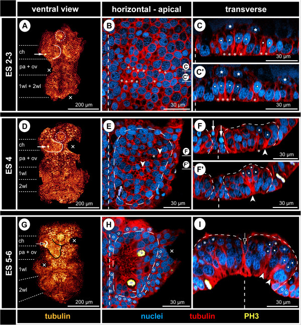
Figure 2.
Early neurogenesis in Pseudopallene sp. (late ES 2 – late ES 5). Ventral overview of germ bands (tubulin) and optical sections through hemi-neuromeres of walking leg segment 1 (tubulin-, PH3- and nuclear labelling). Horizontal sections represent 2D projections of curved composite sections. Stippled circles mark stomodeum or pharynx. White arrows in A and D indicate immigrating cells related to spinning gland development. Black arrow in G marks spinning gland duct. Crosses indicate damaged germ band regions or virtually removed chelifore bud. Vertical dashed lines mark the VMR. (A–C’) Initiation of cell immigration, late ES 2. More intensely tubulin-labelled areas relate to constricting cell cortices (asterisks) of columnar cells with basally displaced nucleus (white spots). CISs are not yet recognizable. The VNE is underlain by scattered entodermal cells (stars). (D–F’) Formation of CISs, ES 4. Stippled outlines (E–F’) mark hemi-neuromere extensions. CISs have become defined in the VNE (tubulin-stained spots in D, selection marked by arrowheads in E). The nuclei lie in about three apico-basal levels, immigrating flask-shaped cells extending far basally (white spots in F’). Pycnotic bodies (white arrows in F) indicate occurrence of cell death. (G–I) Formation of central invagination and hemi-ganglion anlagen, late ES 5. Stippled outlines (H,I) mark hemi-neuromere extensions. CISs are still discernible in pre-cheliforal lobe and the palpal and ovigeral neuromeres (G). Occasionally, cell arrangements reminiscent of CISs are found in the walking leg hemi-ganglion anlagen (white arrowheads and white spots in I). During mitosis, chromosomes and newly forming nuclei are encountered apically within the nucleus-free invagination (H) that is surrounded by smaller epidermal cells (open spots). The medio-lateral extension of the VMR has diminished, its remaining basal cells being often unpaired and wedged between the hemi-ganglion anlagen (open arrow in I). First GCs have detached basally (asterisks in I).

