Abstract
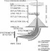
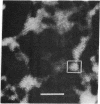
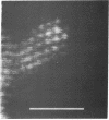
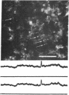
We have shown that a scanning transmission electron microscope with a high brightness field emission source is capable of obtaining better than 3 Å resolution using 30 to 40 keV electrons. Elastic dark field images of single atoms of uranium and mercury are shown which demonstrate this fact as determined by a modified Rayleigh criterion. Point-to-point micrograph resolution between 2.5 and 3.0 Å is found in dark field images of micro-crystallites of uranium and thorium compounds. Furthermore, adequate contrast is available to observe single atoms as light as silver.
Keywords: atom motion, electron optics, single atom visibility, carbon films, field emission source
Full text
PDF

Images in this article
Selected References
These references are in PubMed. This may not be the complete list of references from this article.
- Crewe A. V. The current state of high resolution scanning electron microscopy. Q Rev Biophys. 1970 Feb;3(1):137–175. doi: 10.1017/s0033583500004431. [DOI] [PubMed] [Google Scholar]
- Crewe A. V., Wall J. A scanning microscope with 5 A resolution. J Mol Biol. 1970 Mar;48(3):375–393. doi: 10.1016/0022-2836(70)90052-5. [DOI] [PubMed] [Google Scholar]
- Crewe A. V., Wall J., Langmore J. Visibility of single atoms. Science. 1970 Jun 12;168(3937):1338–1340. doi: 10.1126/science.168.3937.1338. [DOI] [PubMed] [Google Scholar]
- Henkelman R. M., Ottensmeyer F. P. Visualization of single heavy atoms by dark field electron microscopy. Proc Natl Acad Sci U S A. 1971 Dec;68(12):3000–3004. doi: 10.1073/pnas.68.12.3000. [DOI] [PMC free article] [PubMed] [Google Scholar]
- Isaacson M., Johnson D., Crewe A. V. Electron beam excitation and damage of biological molecules; its implications for specimen damage in electron microscopy. Radiat Res. 1973 Aug;55(2):205–224. [PubMed] [Google Scholar]


