Abstract
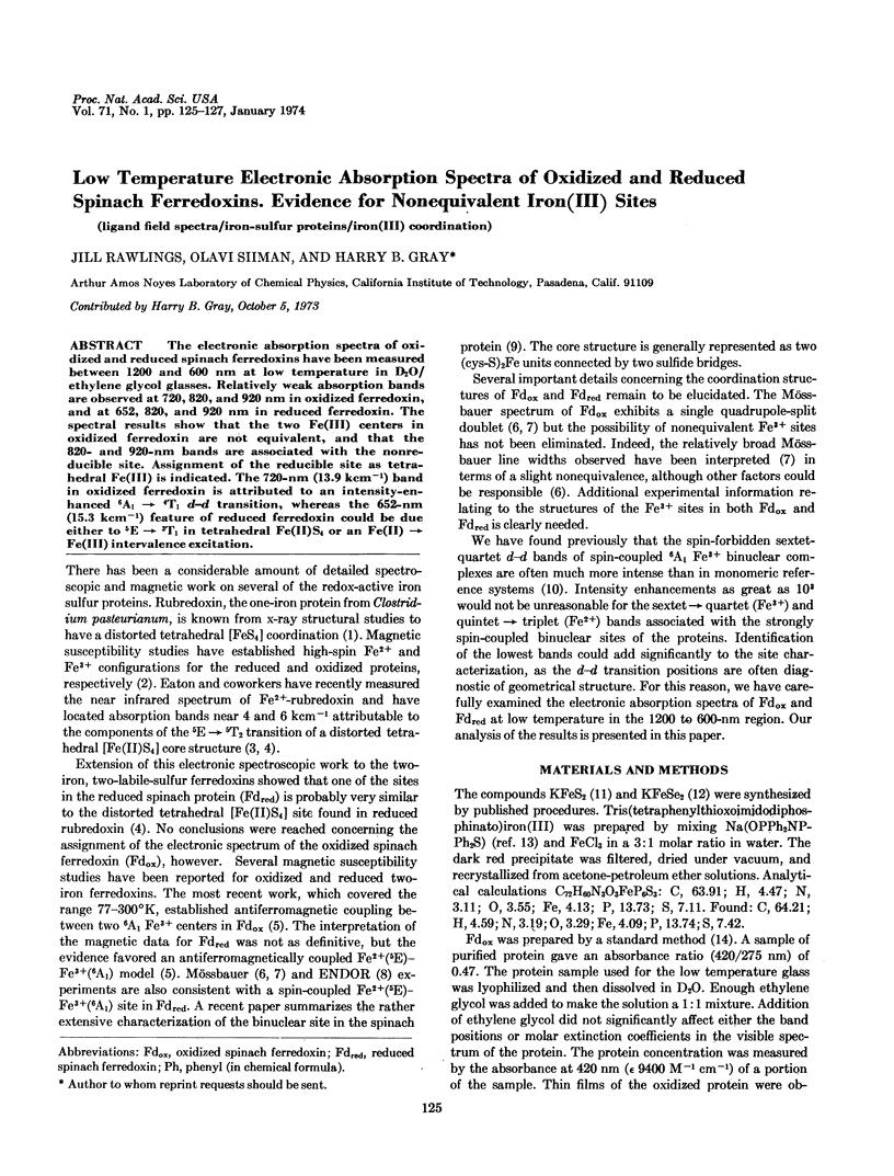
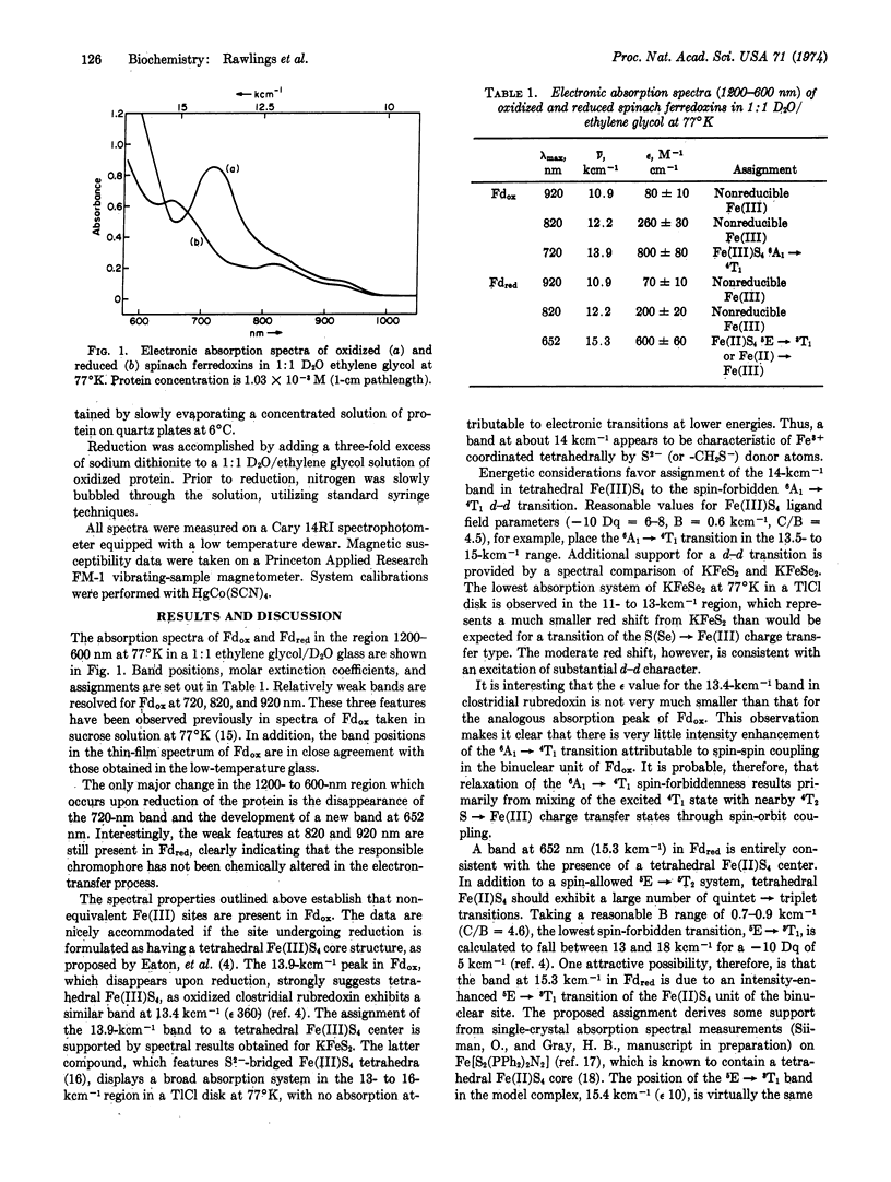
The electronic absorption spectra of oxidized and reduced spinach ferredoxins have been measured between 1200 and 600 nm at low temperature in D2O/ethylene glycol glasses. Relatively weak absorption bands are observed at 720, 820, and 920 nm in oxidized ferredoxin, and at 652, 820, and 920 nm in reduced ferredoxin. The spectral results show that the two Fe(III) centers in oxidized ferredoxin are not equivalent, and that the 820- and 920-nm bands are associated with the nonreducible site. Assignment of the reducible site as tetrahedral Fe(III) is indicated. The 720-nm (13.9 kcm-1) band in oxidized ferredoxin is attributed to an intensity-enhanced 6A1 → 4T1d-d transition, whereas the 652-nm (15.3 kcm-1) feature of reduced ferredoxin could be due either to 5E → 3T1 in tetrahedral Fe(II)S4 or an Fe(II) → Fe(III) intervalence excitation.
Keywords: ligand field spectra, iron-sulfur proteins, iron(III) coordination
Full text
PDF


Selected References
These references are in PubMed. This may not be the complete list of references from this article.
- Dunham W. R., Bearden A. J., Salmeen I. T., Palmer G., Sands R. H., Orme-Johnson W. H., Beinert H. The two-iron ferredoxins in spinach, parsley, pig adrenal cortex, Azotobacter vinelandii, and Clostridium pasteurianum: studies by magnetic field Mössbauer spectroscopy. Biochim Biophys Acta. 1971 Nov 2;253(1):134–152. doi: 10.1016/0005-2728(71)90240-4. [DOI] [PubMed] [Google Scholar]
- Dunham W. R., Palmer G., Sands R. H., Bearden A. J. On the structure of the iron-sulfur complex in the two-iron ferredoxins. Biochim Biophys Acta. 1971 Dec 7;253(2):373–384. doi: 10.1016/0005-2728(71)90041-7. [DOI] [PubMed] [Google Scholar]
- Eaton W. A., Lovenberg W. Near-infrared circular dichroism of an iron-sulfur protein. D leads to d transitions in rubredoxin. J Am Chem Soc. 1970 Dec 2;92(24):7195–7198. doi: 10.1021/ja00727a030. [DOI] [PubMed] [Google Scholar]
- Fritz J., Anderson R., Fee J., Palmer G., Sands R. H., Tsibris J. C., Gunsalus I. C., Orme-Johnson W. H., Beinert H. The iron electron-nuclear double resonance (ENDOR) of two-iron ferredoxins from spinach, parsley, pig adrenal cortex and Pseudomonas putida. Biochim Biophys Acta. 1971 Nov 2;253(1):110–133. doi: 10.1016/0005-2728(71)90239-8. [DOI] [PubMed] [Google Scholar]
- Herriott J. R., Sieker L. C., Jensen L. H., Lovenberg W. Structure of rubredoxin: an x-ray study to 2.5 A resolution. J Mol Biol. 1970 Jun 14;50(2):391–406. doi: 10.1016/0022-2836(70)90200-7. [DOI] [PubMed] [Google Scholar]
- Palmer G., Dunham W. R., Fee J. A., Sands R. H., Iizuka T., Yonetani T. The magnetic susceptibility of spinach ferredoxin from 77-250 degrees K: a measurement of the antiferromagnetic coupling between the two iron atoms. Biochim Biophys Acta. 1971 Aug 6;245(1):201–207. doi: 10.1016/0005-2728(71)90022-3. [DOI] [PubMed] [Google Scholar]
- Petering D. H., Palmer G. Properties of spinach ferredoxin in anaerobic urea solution: a comparison with the native protein. Arch Biochem Biophys. 1970 Dec;141(2):456–464. doi: 10.1016/0003-9861(70)90162-1. [DOI] [PubMed] [Google Scholar]
- Phillips W. D., Poe M., Weiher J. F., McDonald C. C., Lovenberg W. Proton magnetic resonance, magnetic susceptibility and Mössbauer studies of Clostridium pasteurianum rubredoxin. Nature. 1970 Aug 8;227(5258):574–577. doi: 10.1038/227574a0. [DOI] [PubMed] [Google Scholar]
- Rao K. K., Cammack R., Hall D. O., Johnson C. E. Mössbauer effect in Scenedesmus and spinach ferredoxins. The mechanism of electron transfer in plant-type iron-sulphur proteins. Biochem J. 1971 Apr;122(3):257–265. doi: 10.1042/bj1220257. [DOI] [PMC free article] [PubMed] [Google Scholar]
- Wilson D. F. The near infra-red electronic spectra of non-heme iron proteins at minus 196 degrees. Arch Biochem Biophys. 1967 Oct;122(1):254–256. doi: 10.1016/0003-9861(67)90149-x. [DOI] [PubMed] [Google Scholar]


