Abstract
Nuclei are precisely positioned within all cells, and mispositioned nuclei are a hallmark of many muscle diseases. Myonuclear positioning is dependent on Kinesin and Dynein, but interactions between these motor proteins and their mechanisms of action are unclear. We find that in developing Drosophila muscles, Dynein and Kinesin work together to move nuclei in a single direction by two separate mechanisms that are spatially segregated. First, the two motors work together in a sequential pathway that acts from the cell cortex at the muscle poles. This mechanism requires Kinesin-dependent localization of Dynein to cell cortex near the muscle pole. From this location Dynein can pull microtubule minus-ends and the attached myonuclei toward the muscle pole. Second, the motors exert forces directly on individual nuclei independently of the cortical pathway. However, the activities of the two motors on the nucleus are polarized relative to the direction of myonuclear translocation: Kinesin acts at the leading edge of the nucleus, whereas Dynein acts at the lagging edge of the nucleus. Consistent with the activities of Kinesin and Dynein being polarized on the nucleus, nuclei rarely change direction, and those that do, reorient to maintain the same leading edge. Conversely, nuclei in both Kinesin and Dynein mutant embryos change direction more often and do not maintain the same leading edge when changing directions. These data implicate Kinesin and Dynein in two distinct and independently regulated mechanisms of moving myonuclei, which together maximize the ability of myonuclei to achieve their proper localizations within the constraints imposed by embryonic development.
Keywords: Drosophila, Dynein, Kinesin, Nuclear movement, Muscle, Polarity
INTRODUCTION
Although depicted in textbooks as a sphere that occupies the center of the eukaryotic cell, nuclei are highly dynamic and precisely positioned based on developmental stage, cell type and cell behavior. In most systems, nuclear movement is dependent on microtubules (MTs) and the MT motor proteins cytoplasmic Dynein and Kinesin-1; however, different systems require different adaptors and regulators for the two motor proteins (Tsai et al., 2010; Fridolfsson et al., 2010; Lei et al., 2009; Cadot et al., 2012; Wilson and Holzbaur, 2012; Meyerzon et al., 2009). Yet many questions remain, including how the activities of Kinesin and Dynein are regulated, whether the two MT motor proteins work together or in opposition, and whether precise molecular mechanisms are conserved between cell types and over developmental time.
Muscle is an advantageous system in which to study mechanisms of nuclear movement. Myofibers, the cellular units of muscle, are multinucleate and position their myonuclei to maximize internuclear distances (Bruusgaard et al., 2003). Moreover, mispositioned myonuclei correlate with muscle disease (Romero, 2010) and muscle weakness (Metzger et al., 2012), illustrating the functional importance of proper positioning. Little is known regarding the mechanisms of myonuclear positioning, due in part to the lack of appropriate systems for such analysis. Studies in mice have identified some of the factors necessary to position myonuclei (Lei et al., 2009; Puckelwartz et al., 2009; Puckelwartz et al., 2010); however, examination of the dynamics of myonuclear movement in vivo is difficult in mammalian systems. Conversely, cell culture systems have provided dynamic data, such as the speed and dynamics of nuclear movement and the organization of the cytoskeleton (Cadot et al., 2012; Wilson and Holzbaur, 2012; Zhang et al., 2009a), but the myonuclei in in vitro myotubes do not have defined end-point destinations in contrast to their in vivo counterparts. To assess the mechanisms of myonuclear movement in vivo, we utilized the developing Drosophila melanogaster embryo. Similar to mammalian muscles, Drosophila muscle cells are multinucleate, and the myonuclei are positioned to maximize their internuclear distance (Metzger et al., 2012). Moreover, Drosophila embryos are amenable to time-lapse microscopy without perturbing development, such that dynamic movements can be assessed.
Myonuclei in the developing Drosophila embryo undergo several discrete nuclear movements governed by distinct mechanisms that have been best characterized in the lateral transverse (LT) muscles (Folker et al., 2012; Metzger et al., 2012). At the completion of fusion, all of the myonuclei within the muscles reside in a single cluster near the ventral pole of each LT muscle. Shortly after fusion [stage 14, 10:20-11:20 hours after egg laying (AEL)] this single cluster separates into two clusters - one ventral and one dorsal. During stage 15 (11:20-13 hours AEL) and stage 16 (13-16 hours AEL) these clusters move toward their respective poles. Finally, during stage 17 (16-20 hours AEL), the myonuclei become evenly spaced throughout the muscle (Metzger et al., 2012). These different myonuclear movements appear to be mechanistically distinct. For example, embryos that are mutant for the MT-associated protein Ensconsin fail to separate the single post-fusion nuclear cluster into distinct dorsal and ventral clusters. However, Dynein and Kinesin appear to be dispensable for the initial separation of the single post-fusion cluster into two, but are required to move the individual clusters toward the muscle poles (Folker et al., 2012; Metzger et al., 2012). More specifically, Dynein localized to the muscle pole pulls MT minus-ends and attached myonuclei toward the muscle pole (Folker et al., 2012). The role of Kinesin is less clear, and whether Dynein and Kinesin drive distinct aspects of myonuclear movement or work within a common pathway is not known.
We have examined the translocation of myonuclei toward the muscle poles during stage 15 (11:20-13 hours AEL) and find that, in developing Drosophila muscles, Dynein and Kinesin work together to move nuclei in a single direction. This contrasts with other systems, such as interkinetic nuclear migration in mammalian brains and the C. elegans hypodermis, where Kinesin and Dynein move nuclei in opposing directions (Fridolfsson and Starr, 2010; Tsai et al., 2010). As part of this analysis, we find that MTs are bidirectionally oriented throughout most of the developing muscle, unlike in these other in vivo contexts, perhaps promoting the cooperation of the two motors. Furthermore, Dynein and Kinesin cooperate to move nuclei by two separate mechanisms that are spatially segregated. First, the two motors work together in a pathway that acts from the cell cortex at the muscle poles. This mechanism requires Kinesin-dependent localization of Dynein to the cell cortex near the muscle pole. At this location, Dynein can pull MT minus-ends and the attached myonuclei toward the muscle pole. Second, the motors exert forces directly on individual nuclei independently of Dynein activity from the cortex, consistent with recent reports in cell culture (Cadot et al., 2012; Wilson and Holzbaur, 2012). However, we demonstrate that the activities of the two motors on the nucleus are polarized relative to the direction of myonuclear translocation: Kinesin acts at the leading edge of the nucleus, whereas Dynein acts at the lagging edge of the nucleus. Consistent with the activities of Kinesin and Dynein being polarized on the nucleus, nuclei rarely change directions, and those that do, reorient to maintain the same leading edge. Conversely, nuclei in both Kinesin and Dynein mutant embryos change direction more often and do not maintain the same leading edge when changing directions. We hypothesize that these functions increase myonuclear translocation efficiency in dense in vivo environments and enable myonuclei to reach their proper position within the time constraints of development.
RESULTS
A Kinesin- and Dynein-dependent mechanism regulates myonuclear position from the muscle cortex
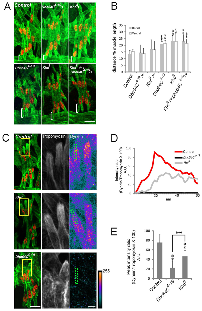
We previously identified Kinesin heavy chain (Khc) (Metzger et al., 2012) and Dynein (Folker et al., 2012) as necessary regulators of myonuclear positioning in Drosophila. To determine whether Kinesin and Dynein regulate the same or distinct aspects of myonuclear positioning, we tested whether they functionally interact to position myonuclei in stage 16 (16 hours AEL) embryos. At this developmental stage, the myonuclei have finished their translocation towards the muscle poles and reside in groups near either of two muscle poles. Embryos that were heterozygous for either Khc8 (null) or Dhc64C4-19 (null) properly positioned their myonuclei. However, Khc8/+; Dhc64C4-19/+ doubly heterozygous embryos displayed mispositioned myonuclei similar to Dhc64C4-19 and Khc8 homozygotes (Fig. 1A,B), suggesting that Kinesin and Dynein position myonuclei through a common pathway.
Fig. 1.
Dynein and Kinesin functionally interact to position myonuclei. (A) Immunofluorescence images of the lateral transverse (LT) muscles (Tropomyosin, green) and their myonuclei (dsRed, red) within a single hemisegment of a stage 16 (16 hours AEL) Drosophila embryo. Brackets indicate the distance between the muscle pole and the nearest nucleus. (B) The position of myonuclei within LT muscles of the indicated genotypes. Values are the distance from the dorsal (gray) or ventral (white) pole to the nearest myonucleus plotted as percentage of muscle length. Error bars indicate s.d. for 30 hemisegments in ten embryos. **P<0.01 compared with controls, with dorsal measurements and ventral measurements considered separately. (C) (Left) Immunofluorescence images of the LT muscles (green) and their nuclei (red) from a single hemisegment. Yellow boxes indicate regions shown at higher magnification to the right. (Right) High-magnification images of the muscle (Tropomyosin) and Dynein near the poles of muscles. Dynein images are shown as heat maps to depict Dynein accumulations. Scale indicating relative intensities is shown bottom right. Green boxes indicate regions used for the linescan analysis shown in D. (D) Intensity profile of Dynein immunofluorescence relative to Tropomyosin immunofluorescence plotted as a function of position. (E) The average peak intensity values for Dynein immunofluorescence in the indicated genotypes. Error bars indicate s.d. for 30 linescans from 15 embryos from three independent experiments. **P<0.01 compared with control, except where indicated by brackets. Scale bars: 10 μm in A, C left; 5 μm in C middle and right. A.U., arbitrary units.
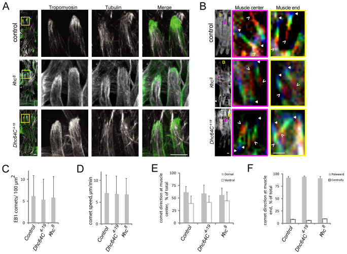
Both Dynein and Kinesin regulate general MT organization (Malikov et al., 2004; Straube et al., 2006), and mispositioned myonuclei correlate with a decreased density of MTs near the muscle poles (Folker et al., 2012). However, in fixed embryos MTs were unaffected, reaching the muscle pole in both Dhc64C4-19 and Khc8 embryos (Fig. 2A). To further examine MT organization, EB1-GFP or EB1-eYFP was expressed in muscle using the UAS/GAL4 system (Brand and Perrimon, 1993). Several characteristics of EB1 comets were measured: the number of comets (number of growing MTs) (Fig. 2C); the velocity of comet movement (MT polymerization rate) (Fig. 2D); and the direction of comets both within the muscle where the central-most nuclei are located (Fig. 2B,E) and at the ends of muscles (gross MT organization) (Fig. 2B,F). Of note, MT organization is different in the regions of the muscle between the muscle pole and the nearest nucleus (Fig. 2F) as compared with all other locations within the muscle (Fig. 2E). Thus, only the nucleus closest to the muscle pole experiences a highly polarized MT array whereas the majority of the nuclei experience MTs that grow in both directions along the dorsal-ventral axis of the muscle. All measurements were similar to control in both Dhc64C4-19 and Khc8 mutants (Fig. 2B-F; supplementary material Movie 1), indicating that the loss of Dynein or Kinesin does not affect myonuclear positioning by disrupting global MT organization.
Fig. 2.
MT organization within LT muscles. (A) (Left) Muscles (Tropomyosin, green) and MTs (tubulin, gray) in the LT muscles of a stage 16 (16 hours AEL) embryo. Yellow boxes indicate regions shown at higher magnification to the right. (Right) Higher magnification images show that MTs reach the muscle poles in each genotype. (B) EB1 tracking in the muscles of late stage 15/early stage 16 embryos of the indicated genotypes. z-stacks were acquired through the entire muscle every 6 seconds and three consecutive frames were summed. Consecutive sums were then pseudocolored red, green, and blue. These images were overlaid to show the direction of comet movement. (Left) Full hemisegment, with pink and yellow boxes indicating the regions of the muscle used to produce the higher magnification views of MT organization near the central-most nucleus and between the poleward-most nucleus and the muscle pole, respectively. (Right) Higher magnification views of individual moving EB1 comets, with their starting point shown in red and indicated by carets and their latest position shown in blue and indicated by arrowheads. (C) The number of EB1 comets per 100 μm2. (D) The speed of EB1 comet movement in the LT muscles. (E) Direction of EB1 comets within the central part of the muscle. (F) The direction of EB1 comets that are detected specifically at the ends of muscles. In C-F error bars are s.d. from >100 EB1 tracks in at least four muscles from each of five embryos. Scale bars: 5 μm in A, B left; 1 μm in B right.
Defects in myonuclear position also correlate with decreased levels of Dynein near the muscle pole (Folker et al., 2012). As Kinesin regulates Dynein localization in other systems (Zhang et al., 2003), we examined Dynein localization in Khc8 embryos and found that Dynein near the muscle pole was reduced (Fig. 1C-E). Hence, Kinesin is required for proper Dynein localization at the muscle poles. However, this does not exclude other roles for Kinesin during myonuclear movement.
Segregated Kinesin and Dynein activities are required for distinct phases of myonuclear movement
If Kinesin contributes to myonuclear translocation only by regulating the localization of Dynein, nuclear movement dynamics should be similarly affected in both Khc8 and Dhc64C4-19 embryos. The speed at which the dorsal cluster and ventral cluster of nuclei separate from one another was assessed by in vivo time-lapse microscopy and found to be similarly reduced in both Dhc64C4-19 and Khc8 embryos. In both genotypes, the clusters separated at 60% of the speed of those in control embryos (Fig. 3A,B). Furthermore, the velocity of individual nuclei was measured. For this measurement, only myonuclei that were actively translocating relative to a stationary landmark within the embryo were assessed. In both Dhc64C4-19 and Khc8 embryos, myonuclear translocation velocity was reduced by more than 50% (Fig. 3C). That the speeds were similarly affected in both Dhc64C4-19 and Khc8 mutants is consistent with a model in which Kinesin regulates Dynein localization to the muscle end, and then Dynein acts to move myonuclei.
Fig. 3.

Nuclear translocation is impaired in both Kinesin and Dynein mutant embryos. (A) Kymographs showing separation of the dorsal and ventral nuclear clusters within a single LT muscle of late stage 15 embryos of the indicated genotypes expressing the apME-NLS::dsRed transgene to visualize myonuclei. Scale bar: 5 μm. (B) The speed at which the dorsal and ventral clusters of nuclei separate. Error bars indicate s.d. from 20 movies from at least five embryos. **P<0.01, *P<0.05, compared with control unless otherwise indicated by brackets. (C) The translocation velocity of individual nuclei in the indicated genotypes. Error bars indicate s.d. from the measurements of 100 individual nuclei from five embryos. **P<0.01, compared with control.
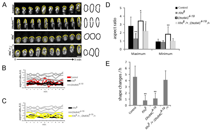
Closer analysis revealed that translocating myonuclei in control embryos had an identifiable front and back and continually changed shape during translocation. Specifically, the leading edge of the nucleus moved forward before the rear of the nucleus advanced to complete a translocation cycle (Fig. 4A). Thus, translocating nuclei shifted between spherical and elongated shapes (Fig. 4A,B,D,E). Conversely, the myonuclei in Khc8 and Dhc64C4-19 mutants did not change shape during translocation. In Khc8 embryos, the myonuclei remained spherical, whereas those in Dhc64C4-19 embryos remained elongated (Fig. 4A,B,D,E).
Fig. 4.
Translocating myonuclei change shape in a Kinesin- and Dynein-dependent manner. (A) Kymographs of individual translocating nuclei in the indicated genotypes with all nuclei moving upward. Yellow solid lines indicate the leading edge of the myonucleus and highlight leading edge dynamics during translocation. Yellow dashed lines complete the perimeter of nuclei at individual time points and are combined with the yellow solid lines to produce the shape illustrations (right) that highlight shape changes over time. Scale bar: 2 μm. (B,C) The aspect ratios for each of five translocating myonuclei were measured and plotted over time for the indicated genotypes for visual comparison. (D) The average maximum and minimum aspect ratio of myonuclei as they translocate for between 20 minutes and 1 hour. (E) The number of shape changes that individual myonuclei undergo. Values were calculated by following individual myonuclei as they moved for between 20 minutes and 1 hour. In D,E error bars represent the s.d. from the measurement of 100 individual nuclei from five embryos. *P<0.05, **P<0.01, compared with control.
These data suggest that Kinesin exerts a forward force on the front of the myonucleus and that Dynein exerts a retractive force, or relieves an adhesive force, on the back of the myonucleus. We next examined nuclear morphology in Khc8/+; Dhc64C4-19/+ doubly heterozygous embryos, hypothesizing that if the shape changes resulted from asymmetric forces exerted by Dynein and Kinesin then they would be maintained when Dynein and Kinesin are similarly reduced. Despite slightly reduced velocity (Fig. 3A-C) and improper positioning by end point analysis (Fig. 1A,B), translocating myonuclei in Khc8/+; Dhc64C4-19/+ embryos continually changed shape during translocation (Fig. 4A,C-E), suggesting that opposing forces exerted by Kinesin and Dynein drive nuclear shape changes. However, the magnitude of the shape changes was reduced in the double heterozygotes, indicating some sensitivity to Kinesin and Dynein levels (Fig. 4C,D).
Distinct leading and lagging edges to moving nuclei are essential for directionality
Consistent with the activities of Kinesin and Dynein being polarized, myonuclei in control embryos moved unidirectionally; fewer than 2% of myonuclei changed direction every hour (Fig. 5A; n=250). Importantly, the translocation of all myonuclei is unidirectional, including that of nuclei most centrally positioned within the myotube, which experience a bipolar orientation of the MT network. However, in both Dhc64C4-19 and Khc8 embryos, 10% of nuclei changed direction every hour (Fig. 5A; n=250), suggesting that Dynein and Kinesin move nuclei and confer directionality to that translocation. Moreover, directionality was maintained in Khc8/+; Dhc64C4-19/+ doubly heterozygous embryos, suggesting that the balance of forces between Dynein and Kinesin, rather than the levels of the two proteins, contributes to directionality (Fig. 5A).
Fig. 5.

Nuclei maintain their leading edge when changing direction in a Kinesin- and Dynein-dependent manner. (A) The percentage of nuclei that change direction in the indicated genotypes. Error bars represent s.d. for 250 nuclei in 20 embryos from three independent experiments. **P<0.01, compared with control unless otherwise indicated by brackets. (B) Kymographs of nuclei that change direction in the indicated genotypes. Arrows indicate puncta that show the nucleus reorienting prior to changing direction (control and Khc8/+; Dhc64C4-19/+ double heterozygotes) or maintaining their relative positions during the direction change (Khc8 and Dhc64C4-19). Asterisks indicate additional nuclei within the field of view. Brackets indicate translocation relative to additional nuclei. White bars indicate the time at which the direction of nuclear movement changes. Scale bar: 2 μm.
Analysis of the nuclei that did change direction further suggested that translocating myonuclei are polarized. In control embryos, the few myonuclei that changed direction reoriented and maintained the same leading edge after changing direction by either rotating (Fig. 5B; supplementary material Movie 2) or by performing U-turns similar to an automobile (supplementary material Movie 3). Conversely, although nuclei changed direction more often in both Dhc64C4-19 and Khc8 embryos (Fig. 5A), they did not reorient to maintain the same leading edge (Fig. 5B; supplementary material Movie 2). Taken together, these data suggest that Dynein and Kinesin activities confer a molecular distinction between the leading and lagging edges of translocating myonuclei. More fundamentally, these observations indicate that the leading edge of the nucleus actively participates in leading the direction of nuclear translocation.
As part of this analysis we noted that nuclei rotate more often than they change direction (compare Fig. 6A,B and Fig. 5A). Most nuclear rotations were either not associated with nuclear translocation or resulted in the nucleus continuing on its previous path. Importantly, in those cases in which a nucleus continues on is previous path, it stops rotating with its leading edge pointing in the same direction as that in which it was previously moving. The lack of a strict correlation between nuclear rotations and direction changes suggests that nuclear rotations are not a means to change direction. Instead, it seems that nuclear rotations occur when the symmetry of the opposing forces normally exerted by Dynein and Kinesin to alter nuclear shape is broken. Thus, rotation is evidence of inefficient force transmission. Consistent with this hypothesis, nuclei that were rotating in either control or Khc8/+; Dhc64C4-19/+ doubly heterozygous embryos were primarily round, with aspect ratios consistently close to one (Fig. 6C). Additionally, the aspect ratio of rotating nuclei in Dynein mutants was significantly reduced compared with that of translocating nuclei, whereas the aspect ratio in Kinesin mutants was similar in rotating and translocating nuclei (Fig. 6C). This indicates that rotating nuclei are not subject to the same forces as translocating nuclei.
Fig. 6.
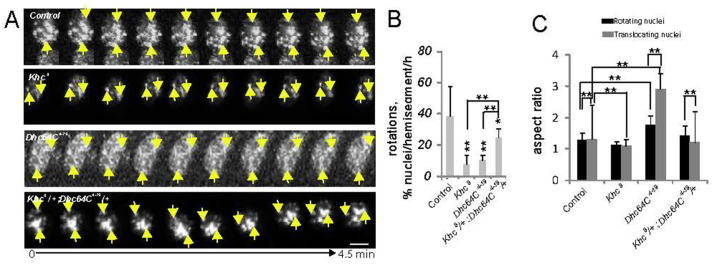
Nuclear rotation is not associated with translocation. (A) Kymographs of stationary myonuclei that express the apME-NLS::dsRed transgene. Arrows indicate puncta that either move relative to one another (control and Khc8/+; Dhc64C4-19/+) or maintain their positions (Khc8 and Dhc64C4-19). Scale bar: 2 μm. (B) The percentage of myonuclei that rotate in the indicated genotypes. (C) The average aspect ratio of nuclei as they either rotate (black) or translocate (gray). For B,C, error bars represent s.d. from the measurement of 250 nuclei from 20 embryos and three independent experiments. **P<0.01, compared with control unless otherwise indicated by brackets. Translocating nuclei in control and Khc8 embryos were compared by a z-test because they had similar averages but different distributions.
Nuclear polarization occurs independently of the cortical mechanism contributing to nuclear movement
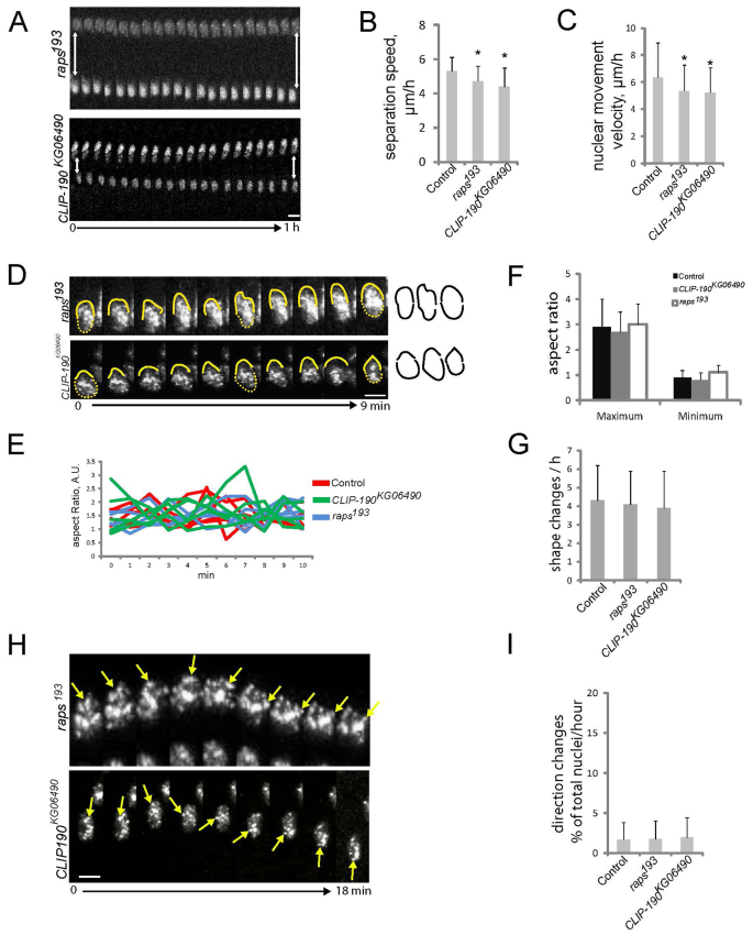
We previously demonstrated that Dynein accumulation near the muscle poles, and the presence of MTs near the muscle pole, were required to properly position myonuclei (Folker et al., 2012). To determine whether cortical Dynein and MT-cortex interactions contribute to the directionality or the shape changes of translocating myonuclei, we examined myonuclear translocation in raps193 and CLIP-190KG06490 mutant embryos, in which cortical Dynein localization and cortical MTs are disrupted, respectively (Folker et al., 2012). Although the myonuclei moved more slowly in each genotype (Fig. 7A-C), they exhibited shape changes with identifiable leading and lagging edges (Fig. 7D-G) and moved unidirectionally (Fig. 7I). Moreover, nuclei in both raps193 and CLIP-190KG06490 mutants that changed direction reoriented and maintained their leading edge (Fig. 7H). These data indicate that the cortical pathway (Folker et al., 2012) (Fig. 1) does not affect the ability of Dynein and Kinesin to alter the shape of translocating myonuclei. Moreover, these data indicate that, although cortical Dynein and MTs are not required to alter the shape of translocating myonuclei, they do contribute to myonuclear translocation.
Fig. 7.
Nuclear shape changes are independent of cortical pulling. (A) Kymographs showing separation of the dorsal and ventral nuclear clusters within a single LT muscle of late stage 15 (13 hours AEL) embryos of the indicated genotypes. (B) The average speed at which the dorsal and ventral clusters of nuclei separate. Error bars are s.d. from 20 movies from at least five embryos. (C) The translocation velocity of individual nuclei in the indicated genotypes. Error bars, s.d. from the measurements of 100 individual nuclei from five embryos. (B,C) *P<0.05, compared with control. (D) Kymographs of individual translocating nuclei in the indicated genotypes with all nuclei moving upward. Yellow solid lines indicate the leading edge of the translocating nucleus and highlight leading edge dynamics. Yellow dashed lines complete the perimeter of nuclei at individual time points and are combined with the yellow solid lines to produce the shape illustrations shown to the right that highlight shape changes over time. (E) The aspect ratios for each of five translocating myonuclei were measured and plotted over time for the indicated genotypes for visual comparison. The nuclei in CLIP-190KG06490 (green) and raps193 (blue) embryos dynamically fluctuate between spherical (aspect ratio ∼1) and elongated (aspect ratio ∼2.5), similar to control (red). (F) The average maximum and minimum aspect ratio of myonuclei as they translocate over a 10-minute period. (G) The number of shape changes that individual myonuclei undergo. Values were calculated by following individual myonuclei as they moved for between 20 minutes and 1 hour. For F,G, error bars represent the s.d. from the measurement of 100 individual nuclei from five embryos. Differences are statistically insignificant. (H) Kymographs of nuclei that change direction in the indicated genotypes. Arrows indicate puncta that show the nucleus reorienting prior to changing direction in both CLIP-190KG06490 and raps193, similar to the control shown in Fig. 5B. (I) The percentage of nuclei that change direction in the indicated genotypes. Error bars represent s.d. for 250 nuclei in 20 embryos from three independent experiments. Differences are statistically insignificant. Scale bars: 5 μm in A; 2 μm in D,H.
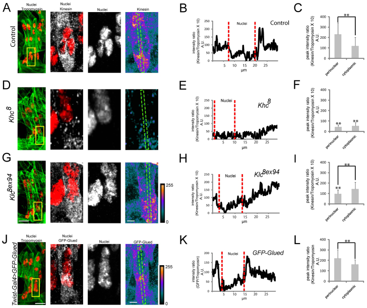
Because the cortical pathway appeared dispensable for nuclear shape dynamics, we hypothesized that nuclear shape changes result from the opposed forces of Dynein and Kinesin acting directly on the nucleus. Consistent with this, in control embryos Kinesin accumulates near myonuclei (Fig. 8A-C). This localization was not seen in Khc8 embryos, indicating that the localization is not the result of nonspecific antibody accumulations (Fig. 8D-F). Additionally, Kinesin failed to localize near the nucleus in Klc8ex94 mutants (Fig. 8G-I), consistent with other systems in which Kinesin light chain regulates interactions between Kinesin-1 and the nucleus (Fridolfsson and Starr, 2010; Meyerzon et al., 2009). Dynein was not detected near myonuclei, however, GFP-Glued, a well-described regulator of Dynein activity that regulates Dynein-nucleus interactions in other systems (Cadot et al., 2012; Fridolfsson and Starr, 2010; Wilson and Holzbaur, 2012), did accumulate near the myonuclei (Fig. 8J-L). Additionally, it often appeared that Glued was more strongly enriched at the back of the myonucleus compared with the front and that Kinesin was more strongly enriched at the front of the myonucleus compared with the back. However, the myonuclei are tightly clustered such that it is impossible to determine with which specific myonucleus the immunofluorescence is associated. Nevertheless, these data are consistent with other systems in which Kinesin and Dynein exert force directly on myonuclei.
Fig. 8.
Kinesin and the Dynactin subunit Glued accumulate near nuclei. (A,D,G) (Left) Immunofluorescence images of the LT muscles (green) and their nuclei (red) in a single hemisegment of a stage 16 embryo. Yellow boxes indicate regions shown at higher magnification to the right. (Right) Higher magnification images of the nuclei and Khc, shown as merged images (red, nuclei; gray, Khc) and as separate signals with Khc shown as a heat map to illustrate regions of accumulation near the nucleus in control embryos and toward the muscle pole in Klc8ex94 embryos. Scale indicating relative intensities is shown bottom right of G. Green boxes indicate regions used for linescan intensity profiles. (B,E,H) Intensity profiles of Khc in control (B), Khc8 (E) and Klc8ex94 (H) embryos. (C,F,I) The average intensity of the Khc immunofluorescence signal both perinuclear and distant from the nucleus in the cytoplasm of control (C), Khc8 (F) and Klc8ex94 (I) embryos. (J) (Left) Immunofluorescence image of the LT muscles (green) and their nuclei (red) in a single hemisegment in a stage 16 embryo expressing GFP-Glued specifically in the mesoderm. Yellow box indicates the region shown at higher magnification to the right. (Right) Higher magnification images of nuclei and GFP-Glued, shown as merged images (red, nuclei; gray, GFP-Glued) and as separate signals with GFP-Glued shown as a heat map to illustrate regions of accumulation. Green box indicates the region used for the linescan intensity profile. (K) Intensity profile of GFP-Glued in a muscle expressing the transgene. (L) The average intensity of GFP-Glued fluorescence signal both perinuclear and distant from the nucleus in the cytoplasm. For C,F,I,L, error bars are the s.d. from 30 muscles in 15 embryos from three independent experiments. **P<0.01, compared with control in C unless otherwise indicated by brackets. Scale bars: 10 μm in A,D,G,J left; 5 μm in A,D,G,J right.
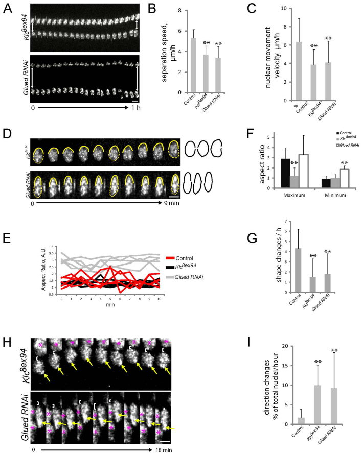
Additionally, myonuclei in Klc8ex94 mutants resembled those in Khc8 mutants: nuclei moved more slowly (Fig. 9A-C), remained spherical (Fig. 9D-G), changed direction more often (Fig. 9I) and did not reorient when changing direction (Fig. 9H), suggesting that Klc mediates Kinesin-nucleus interactions that are necessary to polarize the nucleus. Similarly, the nuclei in embryos depleted of Glued resembled those in Dynein mutants: they moved more slowly than controls (Fig. 9A-C), were persistently elongated (Fig. 9D-G), changed direction more frequently (Fig. 9I) and did not reorient when they changed direction (Fig. 9H). These data suggest that the Dynactin subunit Glued mediates Dynein-nucleus interactions and that these interactions are necessary for Dynein and Kinesin to exert force directly on the nucleus.
Fig. 9.
Klc and Glued are necessary for Kinesin and Dynein to regulate nuclear dynamics, respectively. (A) Kymographs showing separation of the dorsal and ventral nuclear clusters within a single LT muscle of late stage 15 (13 hours AEL) embryos of the indicated genotypes. (B) The average speed at which the dorsal and ventral clusters of nuclei separate. Error bars are s.d. from 20 movies from at least five embryos. (C) The translocation velocity of individual nuclei in the indicated genotypes. (D) Kymographs of individual translocating nuclei in the indicated genotypes with all nuclei moving upward. Yellow solid lines indicate the leading edge of the nucleus and highlight leading edge dynamics. Yellow dashed lines complete the perimeter of nuclei at individual time points and are combined with the yellow solid lines to produce the shape illustrations shown to the right that highlight shape changes over time. (E) The aspect ratios for each of five translocating myonuclei were measured and plotted over time for the indicated genotypes for visual comparison. The nuclei in Klc8ex94 (black) and Glued RNAi (gray) maintain their shapes, unlike those in controls, which dynamically change (red). (F) The average maximum and minimum aspect ratio of myonuclei as they translocate for between 20 minutes and 1 hour. (G) The number of shape changes that individual myonuclei undergo. Values were calculated by following individual myonuclei as they moved for between 20 minutes and 1 hour. For B,C,F,G, error bars represent the s.d. from the measurement of 100 individual nuclei from five embryos; **P<0.01, compared with control. (H) Kymographs of nuclei that change direction in the indicated genotypes. Arrows indicate puncta that maintain their relative positions in Klc8ex94 and Glued RNAi embryos, similar to the Khc8 and Dhc64C4-19 mutants shown in Fig. 5B. Asterisks indicate additional nuclei within the field of view. Brackets indicate translocation relative to additional nuclei. (I) The percentage of nuclei that change direction in the indicated genotypes. Error bars represent s.d. for 250 nuclei in 20 embryos from three independent experiments. **P<0.01, compared with control. Scale bars: 5 μm in A; 2 μm in D,H.
DISCUSSION
The developing muscles of the Drosophila embryo provide unique advantages in determining the mechanisms and impact of nuclear movement. Like their mammalian counterparts, Drosophila muscles are multinucleate and the myonuclei are precisely positioned to maximize internuclear distance. Importantly, improper positioning has been correlated with muscle disease (Romero, 2010) and may contribute to muscle weakness (Metzger et al., 2012). Moreover, Drosophila provides a system amenable to in vivo time-lapse microscopy, in which the subcellular dynamics necessary for proper cellular organization can be assessed without perturbing development. Here we used a combination of genetics, immunolocalization and time-lapse microscopy of live Drosophila embryos to determine that Kinesin and Dynein together move myonuclei by at least two independent mechanisms (Fig. 10).
Fig. 10.
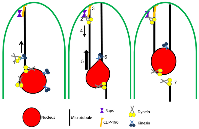
Model for the movement of muscle nuclei toward muscle poles. Myonuclear translocation depends on at least two Kinesin- and Dynein-dependent pathways. The first pathway acts from the cortex and is depicted in steps 1-5. (1) Kinesin transports Dynein to the muscle pole. (2) At the pole, Dynein is anchored by Raps. (3) In a CLIP-190-dependent manner, MTs interact with the muscle pole. (4) Dynein that is anchored at the pole pulls on the CLIP-190-dependent MTs. (5) Because Dynein is stably attached to the muscle pole and the MT minus-ends are attached to the nucleus, the nuclei are moved toward the muscle pole. Additionally, in a second pathway, (6) Kinesin stretches the leading edge of the translocating nucleus and (7) Dynein releases the trailing edge of the nucleus. These functions result in nuclei that dynamically change morphology during translocation, maximizing their ability to move under the temporal constraints of development.
The first mechanism requires the sequential activities of Kinesin and Dynein, in which ultimately Dynein pulls nuclei toward the muscle cortex. Specifically, Kinesin is required to localize Dynein to the muscle poles. Once anchored near the muscle pole, Dynein pulls MT minus-ends and the attached myonuclei toward the muscle pole, as previously suggested (Folker et al., 2012).
The second Kinesin- and Dynein-dependent mechanism involves the two motors generating force directly on individual nuclei, consistent with data from cell culture (Cadot et al., 2012; Wilson and Holzbaur, 2012). These activities of Kinesin and Dynein at the surface of the nucleus are, however, spatially segregated: Kinesin acts at the leading edge and Dynein acts at the lagging edge. This conclusion is based on the distinct behaviors of translocating myonuclei in both Kinesin and Dynein mutant embryos compared with those in control embryos. In controls, myonuclei continually shift between spherical and elongated shapes. However, when Kinesin activity is lost the myonuclei remain spherical, whereas when Dynein activity is lost the myonuclei remain elongated (Fig. 4). Thus, Kinesin provides a pulling force that extends the leading edge of the myonucleus in the direction of nuclear translocation to generate the elongated shape. Conversely, Dynein provides the force that releases the trailing edge of the translocating myonucleus to return that nucleus to a spherical shape (Table 1). It is not clear how Dynein facilitates the retraction of the trailing edge of the myonucleus. One possibility is that MTs emanating from adjacent myonuclei are cross-linked and function as a staple connecting the myonuclei. Dynein might then resolve myonucleus-myonucleus interactions to facilitate the forward translocation of individual myonuclei.
Table 1.
Summary of nuclear dynamics in the tested genotypes

The conclusion that Kinesin and Dynein activities are spatially restricted on the nucleus is significant. At its core, this suggests that the leading and lagging edges of translocating myonuclei are molecularly distinct. This molecular distinction is supported by two findings. First, the translocation of nuclei in this system is unidirectional; fewer than 2% of nuclei change direction (Fig. 5). Second, the nuclei that do change direction reorient to maintain the same leading edge (Fig. 5). Thus, there is a molecular signature to the front of the translocating myonucleus such that it must lead the translocation. Both the unidirectional nature of nuclear translocation and the maintenance of the leading edge during directional changes require both Kinesin and Dynein activities, suggesting that the segregation of Kinesin and Dynein motor activities provides this molecular signature, consistent with their distinct effects on myonuclear shape.
The molecular distinction of the leading and lagging edges of the myonucleus is maintained in both CLIP-190KG06490 and raps193 mutant embryos (Fig. 7). We had previously demonstrated that CLIP-190 is necessary for proper MT organization at muscle poles, that Raps is necessary for Dynein accumulation at muscle poles, and that both of these activities are required for Dynein to generate force from the muscle pole (Folker et al., 2012). Thus, the regulation of nuclear shape changes and directionality occurs independently of Dynein-dependent pulling of nuclei from the cell cortex near the muscle pole and therefore represents a novel mechanism of myonuclear translocation (Table 1).
The mechanism by which the activities of Dynein and Kinesin are polarized is a key avenue for future work. The orientation of the MT cytoskeleton could enable this polarization if most of the MT plus-ends were oriented in a single direction. However, the nuclei positioned furthest from the muscle pole exhibit the same directionality and shape changes as those nuclei nearest to the muscle pole, despite the two nuclei experiencing different MT organizations (Fig. 2). The proximity of adjacent nuclei might produce local asymmetries in MT organization, which then causes Kinesin activity to cluster at the front of the nucleus and Dynein activity to cluster at the back. Indeed, clustering of motors has been proposed as a means to increase efficiency for moving large cargos (Erickson et al., 2011), but this is a question that requires more extensive analysis as it is complicated by the clustered nature of myonuclei during these movements. Alternatively, a signaling gradient from the muscle pole could provide a cue that activates Kinesin, or deactivates Dynein, on the front of the nucleus. Finally, perhaps nuclear protein(s) spatially segregate either motor activity or the ability of motors to transmit their force to the nucleus. Many lines of data suggest that Nesprin, SUN and Lamin proteins (Elhanany-Tamir et al., 2012; Fridolfsson et al., 2010; Lei et al., 2009; Starr et al., 2001; Wilson and Holzbaur, 2012; Zhang et al., 2009b; Meyerzon et al., 2009) could accomplish this goal, but our experiments to date have failed to implicate any of these proteins in myonuclear translocation in the Drosophila embryo.
When combined with previous work (Folker et al., 2012; Metzger et al., 2012), we suggest that at least three distinct mechanisms move myonuclei to the muscle poles in the Drosophila embryo. First, at the completion of fusion, a single cluster of nuclei is broken into distinct dorsal and ventral clusters of myonuclei. The details of how this occurs are not clear, but the MT-associated protein Ensconsin is required for this process, whereas Dynein and Kinesin do not seem to be necessary (Folker et al., 2012; Metzger et al., 2012) (this work). Next, the individual clusters move toward their respective muscle poles via two distinct Kinesin- and Dynein-dependent mechanisms. One mechanism relies on Dynein activity at the muscle pole and a second mechanism depends on Kinesin and Dynein acting directly on the nucleus (Folker et al., 2012) (this work, Fig. 10). That there are multiple distinct mechanisms driving nuclear movement complicates analysis and might explain why some nuclear translocation persists even when individual mechanisms are abrogated.
To fully understand myonuclear translocation, we will need to determine whether these pathways act simultaneously or in series. In this regard it is interesting to consider the Khc8/+; Dhc64C4-19/+ double heterozygotes. Myonuclei in embryos of this genotype exhibit normal directionality and change shape similarly to those in controls. Yet, these nuclei move more slowly than those in control embryos and are mispositioned by end point analysis. Moreover, the speed of myonuclear translocation in Khc8/+; Dhc64C4-19/+ double heterozygotes is more similar to that of raps193 and CLIP-190KG06490 mutant embryos than it is to Dhc64C4-19 or Khc8 embryos. This might suggest that the action of Dynein from the muscle pole is more sensitive to Dynein and Kinesin levels than is the action of Dynein and Kinesin proximal to the nucleus. Furthermore, it might suggest that the Khc8/+; Dhc64C4-19/+ double heterozygotes are specifically impacting the contribution of Dynein at the muscle pole with regards to nuclear translocation. This would suggest that at least these two pathways are working simultaneously, but more analysis must be performed to resolve this question.
The ability of Kinesin and Dynein activities to alter the shape of moving nuclei is likely to be essential for nuclei to navigate the dense space of the developing muscle. Furthermore, overlaying multiple mechanisms that aim to achieve the same end is an elegant way to efficiently regulate cellular and tissue development within strict time constraints. It seems likely that the constraints of development necessitate the layering of mechanisms to make necessary processes plastic, yet robust. Using the same factors in multiple ways allows the cell to conserve energy at the level of gene regulation and provides an environment in which slight variations in the concentrations of individual proteins will not be catastrophic. We have described a biological setting in which this means of regulation enables muscle cells to rapidly respond to environmental pressures such that they can efficiently organize their cytoplasm within the time constraints imposed by development.
MATERIALS AND METHODS
Drosophila genetics
The following stocks were grown under standard conditions: apME-NLS::dsRed (Richardson et al., 2007), Dhc64C4-19 (Gepner et al., 1996), Khc8 (Brendza et al., 1999), CLIP-190KG06490 (Bloomington Drosophila Stock Center, 14493), raps193 (Parmentier et al., 2000), Klc8ex94 (Gindhart et al., 1998), UAS-Glued RNAi (Vienna Drosophila RNAi Center, v3785), UAS-EB1-eYFP and UAS-EB1-GFP (Rogers et al., 2008) and UAS-GFP-Glued(3) (Bloomington Drosophila Stock Center, 29983). Mutants were balanced and identified using CTG (CyO, twi-Gal4, UAS-2xeGFP), TTG (TM3, twi-Gal4, UAS-2xeGFP), TM6B, CyO P[w+wgen11lacZ].
Immunohistochemistry
Stage 16 embryos were collected at 25°C and fixed with 4% EM-grade paraformaldehyde (Polysciences, 00380) diluted in PBS (Roche, 11666789001) with 50% heptane. Stained embryos were mounted in ProLong Gold (Invitrogen). Antibodies were preabsorbed (PA) where noted and used at the following final dilutions: rabbit anti-dsRed (PA, 1:200; Clontech, 632496), rat anti-Tropomyosin (PA, 1:500; Abcam, ab50567), mouse anti-GFP (PA, 1:200; Clontech, 632381), mouse anti-DHC (1:50; Developmental Studies Hybridoma Bank), mouse anti-Tubulin (1:500; Sigma, T9026) and rabbit anti-Khc (1:200; Cytoskeleton, AKIN01). We used Alexa Fluor 488-, Alexa Fluor 555- and Alexa Fluor 647-conjugated fluorescent secondary antibodies (1:200; Invitrogen). Fluorescent images were acquired on a Leica SP5 laser-scanning confocal microscope equipped with a 63× 1.4 NA HCX PL Apochromat oil objective and LAS AF 2.2 software. Maximum intensity projections of confocal z-stacks were rendered using Volocity Visualization software (Improvision) and further processed in ImageJ (NIH).
Immunofluorescence intensity quantification
For Kinesin and GFP-Glued, embryos were collected and fixed with 4% paraformaldehyde diluted 1:1 in heptane for 20 minutes; antibody incubations were performed in PBS supplemented with 1% BSA and 0.3% Triton X-100. For Dynein, embryos were fixed and stained as previously described (Folker et al., 2012). For both sets of data 2 μm stacks were acquired with a 0.25 μm step size, and projections of these stacks were rendered using Volocity Visualization software and further processed and analyzed in ImageJ. The section of the muscle that was imaged was chosen solely by examination of the nuclear signal and was central within the muscle to limit interference from the overlying and underlying tissues. Regions used for linescans were chosen by looking only at the signal from the nuclear immunofluorescence and then transferring that region to the appropriate Dynein, Kinesin or GFP-Glued image for analysis. Histograms indicate average values from 30 linescans that were obtained from 15 embryos from three independent experiments.
Nuclear position and muscle length measurements
Analysis was identical to that described previously (Folker et al., 2012).
Live embryo imaging and analysis
Embryos were harvested at 25°C and dechorionated with 50% bleach for 4 minutes. Embryos were then mounted on a gas-permeable membrane in halocarbon oil (Halocarbon Products, Series 700, 9002-83-9) and imaged on a Leica SP5 confocal microscope using a 1.4 NA HCX PL Apochromat oil objective and LAS AF 2.2 software. For imaging nuclear translocation, shape changes and nuclear rotations, z-stacks were acquired through all of the LT muscles within three hemisegments at a frame rate of 1 every 30 seconds. Embryos for EB1 imaging were prepared similarly and imaged with the same hardware. However, EB1 was imaged within relatively flat portions of the LT muscles such that z-stacks through the entire depth of the muscle could be acquired in the 6-second frame rate.
EB1 quantification
All quantifications of EB1 and nuclear movements and morphology changes were performed by hand using measurement functions in ImageJ. The number of EB1 comets was counted in a field of fixed size. The speed of EB1 comets was determined by tracking the position of individual comets over time (≥1 minute) and determining their displacement per unit time. Only comets that could be tracked for a full minute (ten frames) were used in this analysis. Direction of EB1 comets was determined separately within either the central region of the muscle or the end of the muscle. These data were quantified for 10 minutes per movie for each of five movies from five stage 15 embryos. Trajectory images were made by summing three consecutive frames and then overlaying the resultant images in a red-green-blue sequence using ImageJ.
Nuclear translocation speed
Nuclear separation speed was determined by measuring the distance between the dorsal and ventral clusters of nuclei at t=0 and t=1 hour and determining the increased distance between the two clusters. The speed of individual nuclei was obtained by tracking individual nuclei as they moved relative to a fixed position in the embryo over the course of 10 minutes. One hundred individual nuclei from five embryos were assayed for each genotype.
Nuclear directionality and rotations
For nuclear rotation and direction change frequencies, 250 myonuclei were examined and each myonucleus was counted once. Myonuclei that rotated, stopped, and then rotated again were counted as one rotating myonucleus. A nucleus was judged to have changed direction if it persistently moved a distance of at least one nuclear radius in the direction opposite to its previous direction of translocation. With respect to directionality, the leading edge of a nucleus was defined as the edge of the nucleus furthest in the direction of translocation.
Nuclear shape changes
The aspect ratio of myonuclei was determined by dividing the length of the dorsal-ventral axis by the length of the anterior-posterior axis of a myonucleus using ImageJ. Aspect ratios were considered to change with respect to Fig. 4E, Fig. 7G and Fig. 9G if the ratio changed by ≥0.7. Two hundred and fifty myonuclei were examined and at least 80% exhibited the behavior that is noted.
Statistics
All statistics were performed using Prism 4.0 (Graphpad). Unless otherwise noted, P-values were determined by Student’s t-test.
Supplementary Material
Acknowledgments
We thank the Bloomington Stock Center and the Vienna Stock Center for fly stocks and the Developmental Studies Hybridoma Bank for antibodies. We also thank Bret Judson at the Boston College imaging facility for help and microscope access.
Footnotes
Competing interests
The authors declare no competing financial interests.
Author contributions
E.S.F. and M.K.B. developed the project and designed the experiments. E.S.F. and V.K.S. performed and analyzed the experiments. All authors contributed to data interpretation and writing of the manuscript.
Funding
Our work is supported by the Muscular Dystrophy Association (MDA) and National Institutes of Health (NIH) [GM078318 to M.K.B.]; V.K.S. is supported by an NIH Training Grant in Developmental Biology [T32HD060600]. Deposited in PMC for release after 12 months.
Supplementary material
Supplementary material available online at http://dev.biologists.org/lookup/suppl/doi:10.1242/dev.095612/-/DC1
References
- Brand A. H., Perrimon N. (1993). Targeted gene expression as a means of altering cell fates and generating dominant phenotypes. Development 118, 401–415 [DOI] [PubMed] [Google Scholar]
- Brendza K. M., Rose D. J., Gilbert S. P., Saxton W. M. (1999). Lethal kinesin mutations reveal amino acids important for ATPase activation and structural coupling. J. Biol. Chem. 274, 31506–31514 [DOI] [PMC free article] [PubMed] [Google Scholar]
- Bruusgaard J. C., Liestøl K., Ekmark M., Kollstad K., Gundersen K. (2003). Number and spatial distribution of nuclei in the muscle fibres of normal mice studied in vivo. J. Physiol. 551, 467–478 [DOI] [PMC free article] [PubMed] [Google Scholar]
- Cadot B., Gache V., Vasyutina E., Falcone S., Birchmeier C., Gomes E. R. (2012). Nuclear movement during myotube formation is microtubule and dynein dependent and is regulated by Cdc42, Par6 and Par3. EMBO Rep. 13, 741–749 [DOI] [PMC free article] [PubMed] [Google Scholar]
- Elhanany-Tamir H., Yu Y. V., Shnayder M., Jain A., Welte M., Volk T. (2012). Organelle positioning in muscles requires cooperation between two KASH proteins and microtubules. J. Cell Biol. 198, 833–846 [DOI] [PMC free article] [PubMed] [Google Scholar]
- Erickson R. P., Jia Z., Gross S. P., Yu C. C. (2011). How molecular motors are arranged on a cargo is important for vesicular transport. PLoS Comput. Biol. 7, e1002032 [DOI] [PMC free article] [PubMed] [Google Scholar]
- Folker E. S., Schulman V. K., Baylies M. K. (2012). Muscle length and myonuclear position are independently regulated by distinct Dynein pathways. Development 139, 3827–3837 [DOI] [PMC free article] [PubMed] [Google Scholar]
- Fridolfsson H. N., Starr D. A. (2010). Kinesin-1 and dynein at the nuclear envelope mediate the bidirectional migrations of nuclei. J. Cell Biol. 191, 115–128 [DOI] [PMC free article] [PubMed] [Google Scholar]
- Fridolfsson H. N., Ly N., Meyerzon M., Starr D. A. (2010). UNC-83 coordinates kinesin-1 and dynein activities at the nuclear envelope during nuclear migration. Dev. Biol. 338, 237–250 [DOI] [PMC free article] [PubMed] [Google Scholar]
- Gepner J., Li M., Ludmann S., Kortas C., Boylan K., Iyadurai S. J., McGrail M., Hays T. S. (1996). Cytoplasmic dynein function is essential in Drosophila melanogaster. Genetics 142, 865–878 [DOI] [PMC free article] [PubMed] [Google Scholar]
- Gindhart J. G., Jr, Desai C. J., Beushausen S., Zinn K., Goldstein L. S. (1998). Kinesin light chains are essential for axonal transport in Drosophila. J. Cell Biol. 141, 443–454 [DOI] [PMC free article] [PubMed] [Google Scholar]
- Lei K., Zhang X., Ding X., Guo X., Chen M., Zhu B., Xu T., Zhuang Y., Xu R., Han M. (2009). SUN1 and SUN2 play critical but partially redundant roles in anchoring nuclei in skeletal muscle cells in mice. Proc. Natl. Acad. Sci. USA 106, 10207–10212 [DOI] [PMC free article] [PubMed] [Google Scholar]
- Malikov V., Kashina A., Rodionov V. (2004). Cytoplasmic dynein nucleates microtubules to organize them into radial arrays in vivo. Mol. Biol. Cell 15, 2742–2749 [DOI] [PMC free article] [PubMed] [Google Scholar]
- Metzger T., Gache V., Xu M., Cadot B., Folker E. S., Richardson B. E., Gomes E. R., Baylies M. K. (2012). MAP and kinesin-dependent nuclear positioning is required for skeletal muscle function. Nature 484, 120–124 [DOI] [PMC free article] [PubMed] [Google Scholar]
- Meyerzon M., Fridolfsson H. N., Ly N., McNally F. J., Starr D. A. (2009). UNC-83 is a nuclear-specific cargo adaptor for kinesin-1-mediated nuclear migration. Development 136, 2725–2733 [DOI] [PMC free article] [PubMed] [Google Scholar]
- Parmentier M. L., Woods D., Greig S., Phan P. G., Radovic A., Bryant P., O’Kane C. J. (2000). Rapsynoid/partner of inscuteable controls asymmetric division of larval neuroblasts in Drosophila. J. Neurosci. 20, RC84 [DOI] [PMC free article] [PubMed] [Google Scholar]
- Puckelwartz M. J., Kessler E., Zhang Y., Hodzic D., Randles K. N., Morris G., Earley J. U., Hadhazy M., Holaska J. M., Mewborn S. K., et al. (2009). Disruption of nesprin-1 produces an Emery Dreifuss muscular dystrophy-like phenotype in mice. Hum. Mol. Genet. 18, 607–620 [DOI] [PMC free article] [PubMed] [Google Scholar]
- Puckelwartz M. J., Kessler E. J., Kim G., Dewitt M. M., Zhang Y., Earley J. U., Depreux F. F., Holaska J., Mewborn S. K., Pytel P., et al. (2010). Nesprin-1 mutations in human and murine cardiomyopathy. J. Mol. Cell. Cardiol. 48, 600–608 [DOI] [PMC free article] [PubMed] [Google Scholar]
- Richardson B. E., Beckett K., Nowak S. J., Baylies M. K. (2007). SCAR/WAVE and Arp2/3 are crucial for cytoskeletal remodeling at the site of myoblast fusion. Development 134, 4357–4367 [DOI] [PMC free article] [PubMed] [Google Scholar]
- Rogers G. C., Rusan N. M., Peifer M., Rogers S. L. (2008). A multicomponent assembly pathway contributes to the formation of acentrosomal microtubule arrays in interphase Drosophila cell. Mol. Biol. Cell 19, 3163–3178 [DOI] [PMC free article] [PubMed] [Google Scholar]
- Romero N. B. (2010). Centronuclear myopathies: a widening concept. Neuromuscul. Disord. 20, 223–228 [DOI] [PubMed] [Google Scholar]
- Starr D. A., Hermann G. J., Malone C. J., Fixsen W., Priess J. R., Horvitz H. R., Han M. (2001). unc-83 encodes a novel component of the nuclear envelope and is essential for proper nuclear migration. Development 128, 5039–5050 [DOI] [PubMed] [Google Scholar]
- Straube A., Hause G., Fink G., Steinberg G. (2006). Conventional kinesin mediates microtubule-microtubule interactions in vivo. Mol. Biol. Cell 17, 907–916 [DOI] [PMC free article] [PubMed] [Google Scholar]
- Tsai J. W., Lian W. N., Kemal S., Kriegstein A. R., Vallee R. B. (2010). Kinesin 3 and cytoplasmic dynein mediate interkinetic nuclear migration in neural stem cells. Nat. Neurosci. 13, 1463–1471 [DOI] [PMC free article] [PubMed] [Google Scholar]
- Wilson M. H., Holzbaur E. L. (2012). Opposing microtubule motors drive robust nuclear dynamics in developing muscle cells. J. Cell Sci. 125, 4158–4169 [DOI] [PMC free article] [PubMed] [Google Scholar]
- Zhang J., Li S., Fischer R., Xiang X. (2003). Accumulation of cytoplasmic dynein and dynactin at microtubule plus ends in Aspergillus nidulans is kinesin dependent. Mol. Biol. Cell 14, 1479–1488 [DOI] [PMC free article] [PubMed] [Google Scholar]
- Zhang T., Zaal K. J., Sheridan J., Mehta A., Gundersen G. G., Ralston E. (2009a). Microtubule plus-end binding protein EB1 is necessary for muscle cell differentiation, elongation and fusion. J. Cell Sci. 122, 1401–1409 [DOI] [PMC free article] [PubMed] [Google Scholar]
- Zhang X., Lei K., Yuan X., Wu X., Zhuang Y., Xu T., Xu R., Han M. (2009b). SUN1/2 and Syne/Nesprin-1/2 complexes connect centrosome to the nucleus during neurogenesis and neuronal migration in mice. Neuron 64, 173–187 [DOI] [PMC free article] [PubMed] [Google Scholar]
Associated Data
This section collects any data citations, data availability statements, or supplementary materials included in this article.


