Abstract
A sound mind resides in a sound body. Many individuals with an active lifestyle show sharp mental skills at an advanced age. Regular exercise has been shown to exert numerous beneficial effects on brawn as well as brain. The present study was undertaken to evaluate the influence of swimming on memory of rodents. A specially designed hexagonal water maze was used for the swimming exposures of animals. The learning and memory parameters were measured using exteroceptive behavioral models such as Elevated plus-maze, Hebb-Williams maze and Passive avoidance apparatus. The rodents (rats and mice) were divided into twelve groups. The swimming exposure to the rodents was for 10- minute period during each session and there were two swimming exposures on each day. Rats and mice were subjected to swimming for -15 and -30 consecutive days. Control group animals were not subjected to swimming during above period. The learning index and memory score of all the animals was recorded on 1st, 2nd, 15th, 16th, 30th and 31st day employing above exteroceptive models. It was observed that rodents that underwent swimming regularly for 30- days showed sharp memories, when tested on above behavioral models whereas, control group animals showed decline in memory scores. Those animals, which underwent swimming for 15- days only showed good memory on 16th day, which however, declined after 30-days. These results emphasize the role of regular physical exercise particularly swimming in the maintenance and promotion of brain functions. The underlying physiological mechanism for improvement of memory appears to be the result of enhanced neurogenesis.
Key Points.
Maintaining brain health throughout life is an important public health goal.
Our results point out that integration of exercise schedule into the life style of Alzheimer patients is advantageous and worthwhile.
Exercising regularly as you get older may not only keep your body in shape, but your brains as well.
Key words: Dementia, swimming, exercise, neurogenesis
Introduction
Regular exercise has been shown to exert numerous beneficial effects on brawn (Vijay Kumar and Naidu, 2002) as well as brain (Tong et al., 2001; Ahmadiasl et al., 2003; Woods et al., 2003). The most important favorable effects on the body include enhanced respiration, increased utilization of oxygen by muscles, increased blood flow to vital organs, improved short-term memory and decreased oxidative damage (Teri et al., 1998; Radak et al., 2001; Sim et al., 2004). Moderate aerobic exercise improved cognitive performance but heavy bouts of physical activity interfered with information processing and memory (Dustman et al., 1984; Tomporowski, 2003).
A good deal of evidence is available indicating the usefulness of regular physical activity in the management of Alzheimer’s disease (Berchtold et al., 2001; Teri et al., 2003). Alzheimer’s disease (AD) is a progressive, neurodegenerative, debilitating disorder manifested by loss of memory, impaired judgment, aphasia and apraxia. The slow death of brain cells particularly cholinergic neurons appears to be the main culprit for the development of Alzheimer’s disease (Parle et al., 2004). The treatment of Alzheimer’s disease is still a nightmare in the field of medicine. Therefore, neuroscientists all over the world are busy exploring the usefulness of alternative systems of medicine (e.g. Nature cure, Ayurveda, Homeopathy etc). Exercise probably regulates not only muscular activities but also brain functions. Lack of exercise constitutes one of the main risk factors for age-related diseases like hypertension, diabetes and Alzheimer’s disease. Interestingly, some studies have indicated that jogging is beneficial in preventing AD (Berchtold et al., 2001). In the light of above, the present investigation was undertaken to delineate the effects of swimming on learning and memory of rodents employing various behavioral models.
Methods
Subjects
Male Wistar rats (aged -16 months), weighing around -250 gm and male Swiss mice (aged -9 months), weighing around 25 gm of were used in the present study. Animals were procured from the disease-free animal house of CCS Haryana Agriculture University, Hisar (Haryana, India). The animals had free access to food and water. Food given to the animals consisted of wheat flour kneaded with water and mixed with small amount of refined vegetable oil. The animals were acclimatized to the laboratory conditions for at least 5 days before behavioral experiments with alternating light and dark cycles of 12 h each. Experiments were carried out between 09 AM and 06 PM on all the days. The experimental protocol was approved by the Institutional Animals Ethics Committee (IAEC) and care of laboratory animals was taken as per the guidelines of CPCSEA, Ministry of Forests and Environment, Government of India (Reg. No. 436).
Laboratory models
A specially designed hexagonal water maze was used for the swimming exposures of animals. The water of the swimming pool was changed every day. The learning and memory parameters were measured using exteroceptive behavioral models like Hebb-Williams maze, Elevated plus-maze, and Passive avoidance apparatus.
Hebb-Williams maze
Hebb-Williams maze (Picture 1) is an incentive based exteroceptive behavioural model useful for measuring spatial working memory of rats (Parle and Singh, 2004). It consists of mainly three components. Animal Chamber (or start box), which is attached to the middle chamber (or exploratory area) and a reward chamber at the other end of the maze in which the reward (food) is kept. All the three components are provided with guillotine removable doors. On the first day, the rat was placed in the animal chamber or start box and the door was opened to facilitate the entry of the animal into the next chamber. The door of start box was closed immediately after the animal moved into the next chamber so as to prevent back-entry. Time taken by the animal to reach reward chamber (TRC) from start box on 1st day reflected the learning index. The learning index was noted for each animal. Retention (memory score) of this learning-index was examined 24 h after the first day trial. Each animal was allowed to explore the maze for 3- minutes with all the doors opened before returning to its home cage.
Picture 1.
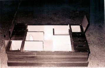
Hebb-Williams maze.
Elevated plus-maze
Elevated plus-maze served as the exteroceptive behavior model to evaluate learning and memory in rats and mice. The procedure, technique and end point for testing learning and memory was followed as per the parameters described by the investigators working in the area of psychopharmacology (Itoh et al, 1990; Reddy and Kulkarni, 1998; Dhingra et al., 2003; Parle and Dhingra, 2003). Briefly, the elevated plus maze apparatus (Picture 2) for rats consisted of a central platform (10 cm2) connected to two open arms (50 cm × 10 cm) and two covered (enclosed) arms (50 cm × 40 cm × 10 cm) and the maze was elevated to a height of 50 cm from the floor (Parle and Singh , 2004). The elevated plus maze apparatus for mice consisted of two open arms (16 cm × 5 cm) and two covered arms (16 cm × 5 cm × 12 cm) extended from a central platform (5 cm × 5 cm), and the maze was elevated to a height of 25 cm from the floor (Dhingra et al., 2004). On the first day, each mouse/rat was placed at the end of an open arm, facing away from the central platform. Transfer latency (TL) was defined as the time taken by the animal to move into one of the enclosed arms with all its four legs. TL was recorded on the first day for each animal. If the animal did not enter into the enclosed arm within 90 seconds, and it was gently pushed into one of the enclosed arms, and the TL was assigned as 90 seconds. The mouse/rat was allowed to explore the maze for another 2 minutes and then returned to its home cage. Retention of this learned-task was examined 24 h after the first day trial.
Picture 2.

Elevated plus maze.
Passive Avoidance Paradigm
Passive Avoidance Behaviour based on negative reinforcement was used to examine the long-term memory (Reddy and Kulkarni, 1998; Dhingra et al., 2004). The apparatus consisted of a box (27 cm × 27 cm × 27 cm) having three walls of wood and one wall of Plexiglass, featuring a grid floor (made up of 3 mm stainless steel rods set 8 mm apart), with a wooden platform (10 cm × 7 cm × 1.7 cm) in the center of the grid floor (Picture 3). The box was illuminated with a 15 W bulb during the experimental period. Electric shock (20 V, A.C.) was delivered to the grid floor. Training was carried out in two similar sessions. Each mouse was gently placed on the wooden platform set in the center of the grid floor. When the mouse stepped-down placing all its paws on the grid floor, shocks were delivered for 15 seconds and the step-down latency (SDL) was recorded. SDL was defined as the time taken by the mouse to step down from the wooden platform to grid floor with all its paws on the grid floor. Animals showing SDL in the range of 2-15 seconds during the first test were used for the second session and the retention test. The second-session was carried out 90 minutes after the first test. When the animals stepped down before 60 seconds, electric shocks were delivered for 15 seconds. During the second test animals were removed from shock free zone, if they did not step down for a period of 60 seconds. Retention was tested after 24 h in a similar manner, except that the electric shocks were not applied to the grid floor observing an upper cut-off time of 300 seconds (Parle et al., 2004).
Picture 3.
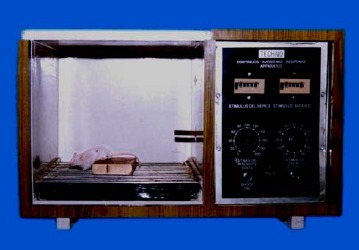
Passive avoidance apparatus.
Swimming protocol
The rodents (rats and mice) were divided into twelve groups and each group comprised of a minimum of 6 animals (Table 1). The swimming exposure to the rodents was for 10- minutes during each session and there were two swimming exposures on each day.
Table 1.
Swimming protocol and group description.
| Group I | Served as control (non-swimming) group of rats. Learning index and memory score was tested employing Hebb-Williams maze |
| Group II | Rats were subjected to swimming for 15 days (Sub-chronic swimming exposure). TRC was tested employing Hebb-Williams maze. |
| Group III | Rats were subjected to swimming for 30 days (Chronic swimming exposure). TRC was tested employing Hebb-Williams maze. |
| Group IV | Served as control (non-swimming) group for mice and transfer latency was tested employing elevated plus maze. |
| Group V | Mice were subjected to swimming for 15 days (Sub-chronic swimming exposure) and transfer latency was tested employing elevated plus maze. |
| Group VI | Mice were subjected to swimming for 30 days (Chronic swimming exposure) and transfer latency was tested employing elevated plus maze. |
| Group VII | Served as control (non-swimming) group of rats and transfer latency was tested employing elevated plus-maze. |
| Group VIII | Rats were subjected to swimming for 15 days (sub-chronic swimming exposure) and transfer latency was tested employing elevated plus maze. |
| Group IX | Rats were subjected to swimming for 30 days (chronic swimming exposure) and transfer latency was tested employing elevated plus maze. |
| Group X | Served as control (non-swimming) group for mice and step-down latency was tested employing passive-avoidance apparatus. |
| Group XI | Mice were subjected to swimming for 15 days (Sub-chronic swimming exposure) and step-down latency was tested employing passive-avoidance apparatus. |
| Group XII | Mice were subjected to swimming for 30 days (Chronic swimming exposure) and step-down latency was tested employing passive-avoidance apparatus. |
The learning index and memory score (as determined by their TRC/TL/SDL) of all the animals was recorded on 1st, 2nd, 15th, 16th, 30th and 31st day.
Statistical analysis
All the results were expressed as Mean ± Standard Error (SEM). Data was analyzed using one-way ANOVA followed by Dunnett’s ‘t’ test and student’s paired ‘t’ test. P < 0.05 was considered as statistically significant.
Results
Effect of swimming exposure on Time taken to reach Reward Chamber (TRC) of rats using Hebb -Williams maze
When the rats were exposed to swimming for 15- days (sub chronic swimming exposure), there was a significant reduction (p < 0.05) in TRC of 15th day as compared to TRC of 1st day in the same group (Figure 1). Furthermore, the TRC of 16th day was also significantly reduced (p < 0.05) when compared to 2nd day TRC in the same group of rats. This indicated that learning index and memory score of animals was remarkably improved after 15- days of swimming exercise. However, when the TRC was measured on 30th day (p < 0.05) and 31st day (p < 0.05) of mice exposed sub chronically to swimming (for 15-days), it was observed that there was significant increase in TRC as compared to corresponding TL of 15th day and 16th day (Figure 1). Rats, which underwent chronic swimming exposure (for 30- days) showed good learning index and sharp memory as reflected by significant reduction of TRC value on 30th day (p < 0.001) and 31st day (p < 0.001), when compared to the corresponding TRC of 1st day and 2nd day in the same group. There was also a significant (p < 0.01) reduction in 30th day TRC of rats exposed chronically to swimming (for 30-days) when compared to 30th day TRC of control group thereby indicating that chronic swimming exposure improved learning index. So also, there was a significant (p < 0.001) reduction in 31st day TRC (denoting memory) of rats exposed chronically to swimming (for 30 days), when compared to 31st day TRC of control group (Figure 1).
Figure 1.
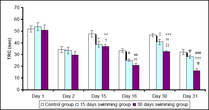
Effect of swimming on TRC (Time taken to reach Reward Chamber) of rats using Hebb Williams Maze.Values represent mean (± SEM).
*, ** and *** denote p < 0.05, 0.01 and 0.001, respectively, compared with 1st day in same group.
#, ## and ### denote p < 0.05, 0.01 and 0.001, respectively, compared with 2nd day in same group.
†, †† and ††† denote p < 0.05, 0.01 and 0.001, respectively, compared with control group on same day.
‡ and ‡‡ denote p < 0.05 and 0.01, respectively, compared with 15th day in same group.
Ψ denotes p < 0.05 compared with 16th day in same group.
Effect of swimming exposure on Transfer Latency (TL) of mice using elevated plus maze
When the mice were exposed to swimming for 15- days (sub-chronic swimming exposure), there was a significant reduction (p < 0.01) in TL of 15th day as compared to TL of 1st day in the same group (Figure 2). Furthermore, the TL of 16th day was also significantly reduced (p < 0.05) when compared with 2nd day TL in the same group of mice. This indicated that learning ability and memory score of animals was remarkably improved after 15- days of swimming exercise. However, the TL on 30th day and 31st day of mice exposed sub-chronically to swimming (for 15-days), was significantly higher as compared to corresponding TL of 15th day and 16th day. Mice, which underwent chronic swimming exposure (for 30- days) showed good learning ability and sharp memory as reflected by significant reduction of TL value on 30th day (p < 0.001) and 31st day (p <0.001), when compared to the corresponding TL of 1st day and 2nd day in the same group (Figure 2). There was also a significant (p < 0.01) reduction in 30th day TL of mice exposed chronically to swimming (for 30-days) when compared to 30th day TL of control group thereby indicating that chronic swimming exposure improved learning ability. So also, there was a significant (p < 0.001) reduction in 31st day TL (denoting memory) of mice exposed chronically to swimming (for 30 days), when compared to 31st day TL of control group.
Figure 2.
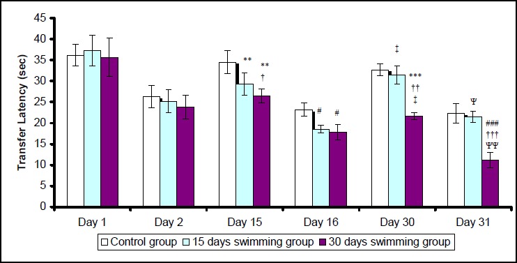
Effect of swimming on Transfer-Latency (TL) of mice using Elevated Plus Maze. Values represent mean (± SEM).
** and *** denote p < 0.01 and 0.001, respectively, compared with 1st day in same group.
# and ### denote p < 0.05 and 0.001, respectively, compared with 2nd day in same group.
†, †† and ††† denote p < 0.05, 0.01 and 0.001, respectively, compared with control group on same day.
‡ denotes p < 0.05 compared with 15th day in same group.
Ψ and ΨΨ denote p < 0.05 and 0.01, respectively, compared with 16th day in same group.
Effect of swimming exposure on Transfer Latency (TL) of rats using elevated plus maze
When the rats were exposed to swimming for 15- days (sub-chronic swimming exposure), there was a significant reduction (p < 0.05) in TL of 15th day as compared to the TL of 1st day (Figure 3). Furthermore, the TL of 16th day was also significantly reduced (p < 0.01) when compared with 2nd day TL in the same group of rats. This indicated that learning ability and retention capacity of animals was remarkably improved after 15- days of swimming exercise. Rats, which underwent chronic swimming exposure (for 30- days) showed good learning ability and sharp memory as reflected by significant reduction of TL value on 30th day (p < 0.001) and 31st day (p < 0.001), when compared to the corresponding TL of 1st day and 2nd day in the same group (Figure 3). There was also a significant (p < 0.001) reduction in 30th day TL of rats exposed chronically to swimming (for 30-days) when compared to 30th day TL of control group, thereby indicating that chronic swimming exposure improved learning ability. So also, there was a significant (p < 0.001) reduction in 31st day TL (denoting memory) of rats exposed chronically to swimming (for 30 days), when compared to 31st day TL of control group.
Figure 3.
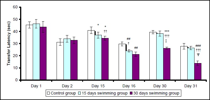
Effect of swimming on Transfer-Latency (TL) of rats using Elevated Plus Maze. Values represent mean (± SEM).
* and *** denote p < 0.05 and 0.001, respectively, compared with 1st day in same group.
## and ### denote p < 0.01 and 0.001, respectively, compared with 2nd day in same group.
†, †† and ††† denote p < 0.05, 0.01 and 0.001, respectively, compared with control group on same day.
‡ denotes p < 0.01 compared with 15th day in same group.
Ψ denotes p < 0.01 compared with 16th day in same group.
Effect of swimming exposure on Step-Down-Latency of mice using passive avoidance paradigm
When the mice were exposed to swimming for 15- days (sub-chronic swimming exposure), there was a significant increase (p < 0.01) in SDL of 16th day as compared to SDL of 2nd day in the same group. This compared to SDL of 2nd day in the same group. This indicated that memory score of animals was remarkably improved after 15- days of swimming exercise. Mice, which underwent chronic swimming exposure (for 30- days) showed sharp memory as reflected by marked enhancement in SDL value of 31st day (p <0.001), when compared to the corresponding SDL of 2nd day in the same group (Figure 4). There was a marked (p < 0.001) increase in 31st day SDL (denoting memory) of mice exposed chronically to swimming (for 30 days), when compared to 31st day SDL of control group.
Figure 4.
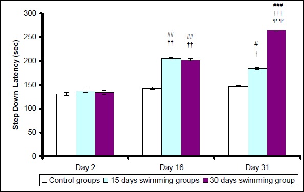
Effect of swimming on Step Down Latency (SDL) of mice using Passive Avoidance Paradigm. Values represent mean (± SEM).
#, ## and ### denote p < 0.05, 0.01 and 0.001, respectively, compared with 2nd day in same group.
†, †† and ††† denote p < 0.05, 0.01 and 0.001, respectively, compared with control group on same day.
ΨΨ denotes p < 0.01compared with 16th day in same group.
Discussion
Memory forms one of the most complex functions of the brain. Time taken by the rat to reach reward chamber (TRC) from the start box on 1st day reflected the learning index, whereas, TRC of the next day (second day) indicated retention capacity (memory score) of animals. In the present study, rats, which underwent chronic swimming exposure (for 30- days) showed good learning index and sharp memory as reflected by significant reduction of TRC value on 30th day and 31st day as compared to the corresponding TRC of 1st day and 2nd day in the same group and TRC of 30th day and 31st day in control group. This observation suggested that regular swimming schedule of 30- days prevented the brain damage possibly caused by neurodegenerative processes and improved learning index and retention capacity (memory) of animals.
Transfer Latency (TL) was defined as the time taken by the animal (rat/mouse) to enter into one of the enclosed arms with all its four legs. TL of the first day reflected learning ability of animals whereas, TL of the next day indicated retention capacity (memory) of animals. SDL was defined as the time taken by the mouse to step down from the wooden platform to grid floor with all its paws. SDL of the second day indicated retention capacity (memory score) of animals. We subjected the rodents to swimming (a pleasant exercise) for a period of 15- and 30- days. The rats as well as mice that underwent swimming regularly for a period of 15- days showed good learning index and memory score as indicated by reduced Transfer Latency and enhanced Step Down Latency, when compared to control animals that did not swim. Interestingly, the learning index as well as memory score deteriorated over next 15- days (i.e. by the end of 30- days), when the swimming exercise was halted suddenly after 15- days. This was perhaps due to the natural physiological process of forgetting, which might have come into play upon halting of swimming exposure to animals after 15- days. These findings suggested that an uninterrupted (regular) exercise schedule such as swimming probably helps in preventing the neurodegenerative damage of brain cells taking place gradually in aged- rats and mice. Above findings underlined the importance of exercise in general and swimming in particular in preventing memory loss. New neurons are continuously being added to certain areas of the brain, such as hippocampus and olfactory bulb in animals (Clayton and Krebs, 1994; Kempermann et al., 1997; Van Praag et al., 1999; Rhodes et al., 2003) as well as humans (Gage et al., 1998; Bruunsgaard et al., 1999). There is a possibility that regular swimming for long periods not only arrested the neurodegenerative processes (responsible for dementia) but also stimulated the process of neurogenesis. This suggestion is in line with the reports available in literature (Neeper et al., 1995; Kempermann et al., 1997). Similarly, voluntary physical activity on a running wheel apparatus doubled the number of surviving new-born hippocampal cells in adult mice (Van Praag et al., 1999; Czurko et al., 1999). Both running and living in an enriched environment doubled the number of surviving newborn cells and improved water maze performance (Russo-Neustadt et al., 1999).
Exercise is known to induce numerous physiological alterations in vital organ systems of the body. Our results point out that integration of exercise schedule into the life style of Alzheimer patients is advantageous and worthwhile. This notion is supported by the studies of Teri et al. (1998), who have shown that gait, flexibility, body strength, and endurance was greatly improved in community-dwelling Alzheimer’s disease subjects and their care givers, when they participated in a structured exercise program. Ahmadiasl et al. (2003) showed that increased physical activity in adult rats enhanced spatial learning performance tested using Morris water maze. Over the past decade, a number of studies on humans have shown the benefits of exercise on brain health, particularly in aging population (Ivy et al., 2001; Teri et al., 2003). Maintaining brain health and plasticity throughout life is an important public health goal. Moreover, regular exercise resulted in a variety of adaptations that may be beneficial in attenuating the process of apoptosis (Sen et al., 1992; Powers et al., 1993; Leeuwenburgh et al., 1997).
Alterations in the levels of various neurochemicals (such as acetylcholine, epinephrine, dopamine, GABA, glutamate etc.) have been found to play a crucial role in the pathogenesis of impaired memory of laboratory animals (Ahmadiasl et al., 2003) and Alzheimer patients (Parle et al., 2004). The redressal of cholinergic deficiency has been the main stay in the treatment of Alzheimer’s disease. For reversing cholinergic deficiency, cholinergic precursors and anti-cholinesterases (such as tacrine, rivastigmine, donepezil, metrifonate etc.) have been successfully employed clinically (Parle et al., 2004). Rats subjected to treadmill exercise scored higher on memory performance probably due to enhanced epinephrine levels in hippocampus (Ahmadiasl et al., 2003). Exercise has also been found to increase the expression of brain neurotrophic factors in rat hippocampus (Russo-Neustadt et al., 2000; Tong et al., 2001; Gomez-Pinilla et al., 2001; Griesbach et al., 2004). Furthermore, release of trophic factors, responsible for progenitor cell survival (Ray et al., 1997), synaptic strength (Schuman, 1999), long-term potentiation (Patterson et al., 1992), and memory (Fischer et al., 1987), were all improved after exercise (Neeper et al., 1995). Interestingly, antidepressant treatment in combination with exercise enhanced exercise-dependent brain derived neurotrophic factor (BDNF) upregulation in the hippocampus (Fujimaki et al., 2000). Oxygen free radicals and other products of oxidative metabolism are reported to be neurotoxic (Sayre et al., 1997). Antioxidant- rich diets improved cerebellar physiology and motor learning in aged rats (Bickford et al., 2000). Regular exercise has been shown to improve cognitive functions and decrease oxidative damage in rat brain (Grealy et al., 1999; Radak et al., 2001). Reasoning ability and working memory was found to be better in men and women who exercised vigorously as compared to sedentary individuals (Clarkson-Smith and Hartley, 1989).
In the present investigation, the learning index as well as memory score of rodents was remarkably enhanced in a group of animals that underwent swimming regularly for 30- days. These research findings reinforce the concept that a well planned exercise programme would greatly help the aged-citizens with or without Alzheimer’s disease in improving their cognitive functions. In any case, this approach would be preferred over undergoing drug-therapy. Exercising regularly as you get older may not only keep your body in shape, but your brains as well.
Conclusions
In conclusion, our results emphasize the role of regular physical exercise particularly swimming in the maintenance and promotion of brain functions. The underlying physiological mechanism for improvement of memory appears to be the result of enhanced neurogenesis.
Acknowledgements
Authors are deeply grateful to Shri Vishnu Bhagwan, I.A.S., Honorable Vice-Chancellor of Guru Jambheshwar University, Hisar for his constant encouragement. We are grateful to Dr. P.K. Kapoor, Scientist Incharge, Disease free animal house, CCS Haryana Agricultural University, Hisar for continuous supply of animals. We are also thankful to Prof. Avinash Dhake, Dean, Faculty of Pharmaceutical Sciences, Guru Jambheshwar University, Hisar, for his keen interest and kind cooperation.
Biographies

Milind PARLE
Employment
Pharmacology Division, Dept. of Pharm. Sciences, Guru Jambheshwar Univ. Technical Univ. of Haryana State, India.
Degrees
M Pharm, PhD
Research interests
Psychopharmacology and behavioural pharmacology.
E-mail: mparle@rediffmail.com

Mani VASUDEVAN
Employment
Doctoral Student, Pharmacology Division, Dept. of Pharm. Sciences, Guru Jambheshwar Univ. Technical Univ. of Haryana State, Hisar, Haryana, India.
Degrees
M. Pharm.
Research interests
Traditional systems of medicine and mental disorders.
E-mail: vasumpharmacol@yahoo.co.uk

Nirmal SINGH
Employment
Lecturer, Department of Pharmaceutical Sciences & Drug Research, Punjabi University, Patiala (Punjab) India.
Degrees
M. Pharm.
Research interests
Animal models for memory
E-mail: nirmal_puru@rediffmail.com
References
- Ahmadiasl N., Alaei H., Hanninen O. (2003) Effect of exercise on learning, memory and levels of Epinephrine in rats Hippocampus. Journal of Sports Science and Medicine 2, 106-109 [PMC free article] [PubMed] [Google Scholar]
- Berchtold N.C., Kesslak J.P., Adlard P.A., Cotman C.W. (2001) Estrogen and exercise interact to regulate brain-derived neurotrophic factor mRNA and protein expression in the hippocampus. The European Journal of Neuroscience, 14, 1992-2002 [DOI] [PubMed] [Google Scholar]
- Bickford P.C., Gould T., Briedcick L., Chadman K., Polloch A., Young D., Shukitt- Hale B., Joseph J. (2000) Antioxidants-rich diets improve cerebellar physiology and motor learning in aged rats. Brain Research 866, 211-217 [DOI] [PubMed] [Google Scholar]
- Bruunsgaard H., Jensen M.S., Schjerling P., Halkjaer- Kristensen J., Ogawa K., Skinhoj P., Pedersen B.K. (1999) Exercise induces recruitment of lymphocytes with an activated phenotype and short telomeres in young and elderly humans. Life Sciences 65, 2623-2633 [DOI] [PubMed] [Google Scholar]
- Clarkson-Smith L., Hartley A.A. (1989) Relationships between physical exercise and cognitive abilities in older adults. Psychology and Aging 4, 183-189 [DOI] [PubMed] [Google Scholar]
- Clayton N.S., Krebs J.R. (1994) Hippocampal growth and attrition in birds affected by experience. Proceedings of the National Academy of Sciences of the United States of America 91, 7410-7414 [DOI] [PMC free article] [PubMed] [Google Scholar]
- Czurko A., Hirase H., Csicsvari J., Buzsaki G. (1999) Sustained activation of hippocampal pyramidal cells by ‘space clamping’ in a running wheel. The European Journal of Neuroscience 11, 344-352 [DOI] [PubMed] [Google Scholar]
- Dhingra D., Parle M., Kulkarni S.K. (2003) Effect of combination of insulin with dextrose, D (-) fructose and diet on learning and memory in mice. Indian Journal of Pharmacology, 35, 151-156 [Google Scholar]
- Dhingra D., Parle M., Kulkarni S.K. (2004) Memory enhancing activity of Glycyrrhiza glabra in mice. Journal of Ethnopharmacology 91, 361-365 [DOI] [PubMed] [Google Scholar]
- Dustman R.E., Ruhling R.O., Russell E.M., Shearer D.E., Bonekat H.W., Shigeoka J.W., Wood J.S., David C. (1984) Aerobic exercise training and improved neuropsychological function of older individuals. Neurobiology of Aging 5, 35-42 [DOI] [PubMed] [Google Scholar]
- Fischer W., Wictorin K., Bjorklund A., Williams L.R., Varon S., Cage F.H. (1987) Amelioration of cholinergic neuron atrophy and spatial memory impairment in aged rats by nerve growth factor. Nature (London) 329, 65-68 [DOI] [PubMed] [Google Scholar]
- Fujimaki K., Morinobu S., Duman R.S. (2000) Administration of a cAMP phosphodiesterage 4 inhibitor enhances antidepressant- induction of BDNF mRNA in rat hippocampus. Neuropsychopharmacology 22, 42-51 [DOI] [PubMed] [Google Scholar]
- Gage F.H., Kempermann G., Palmer T.D., Peterson D.A., Ray J. (1998) Multipotent progenitor cells in the adult dentate gyrus. Journal of Neurobiology, 36, 249-266 [DOI] [PubMed] [Google Scholar]
- Gomez-Pinilla F., So V., Keeslak J.P. (2001) Spatial learning induces neurotrophin receptor and synapsin I in the hippocampus. Brain Research 904, 13-19 [DOI] [PubMed] [Google Scholar]
- Grealy M.A., Johnson D.A., Rushon S.K. (1999) Improving cognitive function after brain injury: The use of exercise and virtual reality. Archives of Physical Medicine and Rehabilitation 80, 661-667 [DOI] [PubMed] [Google Scholar]
- Griesbach G.S., Hovda D.A., Molteni R., Wu A., Gomez-Pinilla F. (2004) Voluntary exercise following traumatic brain injury: brain-derived neurotrophic factor upregulation and recovery of function. Neuroscience 125, 129-139 [DOI] [PubMed] [Google Scholar]
- Itoh J., Nabeshima T., Kameyama T. (1990) Utility of an elevated plus maze for evaluation of nootropics, scopolamine and electro convulsive shock. Psychopharmacology, 101, 27-33 [DOI] [PubMed] [Google Scholar]
- Ivy A.S., Rodriguez F.G., Russo-Neustadt A. (2001) The effects of NE and 5-HT receptor antagonists on the regulation of BDNF expression during physical activity. Society for Neuroscience Abstracts, 253, 213 [Google Scholar]
- Kempermann G., Kuhn H.G., Gage F.H. (1997) More hippocampal neurons in adult mice living in an enriched environment. Nature (London) 386, 493-495 [DOI] [PubMed] [Google Scholar]
- Leeuwenburgh C., Hollander J., Fiebig R., Leichtweis S., Griffith M., Ji L.L. (1997) Adaptations of glutathione antioxidant system to endurance training are tissue and muscle fiber specific. American Journal of Physiology 272, 363-369 [DOI] [PubMed] [Google Scholar]
- Neeper S.A., Gomez-Pinilla F., Choi J., Cotman C. (1995) Exercise and brain neurotrophins. Nature (London) 373, 109. [DOI] [PubMed] [Google Scholar]
- Parle M., Dhingra D. (2003) Ascorbic acid: a promising memory-enhancer in mice. Journal of Pharmacological Sciences 93, 129-135 [DOI] [PubMed] [Google Scholar]
- Parle M., Singh N. (2004) Animal models for testing memory. Asia Pacific Journal of Pharmacology 16101-120 [Google Scholar]
- Parle M., Dhingra D., Kulkarni S.K. (2004) Neurochemical basis of learning and memory. Indian Journal of Pharmaceutical Sciences, 66, 371-376 [Google Scholar]
- Parle M., Dhingra D., Kulkarni S.K. (2004) Neuromodulators of learning and memory. Asia Pacific Journal of Pharmacology 16, 89-99 [Google Scholar]
- Parle M., Dhingra D., Kulkarni S.K. (2004) Improvement of Mouse memory by Myristica fragrans seeds. Journal of Medicinal Food 7, 157-161 [DOI] [PubMed] [Google Scholar]
- Patterson S.L., Grover L.M., Schwartzkroin P.A., Bothwell M. (1992) Neurotrophin expression in rat hippocampal slices: a stimulus paradigm inducing LTP in CAI evokes increases in BDNF and NT-3 mRNAs. Neuron 9, 1081-1088 [DOI] [PubMed] [Google Scholar]
- Powers S.K., Criswell D., Lawler J., Martin D., Lieu F.K., Ji L.L., Herb R.A. (1993) Rigorous exercise training increases superoxide dismutage activity in ventricular myocardium. American Journal of Physiology 265, 2094-2098 [DOI] [PubMed] [Google Scholar]
- Radak Z., Kaneko T., Tahara S., Nakamoto H., Pucsok J., Sasvari M., Nyakas C., Goto S. (2001) Regular exercise improves cognitive function and decreases oxidative damage in rat brain. Neurochemistry International 38, 17-23 [DOI] [PubMed] [Google Scholar]
- Ray J., Palmer T.D., Suhonen J., Takahashi J., Gage F.H. (1997) In: Isolation, Characterization, and Utilization of CNS Stem Cells. Christen Y., Gage F.H.Springer, Berlin: 129-149 [Google Scholar]
- Reddy D.S., Kulkarni S.K. (1998) Possible role of nitric oxide in the nootropic and antiamnesic effect of neurosteroids on aging and dizocilpine induced learning impairment. Brain Research 799, 215-229 [DOI] [PubMed] [Google Scholar]
- Rhodes J.S., Praag H.V., Jeffrey S., Girard I., Mitchell G.S., Garland T., Gage F.H. (2003) Exercise increases hippocampal neurogenesis to high levels but does not improve spatial learning in mice bred for increased voluntary wheel running. Behavioral Neuroscience 117, 1006-1016 [DOI] [PubMed] [Google Scholar]
- Russo-Neustadt A., Beard R.C., Cotman C.W. (1999) Exercise, antidepressant medications, and enhanced brain derived neurotrophic factor expression . Neuropsychopharmacology 21, 679-682 [DOI] [PubMed] [Google Scholar]
- Russo-Neustadt A.A., Beard R.C., Huang Y.M., Cotman C.W. (2000) Physical activity and antidepressant treatment potentiate the expression of specific brain-derived neurotrophic factor transcripts in the rat hippocampus. Neuroscience 101, 305-312 [DOI] [PubMed] [Google Scholar]
- Sayre L.M., Zagorski M.G., Surewicz W.K., Krafft G.A., Perry G. (1997) Mechanisms of neurotoxicity associated with amyloid-beta deposition and the role of free radicals in the pathogenesis of Alzheimer’s disease: a critical appraisal. Chemical Research and Toxicology 336, 1216-1222 [DOI] [PubMed] [Google Scholar]
- Schuman E.M. (1999) Neurotrophin regulation of synaptic transmission. Current Opinion in Neurobiology 9, 105-109 [DOI] [PubMed] [Google Scholar]
- Sen C.K., Marin E., Kretzschmar M., Hanninen O. (1992) Skeletal muscle and liver glutathione homeostasis in response to training, exercise, and immobilization. Journal of Applied Physiology, 73, 1265-1272 [DOI] [PubMed] [Google Scholar]
- Sim Y., Kim S., Kim J., Shin M., Kim C. (2004) Treadmill exercise improves short-term memory by suppressing ischemia-induced apoptosis of neuronal cells in gerbils. Neuroscience Letters 372, 256-261 [DOI] [PubMed] [Google Scholar]
- Teri L., Gibbons L.E., McCurry S.M., Logsdon R.G., Buchner D.M., Barlow W.E., Kukull W.A., Lacroix A.Z., McCormick W., Larson E.B. (2003) Exercise plus behavioral management in patients with Alzheimer’s disease: a randomized controlled trial. JAMA 290, 2015-2022 [DOI] [PubMed] [Google Scholar]
- Teri L., McCurry S.M., Buchner D.M., Logsdon R.G., La Croix A.Z., Kukull W.A., Barlow W.E., Larson E.B. (1998) Exercise and activity level in Alzheimer’s disease: A potential treatment focus. Journal of Rehabilitation Research and Development 35, 411-419 [PubMed] [Google Scholar]
- Tomporowski P.D. (2003) Effects of acute bouts of exercise on cognition. Acta Psychologica 112, 297-324 [DOI] [PubMed] [Google Scholar]
- Tong L., Shen H., Perreau V.M., Balazs R., Cotman C.W. (2001) Effects of exercise on Gene-expression profile in the rat hippocampus. Neurobiology of Disease 8, 1046-1056 [DOI] [PubMed] [Google Scholar]
- Van Praag H., Kempermann G., Gage F.H. (1999) Running increases cell proliferation and neurogenesis in the adult mouse dentate gyrus. Nature Neuroscience 2, 266-270 [DOI] [PubMed] [Google Scholar]
- Vijay Kumar K., Naidu M.U.R. (2002) Effect of oral melatonin on exercise-induced oxidant stress in healthy subjects. Indian Journal of Pharmacology, 34, 256-259 [Google Scholar]
- Woods J.A., Ceddia M.A., Zack M.D., Lowder T.W., Lu Q. (2003) Exercise training increases the naive to memory T cell ratio in old mice. Brain, Behavior and Immunity 17, 384-392 [DOI] [PubMed] [Google Scholar]


