

Abstract
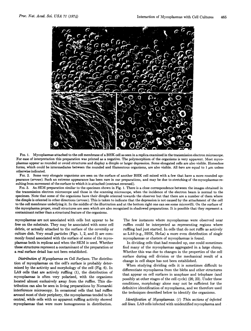
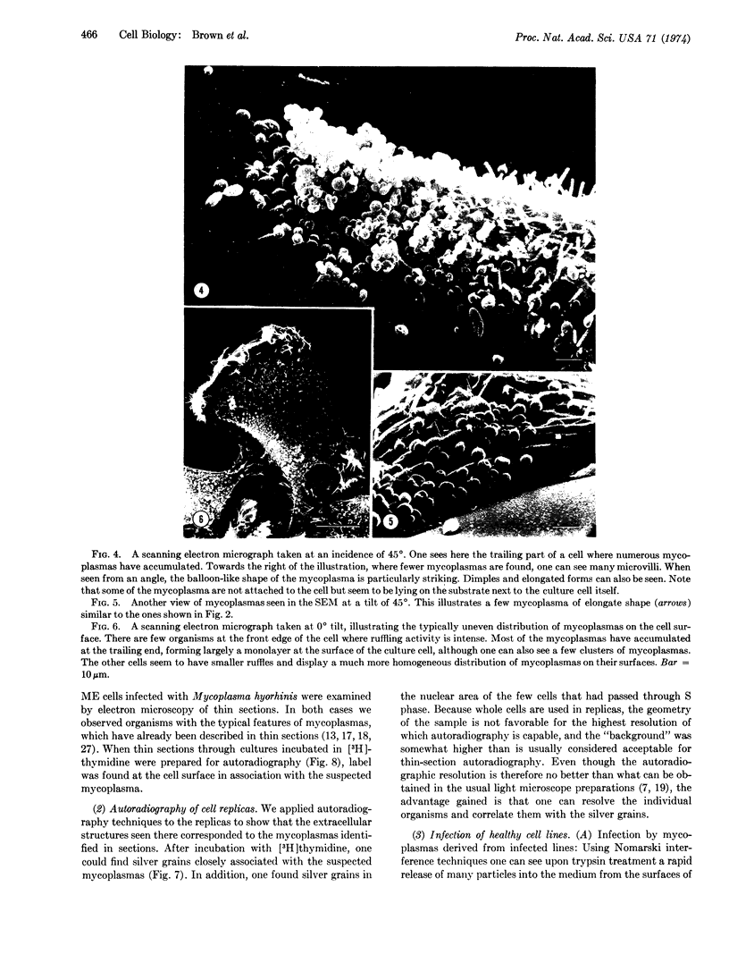
Mycoplasmas were examined on the surfaces of tissue culture cells prepared for transmission and scanning electron microscopy. The pleomorphic bodies seen were proved to be mycoplasmas by the use of thin sections, passage of the infection from one cell line to another, and by autoradiography with [3H]thymidine both on the sections and the replicas. The mycoplasmas were not always evenly distributed over the cell's surface; their arrangement seemed to correlate with the activity or morphology of the cell. The use of replicas and scanning electron microscopy in routine examination of cultures for mycoplasma contamination is discussed.
Keywords: scanning electron microscope, cell-surface replicas, autoradiography
Full text
PDF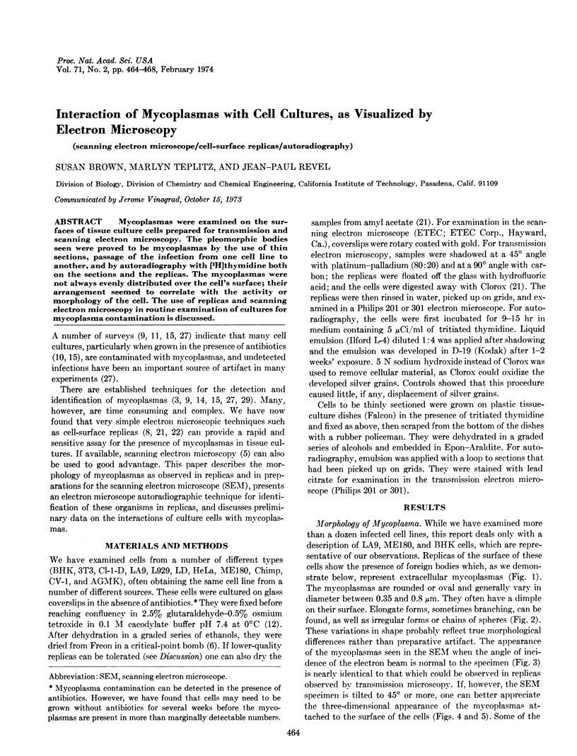
Images in this article
Selected References
These references are in PubMed. This may not be the complete list of references from this article.
- Abercrombie M., Heaysman J. E., Pegrum S. M. The locomotion of fibroblasts in culture. 3. Movements of particles on the dorsal surface of the leading lamella. Exp Cell Res. 1970 Oct;62(2):389–398. doi: 10.1016/0014-4827(70)90570-7. [DOI] [PubMed] [Google Scholar]
- Abercrombie M., Heaysman J. E., Pegrum S. M. The locomotion of fibroblasts in culture. II. "RRuffling". Exp Cell Res. 1970 Jun;60(3):437–444. doi: 10.1016/0014-4827(70)90537-9. [DOI] [PubMed] [Google Scholar]
- Barile M. F., Kern J. Isolation of Mycoplasma arginini from commercial bovine sera and its implication in contaminated cell cultures. Proc Soc Exp Biol Med. 1971 Nov;138(2):432–437. doi: 10.3181/00379727-138-35913. [DOI] [PubMed] [Google Scholar]
- Biberfeld G., Biberfeld P. Ultrastructural features of Mycoplasma pneumoniae. J Bacteriol. 1970 Jun;102(3):855–861. doi: 10.1128/jb.102.3.855-861.1970. [DOI] [PMC free article] [PubMed] [Google Scholar]
- Culp L. A., Black P. H. Release of macromolecules from BALB-c mouse cell lines treated with chelating agents. Biochemistry. 1972 May 23;11(11):2161–2172. doi: 10.1021/bi00761a024. [DOI] [PubMed] [Google Scholar]
- Follett E. A., Goldman R. D. The occurrence of microvilli during spreading and growth of BHK21-C13 fibroblasts. Exp Cell Res. 1970 Jan;59(1):124–136. doi: 10.1016/0014-4827(70)90631-2. [DOI] [PubMed] [Google Scholar]
- Hirsch J. G., Fedorko M. E. Ultrastructure of human leukocytes after simultaneous fixation with glutaraldehyde and osmium tetroxide and "postfixation" in uranyl acetate. J Cell Biol. 1968 Sep;38(3):615–627. doi: 10.1083/jcb.38.3.615. [DOI] [PMC free article] [PubMed] [Google Scholar]
- House W., Waddell A. Detection of mycoplasma in cell cultures. J Pathol Bacteriol. 1967 Jan;93(1):125–132. doi: 10.1002/path.1700930112. [DOI] [PubMed] [Google Scholar]
- Levine E. M., Thomas L., McGregor D., Hayflick L., Eagle H. Altered nucleic acid metabolism in human cell cultures infected with mycoplasma. Proc Natl Acad Sci U S A. 1968 Jun;60(2):583–589. doi: 10.1073/pnas.60.2.583. [DOI] [PMC free article] [PubMed] [Google Scholar]
- Manchee R. J., Taylor-Robinson D. Studies on the nature of receptors involved in attachment of tissue culture cells to mycoplasmas. Br J Exp Pathol. 1969 Feb;50(1):66–75. [PMC free article] [PubMed] [Google Scholar]
- Maniloff J. Ultrastructure of Mycoplasma laidlawii during culture development. J Bacteriol. 1970 May;102(2):561–572. doi: 10.1128/jb.102.2.561-572.1970. [DOI] [PMC free article] [PubMed] [Google Scholar]
- Nardone R. M., Todd J., Gonzalez P., Gaffney E. V. Nucleoside incorporation into strain L cells: inhibition by pleuropneumonia-like organisms. Science. 1965 Sep 3;149(3688):1100–1101. doi: 10.1126/science.149.3688.1100. [DOI] [PubMed] [Google Scholar]
- Porter K., Prescott D., Frye J. Changes in surface morphology of Chinese hamster ovary cells during the cell cycle. J Cell Biol. 1973 Jun;57(3):815–836. doi: 10.1083/jcb.57.3.815. [DOI] [PMC free article] [PubMed] [Google Scholar]
- RODWELL A. W., ABBOT A. The function of glycerol, cholesterol and long-chain fatty acids in the nutrition of Mycoplasma mycoides. J Gen Microbiol. 1961 Jun;25:201–214. doi: 10.1099/00221287-25-2-201. [DOI] [PubMed] [Google Scholar]
- Revel J. P., Wolken K. Electronmicroscope investigations of the underside of cells in culture. Exp Cell Res. 1973 Mar 30;78(1):1–14. doi: 10.1016/0014-4827(73)90031-1. [DOI] [PubMed] [Google Scholar]
- Rubin R. W., Everhart L. P. The effect of cell-to-cell contact on the surface morphology of Chinese hamster ovary cells. J Cell Biol. 1973 Jun;57(3):837–844. doi: 10.1083/jcb.57.3.837. [DOI] [PMC free article] [PubMed] [Google Scholar]
- Smith S. B., Revel J. P. Mapping of concanavalin A binding sites on the surface of several cell types. Dev Biol. 1972 Mar;27(3):434–441. doi: 10.1016/0012-1606(72)90183-2. [DOI] [PubMed] [Google Scholar]
- Sobeslavsky O., Prescott B., Chanock R. M. Adsorption of Mycoplasma pneumoniae to neuraminic acid receptors of various cells and possible role in virulence. J Bacteriol. 1968 Sep;96(3):695–705. doi: 10.1128/jb.96.3.695-705.1968. [DOI] [PMC free article] [PubMed] [Google Scholar]
- Stanbridge E. Mycoplasmas and cell cultures. Bacteriol Rev. 1971 Jun;35(2):206–227. doi: 10.1128/br.35.2.206-227.1971. [DOI] [PMC free article] [PubMed] [Google Scholar]
- Taylor-Robinson D., Manchee R. J. Novel approach to studying relationships between mycoplasmas and tissue culture cells. Nature. 1967 Dec 30;216(5122):1306–1307. doi: 10.1038/2161306a0. [DOI] [PubMed] [Google Scholar]
- Todaro G. J., Aaronson S. A., Rands E. Rapid detection of mycoplasma-infected cell cultures. Exp Cell Res. 1971 Mar;65(1):256–257. doi: 10.1016/s0014-4827(71)80077-0. [DOI] [PubMed] [Google Scholar]