Abstract
Objectives
To evaluate the efficacy and safety of radiofrequency ablation (RFA) versus hepatic resection (HR) for early hepatocellular carcinoma (HCC) meeting the Milan criteria.
Methods
A meta-analysis was conducted, and PubMed, Web of Science, the Cochrane Library, CBM, CNKI and VIP databases were systematically searched through November 2012 for randomized and nonrandomized controlled trials (RCTs and NRCTs). The Cochrane Collaboration's tool and modified MINORS score were applied to assess the quality of RCTs and NRCTs, respectively. The GRADE approach was employed to evaluate the strength of evidence.
Results
Three RCTs and twenty-five NRCTs were included. Among 11,873 patients involved, 6,094 patients were treated with RFA, and 5,779 with HR. The pooled results of RCTs demonstrated no significant difference between groups for 1- and 3-year overall survival (OS), recurrence-free survival (RFS) and disease-free survival (DFS) (p>0.05). The 5-year OS (Relative Risk, RR 0.72, 95% CI 0.60 to 0.88) and RFS (RR 0.56, 95% CI 0.40 to 0.78) were lower with RFA than with HR. The 3- and 5-year recurrences with RFA were higher than with HR (RR 1.48, 95% CI 1.14 to 1.94, and RR 1.52, 95% CI 1.18 to 1.97, respectively), but 1-year recurrence and in-hospital mortality showed no significant differences between groups (p>0.05). The complication rate (RR 0.18, 95% CI 0.06 to 0.53) was lower and hospital stays (Mean difference -8.77, 95% CI −10.36 to −7.18) were shorter with RFA than with HR. The pooled results of NRCTs showed that the RFA group had lower 1-, 3- and 5-year OS, RFS and DFS, and higher recurrence than the HR group (p<0.05). But for patients with very early stage HCC, RFA was comparable to HR for OS and recurrence.
Conclusion
The effectiveness of RFA is comparable to HR, with fewer complications but higher recurrence, especially for very early HCC patients.
Introduction
Cancer is a major component of the global burden of disease (GBD). There were 2.49 billion disability-adjusted life years (DALYs), or 361 DALYs per 1000 population worldwide in 2010[1], and all neoplasms accounted for 7.6% (189 million DALYs) of global DALYs, an increase of 20 million DALYs (11.8%) compared with 2008[1], [2]. A study based on the human development index (HDI) of Bray F et al[3] estimated an increase in the incidence of new cancer cases of all kinds to 22.2 million annually by 2030, and that increases would be proportionally greatest in low-HDI settings compared with high-HDI countries (76% vs. 25%). There were 19.1 million DALYs for liver cancer in 2010, which accounted for 0.8% of the GBD or 10.1% of the DALYS for all neoplasms [1].
Hepatic resection (HR) and liver transplantation (LT) were recommended by the latest guidelines for early hepatocellular carcinoma meeting the Milan criteria, with the 5-year survival rate potentially reaching 50 to 75% [4], [5]. However, only 20–35% of patients are suitable for liver resection because of the low diagnosis rate for early HCC and to poor liver function [6]. In addition, few patients can be treated with liver transplantation because of the strict inclusion criteria, high cost, and limited donor liver resources.
Radiofrequency ablation (RFA) has higher efficacy and is associated with fewer complications and shorter hospital stays. RFA can also be administered repeatedly. Although RFA may gradually reach acceptability as an alternative treatment, the long-term efficacy and safety should still be evaluated systematically. Our previous study demonstrated that the overall quality of previously published systematic reviews and meta-analyses comparing RFA and HR for small hepatocellular carcinoma was poor, with an inadequate base of evidence[7]. Therefore, physicians may make an incorrect decision using these recommendations as best evidence without any additional quality evaluations to guide their clinical practice.
The purpose of this study was to retrieve the best available evidence and produce a meta-analysis comparing the long-term results of RFA and HR for early hepatocellular carcinoma to reduce research bias and improve the quality of evidence.
Materials and Methods
Inclusion criteria
The PICOS approach was used for eligibility criteria[8]:
Population. Patients met the Milan criteria (single HCC smaller than 5 cm in diameter or up to 3 nodules that were each smaller than 3 cm in diameter) or the UCSF criteria (single tumor smaller than 6.5 cm in diameter or up to 3 nodules that were each smaller than 4.5 cm in diameter and 8 cm in total diameter) with liver function Child-Pugh class A or B (the number of patients with Child-Pugh C was no more than 10%). Patients were without major vascular invasion and lymphatic spread or extrahepatic metastasis, and had no previous treatment of HCC with any anti-cancer treatment [Transcatheter Arterial Chemoembolization (TACE), Percutaneous Ethanol Injection (PEI), and Microwave Ablation (MWA)].
Intervention. RFA
Comparison. HR
Outcome. Efficacy: O1, overall survival rate (1-, 3-, 5-years), recurrence-free survival (RFS) (1-, 3-, 5-years), disease-free survival (DFS) (1-, 3-, 5-years).
Safety: O2, mortality; recurrence rate (1-, 3-, 5-years); complication rate
Study design. Randomized controlled trials (RCTs), non-randomized controlled trials (NRCTs), retrospective clinical or cohort study
DFS is defined as the time from randomization until recurrence of tumor or death form any cause[9]. In the case of HCC, the definition of DFS was identical as RFS where both recurrence and death form any causes are events[10]. However, the two terms were not clearly distinguished in the included RCTs or NRCTs, so we reported the two indicators respectively.
Exclusion criteria
Conference abstracts, reviews, letters, systematic reviews or case reports were excluded. Metastatic liver cancer (i.e., colorectal liver metastases) or recurrent hepatocellular carcinoma after resection was excluded. Those studies that mixed other effective interventions in either treatment group or control group (i.e., TACE, PEI, LITT) as well as any that had a length of follow-up of less than one year were also excluded.
Data sources and search strategy
We systematically searched 19 systematic reviews (SRs) comparing RFA with HR for small hepatocellular carcinoma in previously published studies and tracked the 39 primary studies included in these SRs. The six databases of PubMed, Web of Science, the Cochrane Library, CBM, CNKI, and VIP were systematically searched through November 2012. The following MeSH terms or free-words were used: (“hepatic resection[Title/Abstract]” OR “surgical resection[Title/Abstract]” OR “liver resection[Title/Abstract]” OR hepatectomy[MeSH Terms]) AND (radiofrequency[Title/Abstract] OR “radiofrequency ablation[Title/Abstract]” OR “catheter ablation[MeSH Terms]” OR “RFA[Title/Abstract]”) AND (“hepatocellular carcinoma[Title/Abstract]” OR “liver neoplasm[MeSH Terms]” OR “liver tumor[Title/Abstract]” OR “liver cancer[Title/Abstract]”). We did not restrict the language of publication.
Review selection and data extraction
The PRISMA statement was followed when searching and screening the literature[8]. Two reviewers (WYQ, LQQ) selected articles independently by browsing titles and abstracts according to predefined inclusion and exclusion criteria. If necessary, judgment was made by viewing the full text. The two reviewers extracted data using standardized forms independently if the publication met the inclusion criteria. Discrepancies between the two reviewers were resolved by discussion or by a third person (LYP).The extracted contents included first author, publication year, type of study, inclusion criteria, number of participants, age, gender, number of nodules, tumor size, the Child-Pugh score, length of follow-up, and clinical outcomes.
Quality assessment
Two reviewers (WYQ, LQQ) evaluated the methodological quality of included studies independently. The tool for evaluating the risk of bias in the Cochrane Handbook for Systematic Reviews of Interventions[11] and the modified MINORS scores questionnaire[12] were applied to assess the quality of RCTs and NRCTs, respectively. GRADE profiler 3.6 was employed to evaluate the strength of the evidence.
Statistical analysis
Meta-analysis was conducted using RevMan 5.1 software. For dichotomous variables, the relative risk (RR) and the odds ratio (OR) with 95% confidence intervals (CIs) were used for RCTs and NRCTs, respectively, For continuous variables, mean difference (MD) with 95% CI was applied. The fixed-effect model (Mantel-Haenszel) was used if the result of the heterogeneity test was p>0.05 and if I2<50%. Otherwise, the random-effect model was applied. A p value <0.05 was considered significant.
Subgroup analysis
Subgroup analysis was applied based on predetermined subgroup factors (i.e., tumor size, number of nodules, and Child-Pugh class).
Sensitivity analysis
We excluded the studies that might cause heterogeneity and re-estimated the combined effect of values, and then compared the results with the primary outcomes to verify the robustness of the outcomes.
Publication bias
The funnel plot and Egger's test were applied by Revman 5.1 and Stata 10.0 software, respectively. If the result of the test was p<0.05, it suggested that potential publication bias may exist. Otherwise, publication bias was considered absent.
Results
Search results
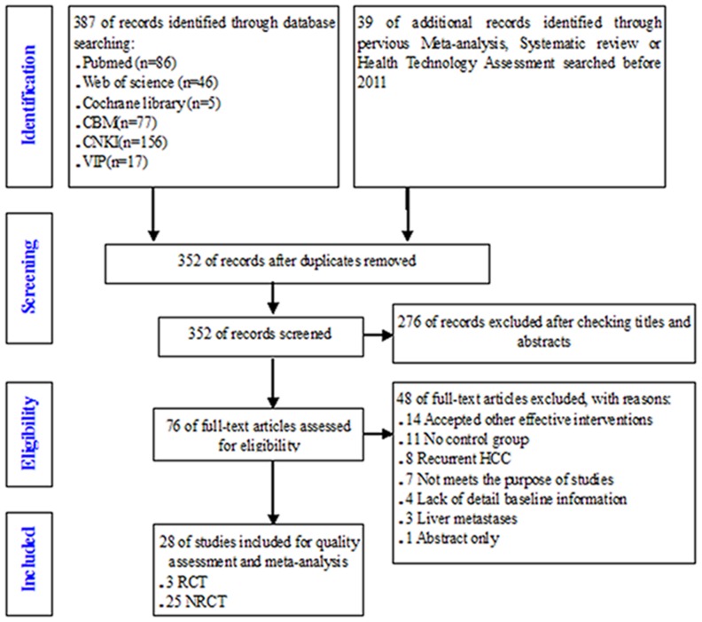
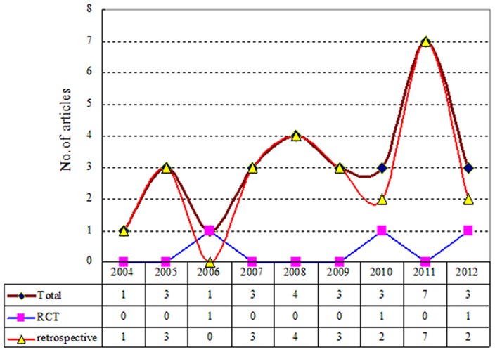
After initial screening, 76 studies were identified. Of these, 48 studies were removed after viewing full-texts for various reasons: a) mixed with other effective interventions (i.e., TACE, PEI) in either intervention group or control group (14 articles), b) without control group (11 articles), c) recurrent HCC (8 articles), d) did not meet the purpose of this study for other reasons (7 articles), e) lack of detailed baseline information (4 articles), f) metastatic liver cancer (3 articles), and g) abstract only (1 article). Finally, 28 studies including 3 RCTs [10], [13], [14] and 25 NRCTs [6], [15]–[38] published between 2004 and 2012 were included (see Figure 1, Figure 2).
Figure 1. PRISMA flowchart of searching and selecting guidelines.
Figure 2. Bibliometric map of included studies.
Baseline characteristics of included studies
A total of 11,873 patients with primary HCC were included in the 28 studies. Among those, 8,567 cases (72%) were males, and 6,094 cases were treated with RFA and 5,779 cases with hepatic resection. There were 11,251 cases (94.8%) with Child-Pugh class A, 1,632 cases (13.7%) with Child-Pugh class B, and only 17 cases (0.1%) with Child-Pugh class C. Patients had an average age of 50 years, with varying degrees of HBsAg-positivity and cirrhosis. The mean length of follow-up ranged from 10 to 60 months (Table 1).
Table 1. Baseline characteristics of RCTs and NRCTs.
| Author, year | Study types | Included criteria | Group | Pts | Men % | Age, year (Mean±SD) | Solitary/Multiple | HBsAg(+), % | Cirrhosis, % | Child-Pugh (A/B/C) | Tumor size (Mean±SD) | Follow-up(mo) (Mean±SD) | Quality* |
| Feng K,2012[13] | RCT | HCC tumor≤4 cm,nodels ≤2,Child A/B | RFA | 84 | 94.0 | 51.0(24–83)‡ | 48/36 | _ | 71.4 | 39/45/0 | 2.4±0.6 | 36.0# | B |
| HR | 84 | 89.3 | 47.0(18–76)‡ | 52/32 | _ | 76.2 | 43/41/0 | 2.6±0.8 | 36.0# | ||||
| Huang JW,2010[10] | RCT | Milan criteria, Child A/B | RFA | 115 | 68.7 | 56.6±14.3 | 84/31 | 87.8 | 58.3 | 110/5/0 | ≤3 cm(57),>3 cm(27)£ | 3.1(0.5–5)‡ | B |
| HR | 115 | 73.9 | 55.9±12.7 | 89/26 | 90.4 | 65.2 | 106/9/0 | ≤3 cm(45),>3 cm(44)£ | 3.87(0.1–5)‡ | ||||
| Chen MS,2006[14] | RCT | Solitary HCC≤5 cm,Child A | RFA | 71 | 78.9 | 51.9±11.2 | 71/0 | _ | _ | 71/0/0 | ≤3 cm(37);3.1–5 cm(34)£ | 27.9±10.6 | B |
| HR | 90 | 83.3 | 49.4±10.9 | 90/0 | _ | _ | 90/0/0 | ≤3 cm(42);3.1–5 cm(48)£ | 29.2±11.9 | ||||
| Wang JH,2012[15] | Retro- | BCLC very early stage HCC | RFA | 52 | 67.3 | ≤60(29) | 52/0 | 61.5 | _ | 52/0/0 | _ | 2.5(1.4–4.1)‡ | 18 |
| HR | 52 | 73.1 | ≤60(35)£ | 52/0 | 65.4 | _ | 52/0/0 | _ | 2.3(1.5–3.7)‡ | ||||
| BCLC early stage HCC | RFA | 254 | 63.4 | ≤60(85)£ | 173/81 | 36.6 | _ | 191/63/0 | ≤2 cm(60)£ | 2.4(1.5–3.6)‡ | |||
| HR | 208 | 80.8 | ≤60(113)£ | 189/19 | 54.3 | _ | 205/3/0 | ≤2 cm(6)£ | 1.4(1.4–3.4)‡ | ||||
| Peng ZW,2012[16] | Retro- | Solitary HCC tumor ≤2 cm,Child A | RFA | 71 | 88.7 | 53.1±12.1 | 71/0 | 97.2 | 95.8 | 58/0/0 | 1.2±0.6 | 59.0±23.2 | 18 |
| HR | 74 | 87.8 | 51.5±12.1 | 74/0 | 94.6 | 83.8 | 62/0/0 | 1.1±0.5 | 57.5±20.0 | ||||
| Kong WT,2011[19] | Pro- | HCC tumor≤5 cm,and nodules≤3,Child A/B | RFA | 47 | 78.7 | 57.0±14.0 | 40/7 | 83.0 | 72.3 | 40/7/0 | <2 cm(15),2∼5 cm(40)£ | 6–69# | 18 |
| HR | 40 | 87.5 | 53.0±13.0 | 38/2 | 65.0 | 75.0 | 37/3/0 | <2 cm(9),2∼5 cm(34)£ | 6–69# | ||||
| Hung HH,2011[22] | Retro- | HCC tumor≤5 cm,and nodules≤3;Child A/B | RFA | 190 | 63.7 | 67.4±11.5 | 152/38 | 46.3 | _ | _/_/_ | 2.4±0.9 | 42.1±23.5 | 17 |
| HR | 229 | 80.3 | 60.1±12.6 | 181/48 | 59.8 | _ | _/_/_ | 2.9±1.1 | 42.1±23.5 | ||||
| Cho CM,2005[37] | Retro- | HCC tumor≤3 cm, and nodels≤3;Child A | RFA | 99 | 76.8 | 58.0¶ | _/_ | 69.7 | _ | 99/0/0 | 3.1±0.8 | 23.0±9.4 | 17 |
| HR | 61 | 78.7 | 57.0¶ | _/_ | 82.0 | _ | 61/0/0 | 3.4±1.0 | 21.9±9.8 | ||||
| Fu J,2011[18] | Retro- | Solitary≤5 cm, or tumor≤4,and each≤3 cm,Child A/B, aged>60 | RFA | 76 | 77.6 | 69.2±8.7 | 69/7 | 72.4 | 69.7 | 64/12/0 | <3 cm(34),3∼5 cm(42)£ | _ | 16 |
| HR | 52 | 76.9 | 70.5±9.3 | 46/5 | 86.5 | 71.2 | 46/5/0 | <3 cm(23),3∼5 cm(28)£ | _ | ||||
| Nishikawa H,2011[20] | Retro- | Solitary HCC≤3 cm | RFA | 162 | 58.6 | 67.4±9.7 | 162/0 | 5.6 | _ | 102/22/3 | 2.0±0.6 | 3.1(0.2–7)‡ | 16 |
| HR | 69 | 72.5 | 68.4±8.7 | 69/0 | 11.6 | _ | 45/5/0 | 2.7±0.5 | 3.3(0.7–7)‡ | ||||
| Ikeda K,2011[21] | Retro- | Small HCC≤3 cm;Child A/B | RFA | 236 | 61.4 | 67.0(38–87)‡ | 195/41 | 10.2 | _ | _/_/_ | 1.8(0.8–3)‡ | 3.7(0.1–9.9)‡ | 16 |
| HR | 138 | 73.2 | 62.5(29–80)‡ | 114/24 | 33.3 | _ | _/_/_ | 2.0(0.5–3)‡ | 4.5(0.1–10.0)‡ | ||||
| Huang JW,2011[23] | Retro- | HCC tumor≤5 cm,and nodules≤3;Child A; | RFA | 413 | 87.4 | 54.67±12.18 | 313/100 | 94.7 | 100.0 | 413/0/0 | 4.0±1.2 | 36.1±12.4 | 16 |
| HR | 648 | 75.5 | 46.13±16.89 | 507/141 | 92.3 | 100.0 | 648/0/0 | 3.6±1.5 | 33.7±17.4 | ||||
| Guo WX,2010[24] | NRCT | HCC tumor≤5 cm, and 2 or 3 nodules. Child A/B | RFA | 86 | 73.3 | 52.5(26–80)‡ | 0/86 | 91.9 | 86.0 | 84/2/0 | 3.2(1.5–5)‡ | 27.0(9–72)‡ | 16 |
| HR | 73 | 78.1 | 50.0(17–68)‡ | 0/73 | 97.3 | 91.8 | 71/2/0 | 3.5(1.7–5)‡ | 30.0(7–84)‡ | ||||
| Ueno S,2009[26] | Retro- | Milan criteria, | RFA | 155 | 64.5 | 66.0(40–79)‡ | 101/54 | 16.1 | _ | 52/92/11 | 2.0±0.1 | 35.0±1.7 | 16 |
| HR | 123 | 66.7 | 67.0(28–85)‡ | 110/13 | 17.9 | _ | 91/31/1 | 2.7±0.1 | 36.8±1.5 | ||||
| Santambrogio R,2009[27] | Retro- | Solitary tumor≤5 cm,Child A | RFA | 74 | 79.7 | 68.0±7.0 | 59/15 | 16.2 | 100.0 | 74/0/0 | 2.6±1.1 | 38.2±28.4 | 16 |
| HR | 78 | 70.5 | 68.0±8.0 | 68/6 | 12.8 | 100.0 | 78/0/0 | 2.9±1.2 | 36.2±23.5 | ||||
| Kobayashi M,2009[6] | Retro- | HCC tumor≤3 cm,and nodels≤3;Child A | RFA | 209 | 65.6 | 67.0(38–87)‡ | 169/40 | 11.0 | _ | 209/0/0 | 1.8(0.8–3)‡ | 3.3(0.1–12.2)‡ | 16 |
| HR | 199 | 73.4 | 62.0(29–80)‡ | 168/31 | 31.7 | _ | 199/0/0 | 2.0(0.9–3)‡ | 3.3(0.1–12.2)‡ | ||||
| Zhou T,2007[32] | Retro- | HCC tumor≤5 cm,and nodels≤3;Child A/B | RFA | 47 | 78.7 | 57.0±14.0 | 40/7 | 83.0 | 72.3 | 40/7/0 | ≤2 cm(8);2–5 cm(39)£ | _ | 16 |
| HR | 40 | 87.5 | 53.0±13.0 | 38/2 | 65.0 | 75.0 | 37/3/0 | ≤2 cm(7);2–5 cm(33)£ | _ | ||||
| Lupo L,2007[34] | Retro- | Single nodule range 3–5 cm;Child A/B | RFA | 60 | 78.3 | 68.0(42–85)‡ | 60/0 | 20.0 | 100.0 | 44/16/0 | 3.7(3–5)‡ | 27.0±18.7 | 16 |
| HR | 42 | 78.6 | 67.0(28–80)‡ | 42/0 | 23.8 | 100.0 | 28/14/0 | 4.0(3–5)‡ | 31.3±24.3 | ||||
| Bu XY,2010[25] | Retro- | HCC tumor≤5 cm;Child A/B | RFA | 46 | 87.0 | 55.9±7.4 | 38/8 | _ | 84.8 | 25/21/0 | ≤3 cm(20),3–5 cm(26)£ | 36.2(9–67)‡ | 15 |
| HR | 42 | 85.7 | 53.9±10.7 | 38/4 | _ | 90.5 | 36/6/0 | ≤3 cm(14),3–5 cm(28)£ | 32.3(6–72)‡ | ||||
| Liu H,2011[17] | Retro- | Solitary HCC≤5 cm;Child A | RFA | 32 | 81.3 | 46.1±24.1 | 32/0 | _ | _ | 32/0/0 | <3 cm(14),3∼5 cm(18)£ | _ | 15 |
| HR | 35 | 82.9 | 48.2±15.6 | 35/0 | _ | _ | 35/0/0 | <3 cm(12),3∼5 cm(19)£ | _ | ||||
| Hiraoka A,2008[28] | Retro- | Solitary HCC≤3 cm;Child A/B | RFA | 105 | 72.4 | 69.4±9.1 | 105/0 | _ | _ | 79/26/0 | 2.0±0.5 | 847.4±700.3† | 15 |
| HR | 59 | 74.6 | 62.4±10.6 | 59/0 | _ | _ | 54/5/0 | 2.3±0.6 | 927.1±698.4† | ||||
| Hasegawa K,2008[29] | Retro- | HCC tumor≤3 cm,and nodels≤3;Child A/B | RFA | 3022 | 64.1 | 69.0(52–80)‡ | 2189/833 | _ | _ | 2288/734/0 | 2.0(1–3)‡ | 10.4(4.8–16.7)‡ | 15 |
| HR | 2857 | 74.0 | 67.0(48–77)‡ | 2410/447 | _ | _ | 2570/287/0 | 2.2(1.2–3)‡ | 10.4(4.8–16.7)‡ | ||||
| Guglielmi A,2008[30] | Retro- | HCC ≤6 cm,and single or multiple tumor≤3; Child A/B | RFA | 109 | 80.7 | ≤65(38)£ >65(65)£ | 65/44 | 12.8 | _ | 64/45/0 | ≤3 cm(32),3–6 cm(77)£ | 23.0(3–92)‡ | 15 |
| HR | 91 | 80.2 | ≤65(47)£ >65(44)£ | 69/22 | 11.0 | _ | 69/22/0 | ≤3 cm(31),3–6 cm(66)£ | 32.0(3–120)‡ | ||||
| Montorsi M,2005[35] | Pro- | Solitary HCC ≤5 cm;Child A/B | RFA | 58 | 74.1 | 67.0±6.0 | 58/0 | _ | 100.0 | 40/18/0 | _ | 25.7±17.5 | 15 |
| HR | 40 | 82.5 | 67.0±9.0 | 40/0 | _ | 100.0 | 32/8/0 | _ | 22.4±16.7 | ||||
| Hong SN,2005[36] | Retro- | Solitary HCC≤4 cm, Child-Pugh score 5 or those without cirrhosis | RFA | 55 | 74.5 | 59.1±9.6 | 55/0 | 72.7 | 87.3 | 55/0/0 | 2.4±0.6 | 22.7(15–57)‡ | 15 |
| HR | 93 | 74.2 | 49.2±9.9 | 93/0 | 87.1 | 80.6 | 93/0/0 | 2.5±0.8 | 25.5(5–57)‡ | ||||
| Vivarelli M,2004[38] | Retro- | HCC tumor ≤3 cm, cirrhosis | RFA | 79 | 84.8 | 67.8±8.7 | 46/33 | 16.5 | 84.8 | 43/36/0 | ≤3 cm(22);>3 cm(57)£ | 15.6±11.7 | 15 |
| HR | 79 | 72.2 | 65.2±8.2 | 66/13 | 25.3 | 72.2 | 70/9/0 | ≤3 cm(21);>3 cm(58)£ | 28.9±17.9 | ||||
| Abu-Hilal M,2008[31] | Retro- | Milan criteria | RFA | 34 | 79.4 | 65.0 | 34/0 | _ | _ | 27/7/0 | 3.0(2–5)‡ | 30.0(0–60)‡ | 14 |
| HR | 34 | 76.5 | 67.0 | 34/0 | _ | _ | 25/9/0 | 3.8(1.3–5)‡ | 43.0(2–129)‡ | ||||
| Gao W,2007[33] | Retro- | Small HCC≤3 cm | RFA | 53 | 77.4 | 57.1(31–81)‡ | 29/24 | _ | _ | 40/11/2 | 2.5±0.4 | 28.4(4–70)‡ | 14 |
| HR | 34 | 82.4 | 51.5(38–67)‡ | 32/2 | _ | _ | 33/1/0 | 2.6±0.4 | 25.2(5–66)‡ |
*The quality of RCTs was assessed by Cochrane handbook for intervention of systematic, which these of NRCTs were scored by MINORS checklist (Total score of 18);
: Mean;
: Median (range);
: Mean days of follow-up;
#:Intervals or range of follow-up;
:Tumor size or Age (No. of patient)
Blinding was not performed in the three RCTs, and the overall quality of evidence level was grade B [10], [13], [14]. The quality of the NRCTs was moderate, with an estimated mean MINORS score (18 of total) of 15.8 (95% CI, 15.4–16.3). Only 15 (60%) studies were scored ≥16 [6], [15]–[38] (Table 1).
Clinical outcome of HCC patients with tumor size smaller than 5 cm
Overall Survival
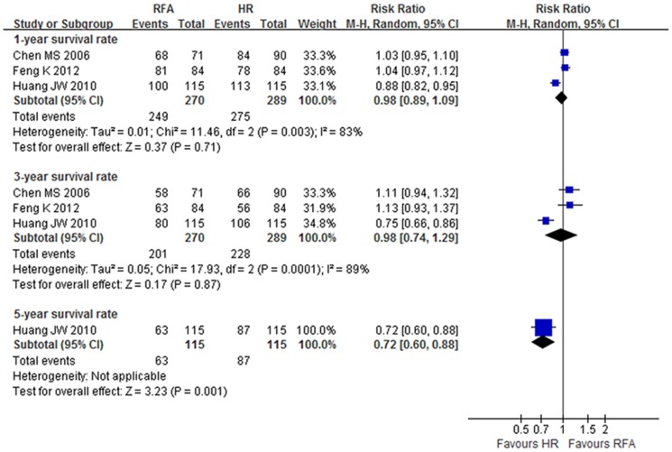
The pooled meta-analysis from the three RCTs [10], [13], [14] showed no significant difference of 1- and 3-year survival rates between groups (RR 0.98, 95% CI: 0.89–1.09, NNH = 33.3, p = 0.71; and RR 0.98, 95% CI: 0.74–1.29, NNH = 22.2, p = 0.87, respectively) (level of evidence: moderate). Only Huang et al[10] demonstrated that the 5-year survival rate in the RFA group was lower than in the HR group (RR 0.75, 95% CI: 0.60–0.88, NNH = 4.8, p = 0.001) (level of evidence: high) (Figure 3, Table 2).
Figure 3. Overall survival (OS) at 1-, 3- and 5-year in RCTs.
Table 2. Summary of finding table for HCC patients with tumor less than 5.
| Event,% | Effect estimate# | Illustrative comparative risks(95%CI)Ψ | |||||||||
| Indicators | Years | No. of Participants (studies) | RFA | HR | OR,95% CI | I2 (%) | P value | NNT or NNH | Assumed risk HR(control)(per 1000) | Corresponding risk RFA(per 1000) | Quality of evidence (GRADE) |
| Overall survival | 1-y | 559(3) | 92.2 | 95.2 | 0.98[0.89,1.09] | 83 | 0.71 | 33.3† | 933 | 914 (830 to 1000) | ⊕⊕⊕⊖ Moderate |
| 3-y | 559(3) | 74.4 | 78.9 | 0.98[0.74,1.29] | 89 | 0.87 | 22.2† | 733 | 718 (542 to 946) | ⊕⊕⊕⊖ Moderate | |
| 5-y | 230(1) | 54.8 | 75.7 | 0.72[0.60,0.88] | — | — | 4.8† | 757 | 545 (454 to 666) | ⊕⊕⊕⊕High | |
| Recurrence-free survival | 1-y | 398(2) | 85.4 | 85.4 | 1.01[0.92,1.11] | 29 | 0.88 | _ | 855 | 864 (787 to 949) | ⊕⊕⊕⊕High |
| 3-y | 398(2) | 52.3 | 56.3 | 0.95[0.60,1.52] | 84 | 0.85 | 25.0† | 554 | 526 (332 to 842) | ⊕⊕⊕⊖ Moderate | |
| 5-y | 230(1) | 28.7 | 51.3 | 0.56[0.40,0.78] | — | — | 4.4† | 513 | 287 (205 to 400) | ⊕⊕⊕⊕High | |
| Disease-free survival | 1-y | 180(1) | 91.1 | 86.7 | 1.05[0.95,1.17]& | — | 0.34 | 22.7‡ | 867 | 910 (824 to 1000) | ⊕⊕⊕⊖Moderate |
| 2-y | 180(1) | 68.9 | 76.7 | 0.90[0.75,1.08]& | — | 0.24 | 12.8† | 767 | 690 (575 to 828) | ⊕⊕⊕⊖Moderate | |
| 3-y | 180(1) | 60.0 | 68.9 | 0.87[0.70,1.08]& | — | 0.22 | 11.2† | 689 | 599 (482 to 744) | ⊕⊕⊕⊖Moderate | |
| 4-y | 180(1) | 47.8 | 51.1 | 0.93[0.70,1.26]& | — | 0.65 | 30.3† | 511 | 475 (358 to 644) | ⊕⊕⊕⊖Moderate | |
| Recurrence | 1-y | 398(2) | 15.6 | 11.1 | 1.41[0.85,2.35]& | 0 | 0.19 | 22.2† | 109 | 154 (93 to 256) | ⊕⊕⊕⊖ Moderate |
| 3-y | 398(2) | 43.2 | 29.1 | 1.48[1.14,1.94]& | 0 | 0.004* | 7.1† | 304 | 450 (347 to 590) | ⊕⊕⊕⊖ Moderate | |
| 5-y | 230(1) | 63.5 | 41.7 | 1.52[1.18,1.97]& | — | 0.001* | 4.6† | 417 | 634 (492 to 821) | ⊕⊕⊕⊖ Moderate | |
| Local recurrence | 398(2) | 20.6 | 14.1 | 1.46[0.97,2.21]& | 0 | 0.07 | 15.4† | 159 | 232 (154 to 351) | ⊕⊕⊕⊖ Moderate | |
| Distant recurrence | 398(2) | 5.0 | 4.0 | 1.25[0.50,3.10]& | 0 | 0.63 | 100.0† | 40 | 50 (20 to 124) | ⊕⊕⊕⊖ Moderate | |
| In-hospital Mortality | 559(3) | 0.0 | 0.3 | 0.42[0.02,10.19]& | — | 0.59 | 333.3‡ | 0 | 0 (0 to 0) | ⊕⊕⊕⊖ Moderate | |
| Complication rate | 559(3) | 5.9 | 34.6 | 0.18[0.06,0.53] | 74 | 0.002* | 3.5‡ | 278 | 50 (17 to 147) | ⊕⊕⊕⊖ Moderate | |
| Hospital stay | 559(3) | — | — | -8.77[−10.36,−7.18]△ | 83 | <0.001* | _ | _ | RFA group was 8.77 lower(10.36 to 7.18 lower) | ⊕⊕⊕⊖ Moderate | |
Ψ: The basis for the assumed risk (e.g. the median control group risk across studies) is provided in footnotes. The corresponding risk (and its 95% confidence interval) is based on the assumed risk in the comparison group and the relative effect of the intervention (and its 95%CI);
#, random-effect model;
&, fixed-effect model;
△: Mean Difference (IV, Random, 95% CI).
,NNH(Number needed to harm);
‡,NNT(Number needed to treat);
*:p<0.05
The pooled meta-analysis from the NRCTs showed that the 1-, 3- and 5-year survival rates in the RFA group were significantly lower than in the HR group (OR 0.78, 95% CI: 0.63–0.97, NNH = 166.7; OR 0.67, 95% CI: 0.52–0.85, NNH = 12.5; and OR 0.58, 95% CI: 0.36–0.94, NNH = 10.6, respectively) (level of evidence: very low) (Table 3).
Table 3. Summary of finding table for HCC patients with tumor size less than 5.
| Event,% | Effect estimate# | Illustrative comparative risks(95%CI)Ψ | ||||||||||
| Indicators | subgroup | Years | No. of Participants (studies) | RFA | HR | (OR,95% CI) | I2 (%) | P value | NNT or NNH | Assumed risk HR(Control)(Per 1000) | Corresponding risk RFA(Per 1000) | Quality of evidence (GRADE) |
| Overall Survival | All | 1-y | 10621(23) | 96.2 | 96.8 | 0.78[0.63,0.97] | 35 | 0.02* | 166.7† | 941 | 926 (909 to 939) | ⊕⊖ ⊖ ⊖ very low |
| 3-y | 5115(23) | 75.4 | 83.4 | 0.67[0.52,0.85]& | 57 | 0.0008* | 12.5† | 774 | 696 (640 to 744) | ⊕⊖ ⊖ ⊖ very low | ||
| 5-y | 4278(15) | 63.7 | 73.1 | 0.68[0.48,0.97]& | 82 | 0.03* | 10.6† | 731 | 649 (566 to 725) | ⊕⊖ ⊖ ⊖ very low | ||
| Child A | 1-y | 2460(11) | 93.2 | 95.4 | 0.58[0.36,0.94] | 42 | 0.002* | 45.5† | 979 | 964 (950 to 975) | ⊕⊖ ⊖ ⊖ very low | |
| 3-y | 2527(12) | 74.9 | 84.7 | 0.58[0.36,0.94]& | 71 | 0.03* | 10.2† | 844 | 758 (661 to 836) | ⊕⊖ ⊖ ⊖ very low | ||
| 5-y | 2076(7) | 63.1 | 76.7 | 0.59[0.36,0.99]& | 75 | 0.05* | 7.4† | 789 | 688 (574 to 787) | ⊕⊖ ⊖ ⊖ very low | ||
| Recurrence-free survival | All | 1-y | 2388(8) | 75.8 | 79.7 | 0.78[0.64,0.95] | 0 | 0.01* | 25.6† | 782 | 737 (697 to 773) | ⊕⊖ ⊖ ⊖ very low |
| 3-y | 2388(8) | 41.9 | 53.6 | 0.67[0.56,0.79] | 38 | <0.001* | 8.5† | 528 | 428 (385 to 469) | ⊕⊖ ⊖ ⊖ very low | ||
| 5-y | 1845(4) | 27.8 | 41.1 | 0.63[0.40,1.00]& | 73 | 0.05* | 7.5† | 398 | 294 (209 to 398) | ⊕⊖ ⊖ ⊖ very low | ||
| Child A | 1-y | 1922(5) | 75.9 | 79.6 | 0.80[0.64,1.00] | 0 | 0.05* | 27.0† | 763 | 720 (673 to 763) | ⊕⊖ ⊖ ⊖ very low | |
| 3-y | 1922(5) | 43.7 | 54.6 | 0.67[0.56,0.81] | 29 | <0.001* | 9.2† | 548 | 448 (404 to 495) | ⊕⊖ ⊖ ⊖ very low | ||
| 5-y | 1614(3) | 30.2 | 42.2 | 0.64[0.35,1.17]& | 82 | 0.15 | 8.3† | 429 | 325 (208 to 468) | ⊕⊖ ⊖ ⊖ very low | ||
| Disease-free survival | All | 1-y | 2766(12) | 70.1 | 83.9 | 0.46[0.38,0.55] | 46 | <0.001* | 7.2† | 830 | 692 (650 to 729) | ⊕⊕⊖ ⊖ low |
| 3-y | 2698(11) | 37.6 | 57.3 | 0.49[0.34,0.69]& | 67 | <0.001* | 5.1† | 559 | 383 (301 to 467) | ⊕⊖ ⊖ ⊖ very low | ||
| 5-y | 2549(9) | 21.7 | 38.6 | 0.52[0.32,0.84]& | 74 | 0.007* | 5.9† | 286 | 172 (114 to 252) | ⊕⊖ ⊖ ⊖ very low | ||
| Child A | 1-y | 1310(4) | 78.7 | 87.9 | 0.51[0.38,0.69] | 45 | <0.001* | 10.9† | 886 | 799 (747 to 843) | ⊕⊕⊖ ⊖ low | |
| 3-y | 1310(4) | 47.0 | 63.9 | 0.50[0.40,0.63] | 0 | <0.001* | 5.9† | 627 | 457 (402 to 514) | ⊕⊕⊕⊖ Moderate | ||
| 5-y | 1165(2) | 32.5 | 43.4 | 0.62[0.49,0.80] | 4 | 0.0002* | 9.2† | 420 | 310 (262 to 367) | ⊕⊕⊖ ⊖ low | ||
| Recurrence | All | 1-y | 7019(6) | 25.2 | 16.9 | 1.50[1.03,2.19]& | 61 | 0.03* | 12.4† | 171 | 260 (237 to 283) | ⊕⊖ ⊖ ⊖ very low |
| 3-y | 1140(5) | 57.1 | 41.4 | 1.87[1.23,2.84]& | 60 | 0.004* | 6.4† | 406 | 585 (524 to 642) | ⊕⊖ ⊖ ⊖ very low | ||
| 5-y | 945(3) | 75.0 | 58.0 | 2.34[1.76,3.11] | 0 | <0.001* | 5.9† | 590 | 771 (717 to 817) | ⊕⊕⊖ ⊖ low | ||
| Child A | 1-y | 335(3) | 21.5 | 19.0 | 1.18[0.69,2.03] | 0 | 0.54 | 40.0† | 180 | 206 (132 to 308) | ⊕⊖ ⊖ ⊖ very low | |
| 3-y | 335(3) | 50.6 | 46.6 | 1.06[0.38,3.00]& | 81 | 0.91 | 25.0† | 423 | 437 (218 to 687) | ⊕⊖ ⊖ ⊖ very low | ||
| 5-y | 268(2) | 68.6 | 67.2 | 1.04[0.18,6.04]& | 90 | 0.96 | 71.4† | 684 | 692 (280 to 929) | ⊕⊖ ⊖ ⊖ very low | ||
| In-hospital Mortality | — | 3061(14) | 0.1 | 0.3 | 0.47[0.13,1.76] | 0 | 0.26 | 500.0‡ | 0 | 0 (0 to 0) | ⊕⊕⊖ ⊖ low | |
| Complication rate | All | 3073(15) | 8.3 | 17.7 | 0.37[0.29,0.47] | 34 | <0.001* | 10.6‡ | 192 | 81 (64 to 100) | ⊕⊕⊖ ⊖ low | |
| Child A | 1585(5) | 7.7 | 16.7 | 0.35[0.25,0.50] | 0 | <0.001* | 11.1‡ | 314 | 138 (103 to 186) | ⊕⊕⊖ ⊖ low | ||
| Hospital stay | — | 1565(4) | — | — | -6.74[−11.33,−2.14]△ | 98 | 0.004* | - | - | RFA was 6.74 lower (2.14 to11.33) | ⊕⊖ ⊖ ⊖ very low | |
Ψ: The basis for the assumed risk (e.g. the median control group risk across studies) is provided in footnotes. The corresponding risk (and its 95% confidence interval) is based on the assumed risk in the comparison group and the relative effect of the intervention (and its 95%CI);
#, random-effect model; &, fixed-effect model;
△: Mean Difference (IV, Random, 95% CI).
,NNH(Number needed to harm);‡,NNT(Number needed to treat);
*:p<0.05
Recurrence-free survival
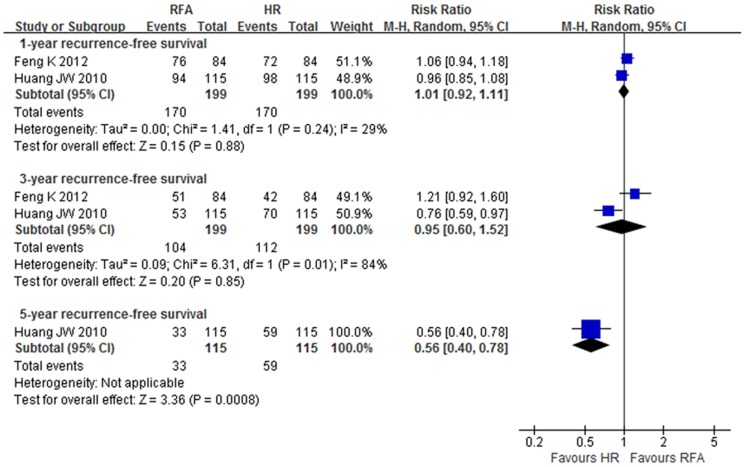
The meta-analysis from the RCTs showed no significant difference of 1- and 3-year recurrence-free survival rates between groups (level of evidence: moderate to high). The 5-year survival rate in the RFA group, however, was lower than in the HR group (RR 0.56, 95% CI: 0.40 to 0.78, NNH = 4.4) (level of evidence: high) (Figure 4, Table 2).
Figure 4. Recurrence-free survival rate (RFS) at 1-, 3- and 5-year in RCTs.
The pooled results from the NRCTs demonstrated that the 1-, 3- and 5-year recurrence-free survival rates in the RFA group were lower than in the HR group (OR 0.78, 95% CI: 0.64–0.95, NNH = 25.6; OR 0.67, 95% CI: 0.56–0.79, NNH = 8.5; and OR 0.63 95% CI: 0.40–1.00, NNH = 7.5, respectively) (level of evidence: very low). However, there was no significant difference between groups of 5-year recurrence-free survival rates for patients with Child-Pugh class A (OR 0.64, 95% CI: 0.35–1.17; NNH = 8.3) (level of evidence: very low) (Table 3).
Disease-free survival
Only one RCT [14] reported the disease-free survival rate, showing no significant difference between groups (level of evidence: moderate).
The pooled meta-analysis from the NRCTs showed that the 1-, 3- and 5-year disease-free survival rates in the RFA group were significantly lower than in the HR group (OR 0.46, 95% CI: 0.38–0.55, NNH = 7.2; OR 0.49, 95% CI: 0.34–0.69, NNH = 5.1; and OR 0.52, 95% CI: 0.32–0.84, NNH = 5.9, respectively) (level of evidence: very low to low) (Table 3).
Recurrence
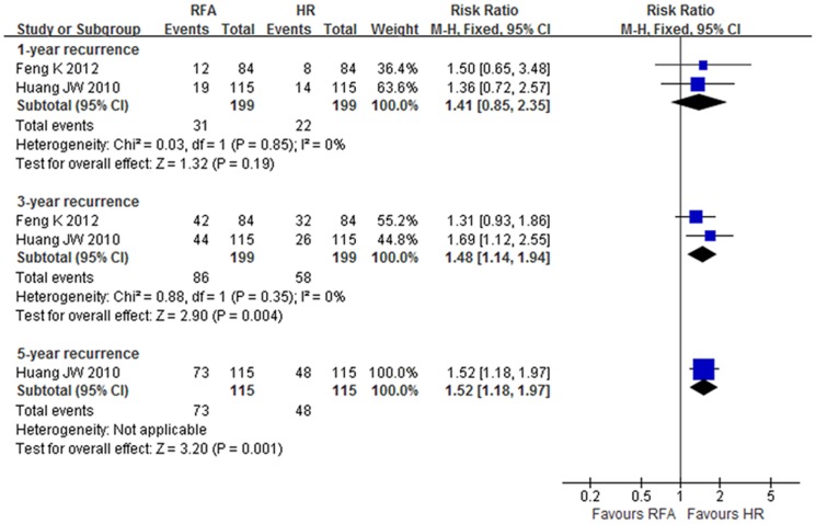
The pooled results from the RCTs showed that the 3- and 5-year recurrence rates in the RFA group were significantly higher than in the HR group (RR 1.48, 95% CI: 1.14–1.94, NNH = 7.1; and RR 1.52, 95% CI: 1.18–1.97, NNH = 4.6, respectively) (level of evidence: moderate). However, there were no significant differences of 1-year recurrence rates or of local or distant recurrence rates between groups (level of evidence: moderate) (Figure 5, Table 2).
Figure 5. Recurrence rate at 1-, 3- and 5-year in RCTs.
The results of meta-analysis for the NRCTs showed that the 1-, 3- and 5-year recurrence rates in the RFA group were higher than in the HR group (OR 1.5, 95% CI: 1.03–2.19, NNH = 12.4; OR 1.87, 95% CI: 1.23–2.84, NNH = 6.4; and OR 2.34, 95% CI: 1.76–3.11, NNH = 5.9, respectively) (level of evidence: very low to low). However, there were no significant differences between groups for patients with Child-Pugh class A (level of evidence: very low) (Table 3).
In-hospital mortality, complication rate and length of hospital stay
The pooled results of the RCTs showed no significant differences of in-hospital mortality between groups, but the complication rate in the RFA group was lower than in the HR group (RR 0.18, 95% CI: 0.06–0.53, NNT = 3.5) (level of evidence: moderate). Length of hospital stay in the RFA group was 8.77 days fewer than in the HR group (95% CI: 10.36 to 7.18 lower) (level of evidence: moderate) (Figure 6, Table 2).
Figure 6. Complication rate of RCTs.
The pooled results of the NRCTs for in-hospital mortality and complication rate were similar to those of the RCTs, but the hospital length of stay in the RFA group was 6.74 days fewer than in the HR group (95% CI: 11.33 to 2.14 lower) (level of evidence: very low) (Table 3).
Clinical outcome of HCC patients with tumor size between 3 and 5 cm
The pooled results of the RCTs showed no significant difference of overall survival rates between the groups for single HCC patients with tumor size ranging from 3 to 5 cm in diameter (level of evidence: low to moderate) (Table 4).
Table 4. Summary of finding table for Solitary HCC with tumor size≤3 cm and 3–5 cm in RCTs.
| Event,% | Effect estimate# | Illustrative comparative risks(95%CI)Ψ | ||||||||||
| Indicators | Subgroup | Years | No. of Participants (studies) | RFA | HR | OR,95% CI | I2 (%) | P value | NNH | Assumed risk HR(control)(per 1000) | Corresponding risk RFA(per 1000) | Quality of evidence (GRADE) |
| Overall survival | Tumor size≤3 cm | 1-y | 181(2) | 93.6 | 97.7 | 0.49[0.03,8.45] | 56 | 0.62 | 24.4 | 976 | 952 (550 to 997) | ⊕⊕⊖ ⊖ low |
| 3-y | 181(2) | 74.5 | 81.6 | 0.65[0.32,1.33]& | 49 | 0.24 | 14.1 | 814 | 743 (512 to 891) | ⊕⊕⊕⊖ Moderate | ||
| 4-y | 181(2) | 74.5 | 78.2 | 0.80[0.20,3.22] | 73 | 0.76 | 27.0 | 779 | 738 (413 to 919) | ⊕⊕⊖ ⊖ low | ||
| Tumor size 3∼5 cm | 1-y | 153(2) | 91.8 | 96.7 | 0.38[0.03,4.85] | 54 | 0.46 | 20.4 | 969 | 928 (775 to 981) | ⊕⊕⊖ ⊖ low | |
| 3-y | 153(2) | 62.3 | 83.7 | 0.26[0.05,1.36] | 69 | 0.11 | 4.7 | 842 | 637 (444 to791) | ⊕⊕⊖ ⊖ low | ||
| 4-y | 153(2) | 57.4 | 72.8 | 0.51[0.25,1.03]& | 3 | 0.06 | 6.5 | 733 | 583 (407 to737) | ⊕⊕⊕⊖ Moderate | ||
Ψ: The basis for the assumed risk (e.g. the median control group risk across studies) is provided in footnotes. The corresponding risk (and its 95% confidence interval) is based on the assumed risk in the comparison group and the relative effect of the intervention (and its 95%CI);
# Statistical method (M-H, random-effect model).
*:p<0.05
The results from the NRCTs showed that the 5-year survival rate in the RFA group was lower than in the HR group (OR 0.43, 95% CI: 0.25–0.73, NNH = 5.3), but there were no significant differences of 1-and 3-year survival rates between groups (level of evidence: very low). The 1-, 3- and 5-year disease-free survival rates in the RFA group were lower than in the HR group (OR 0.47, 95% CI: 0.26–0.83, NNH = 6.9; OR 0.35, 95% CI: 0.18–0.67, NNH = 6.1; and OR 0.18, 95% CI: 0.05–0.61, NNH = 9.9, respectively) (level of evidence: very low to low) (Table 5).
Table 5. Summary of finding table for HCC patients with tumor size between 3 and 5.
| Event,% | Effect estimate# | Illustrative comparative risks(95%CI)Ψ | |||||||||
| Indicators | Years | No. of Participants (studies) | RFA | HR | OR,95% CI | I2 (%) | P value | NNT or NNH | Assumed risk HR(control)(per 1000) | Corresponding risk RFA(per 1000) | Quality of evidence (GRADE) |
| Overall survival | 1-y | 243(3) | 95.4 | 92.9 | 1.62[0.54,4.84] | 0 | 0.39 | 40.0‡ | 907 | 940 (840 to 979) | ⊕⊖ ⊖ ⊖ very low |
| 3-y | 243(3) | 56.2 | 61.9 | 0.80[0.48,1.34] | 0 | 0.40 | 17.5† | 571 | 516 (390 to 641) | ⊕⊖ ⊖ ⊖ very low | |
| 5-y | 243(3) | 26.2 | 45.1 | 0.43[0.25,0.73] | 24 | 0.002* | 5.3† | 464 | 271 (178 to 387) | ⊕⊖ ⊖ ⊖ very low | |
| Disease-free survival | 1-y | 243(3) | 55.4 | 69.9 | 0.47[0.26,0.83] | 46 | 0.01* | 6.9† | 738 | 570 (423 to 700) | ⊕⊖ ⊖ ⊖ very low |
| 3-y | 243(3) | 13.8 | 30.1 | 0.35[0.18,0.67] | 34 | 0.002* | 6.1† | 321 | 142 (78 to 241) | ⊕⊖ ⊖ ⊖ very low | |
| 5-y | 243(3) | 2.3 | 12.4 | 0.18[0.05,0.61] | 32 | 0.006* | 9.9† | 143 | 29 (8 to 92) | ⊕⊕⊖ ⊖ low | |
Ψ: The basis for the assumed risk (e.g. the median control group risk across studies) is provided in footnotes. The corresponding risk (and its 95% confidence interval) is based on the assumed risk in the comparison group and the relative effect of the intervention (and its 95%CI);
# Statistical method (M-H, random-effect model).
,NNH(Number needed to harm);
‡,NNT(Number needed to treat);
*:p<0.05
Clinical outcome of HCC patients with tumor size smaller than 3 cm
The pooled results of RCTs showed no significant differences of the overall survival rates between groups for patients with a single HCC and tumor size ≤3 cm in diameter (level of evidence: low to moderate) (Table 4).
The pooled meta-analysis from NRCTs showed no significant differences of 1- and 3-year survival rates between groups, but the 5-year survival rate in the RFA group was lower than in the HR group (OR 0.62, 95% CI: 0.43–0.90, NNH = 4.5; p = 0.01) (level of evidence: very low). The 1-, 3- and 5-year disease-free survival rates in the RFA group were lower than in the HR group, but the 5-year disease-free survival rates for patients with a single HCC showed no significant difference between groups (OR 0.69, 95% CI: 0.35–1.36). The 1-, 3- and 5-year recurrence rates also showed no significant differences between groups, but the 5-year recurrence rate in the RFA group was lower than in the HR group for patients with Child-Pugh class A (OR 0.42, 95% CI: 0.19–0.93, NNT = 5.1) (level of evidence: moderate). The complication rate in the RFA group was lower than in the HR group (OR 0.44, 95% CI: 0.29–0.65, NNT = 11.2) (level of evidence: low), but there was no significant difference between groups for patients with Child-Pugh class A (Table 6).
Table 6. Summary of finding table for HCC patients with tumor size ≤3cm in NRCTs.
| Event,% | Effect estimate# | Illustrative comparative risks(95%CI)Ψ | ||||||||||
| Indicators | Subgroup | Years | No. of Participants (studies) | RFA | HR | OR,95% CI | I2 (%) | P value | NNT or NNH | Assumed risk HR(control)(per 1000) | Corresponding risk RFA(per 1000) | Quality of evidence (GRADE) |
| Overall survival | All | 1-y | 2782(14) | 95.1 | 97.2 | 0.72[0.50,1.04] | 17 | 0.08 | 47.6† | 967 | 955 (936 to 968) | ⊕⊖ ⊖ ⊖ very low |
| 3-y | 3156(15) | 80.5 | 86.4 | 0.71[0.48,1.05]& | 68 | 0.09 | 16.9† | 811 | 753 (673 to 818) | ⊕⊖ ⊖ ⊖ very low | ||
| 5-y | 2636(12) | 71.1 | 93.3 | 0.62[0.43,0.90]& | 67 | 0.01* | 4.5† | 764 | 667 (582 to 744) | ⊕⊖ ⊖ ⊖ very low | ||
| Child A | 1-y | 1632(9) | 96.0 | 95.8 | 0.94[0.58,1.53] | 22 | 0.81 | 500.0‡ | 981 | 980 (968 to 972) | ⊕⊕⊖ ⊖ low | |
| 3-y | 1632(9) | 81.5 | 86.5 | 0.66[0.33,1.32]& | 77 | 0.24 | 20.0† | 881 | 830 (710 to 907) | ⊕⊖ ⊖ ⊖ very low | ||
| 5-y | 1386(6) | 72.4 | 80.9 | 0.64[0.38,1.08]& | 65 | 0.09 | 11.8† | 824 | 750 (640 to 835) | ⊕⊖ ⊖ ⊖ very low | ||
| Single HCC | 1-y | 1516(8) | 94.7 | 95.7 | 0.67[0.42,1.09] | 9 | 0.11 | 100.0† | 982 | 973 (958 to 983) | ⊕⊕⊖ ⊖ low | |
| 3-y | 1516(8) | 80.4 | 87.2 | 0.59[0.32,1.09]& | 70 | 0.09 | 14.7† | 898 | 839 (738 to 906) | ⊕⊖ ⊖ ⊖ very low | ||
| 5-y | 1516(8) | 64.3 | 77.9 | 0.52[0.31,0.87]& | 74 | 0.01* | 7.4† | 773 | 639 (514 to 748) | ⊕⊖ ⊖ ⊖ very low | ||
| Single + Child A | 1-y | 1009(6) | 94.6 | 95.2 | 0.83[0.48,1.46] | 43 | 0.52 | 166.7† | 990 | 988 (979 to 993) | ⊕⊕⊖ ⊖ low | |
| 3-y | 1009(6) | 79.5 | 88.5 | 0.38[0.12,1.16]& | 80 | 0.09 | 11.1† | 935 | 845 (633 to 943) | ⊕⊕⊖ ⊖ low | ||
| 5-y | 978(5) | 71.3 | 81.7 | 0.58[0.28,1.19]& | 69 | 0.14 | 9.6† | 840 | 753 (595 to 862) | ⊕⊖ ⊖ ⊖ very low | ||
| Disease-free survival | All | 1-y | 1722(9) | 71.6 | 85.2 | 0.43[0.34,0.55] | 45 | <0.00001* | 7.4† | 854 | 716 (665 to 763) | ⊕⊕⊖ ⊖ low |
| 3-y | 1722(9) | 43.1 | 64.3 | 0.45[0.31,0.64]& | 54 | <0.0001* | 4.7† | 604 | 407 (321 to 494) | ⊕⊖ ⊖ ⊖ very low | ||
| 5-y | 1592(7) | 24.8 | 46.2 | 0.50[0.26,0.97]& | 83 | 0.04* | 4.7† | 357 | 217 (126 to 350) | ⊕⊕⊖ ⊖ low | ||
| Child A | 1-y | 772(4) | 76.9 | 86.6 | 0.52[0.36,0.75] | 45 | 0.0006* | 10.3† | 876 | 771 (700 to 829) | ⊕⊕⊖ ⊖ low | |
| 3-y | 772(4) | 57.7 | 71.7 | 0.55[0.40,0.74] | 0 | 0.0001* | 7.1† | 649 | 504 (425 to 578) | ⊕⊕⊖ ⊖ low | ||
| 5-y | 627(2) | 42.8 | 53.7 | 0.66[0.48,0.91] | 0 | 0.01* | 9.2† | 482 | 380 (309 to 459) | ⊕⊕⊖ ⊖ low | ||
| Single HCC | 1-y | 1024(5) | 78.7 | 86.4 | 0.54[0.39,0.75] | 38 | 0.0003* | 13.0† | 859 | 767 (704 to 820) | ⊕⊕⊖ ⊖ low | |
| 3-y | 1024(5) | 53.5 | 68.7 | 0.55[0.43,0.72] | 0 | <0.0001* | 6.6† | 615 | 468 (407 to 535) | ⊕⊕⊖ ⊖ low | ||
| 5-y | 1024(5) | 32.3 | 46.7 | 0.69[0.35,1.36]& | 78 | 0.28 | 6.9† | 404 | 319 (192 to 480) | ⊕⊖ ⊖ ⊖ very low | ||
| Single + Child A | 1-y | 717(3) | 75.9 | 87.0 | 0.48[0.33,0.71] | 38 | 0.0003* | 9.0† | 898 | 809 (744 to 862) | ⊕⊕⊕⊖ moderate | |
| 3-y | 717(3) | 79.5 | 88.5 | 0.55[0.40,0.75] | 80 | 0.09 | 11.1† | 712 | 576 (497 to 650) | ⊕⊕⊖ ⊖ low | ||
| 5-y | 627(2) | 42.8 | 53.7 | 0.66[0.48,0.91] | 0 | 0.01* | 9.2† | 482 | 380 (309 to 459) | ⊕⊕⊖ ⊖ low | ||
| Recurrence-free survival | All | 1-y | 1467(5) | 78.8 | 77.6 | 1.02[0.79,1.31] | 0 | 0.89 | 83.3‡ | 757 | 761 (711 to 803) | ⊕⊕⊖ ⊖ low |
| 3-y | 1467(5) | 44.5 | 50.6 | 0.85[0.69,1.05] | 17 | 0.13 | 16.4† | 493 | 453 (402 to 505) | ⊕⊕⊖ ⊖ low | ||
| 5-y | 1307(4) | 29.2 | 37.4 | 0.76[0.40,1.43]& | 83 | 0.39 | 12.2† | 368 | 307 (189 to 454) | ⊕⊖ ⊖ ⊖ very low | ||
| Child A | 1-y | 1236(4) | 77.8 | 76.7 | 1.05[0.80,1.38] | 0 | 0.71 | 90.9‡ | 748 | 757 (725 to 820) | ⊕⊕⊖ ⊖ low | |
| 3-y | 1236(4) | 46.2 | 50.9 | 0.88[0.70,1.11] | 27 | 0.27 | 21.3† | 514 | 482 (425 to 540) | ⊕⊕⊖ ⊖ low | ||
| 5-y | 1076(3) | 32.9 | 38.7 | 0.81[0.36,1.83]& | 89 | 0.61 | 17.2† | 370 | 322 (175 to 518) | ⊕⊖ ⊖ ⊖ very low | ||
| Single HCC | 1-y | 899(3) | 78.2 | 76.0 | 1.03[0.74,1.42] | 0 | 0.87 | 45.5 | 757 | 762 (697 to 816) | ⊕⊕⊖ ⊖ low | |
| 3-y | 899(3) | 49.0 | 52.2 | 0.96[0.73,1.26] | 26 | 0.79 | 31.3† | 521 | 511 (443 to 578) | ⊕⊕⊖ ⊖ low | ||
| 5-y | 899(3) | 34.8 | 37.7 | 1.05[0.79,1.39] | 37 | 0.75 | 34.5† | 370 | 381 (317 to 449) | ⊕⊕⊖ ⊖ low | ||
| Single + Child A | 1-y | 668(2) | 76.0 | 74.3 | 1.09[0.76,1.56] | 0 | 0.63 | 58.8‡ | 748 | 764 (693 to 822) | ⊕⊕⊖ ⊖ low | |
| 3-y | 668(2) | 55.1 | 53.0 | 1.07[0.78,1.46] | 0 | 0.68 | 47.6‡ | 544 | 561 (482 to 635) | ⊕⊖ ⊖ ⊖ very low | ||
| 5-y | 668(2) | 44.5 | 39.7 | 1.17[0.86,1.61] | 0 | 0.32 | 20.8‡ | 442 | 481 (405 to 560) | ⊕⊖ ⊖ ⊖ very low | ||
| Recurrence | All | 1-y | 490(2) | 12.9 | 15.4 | 0.83[0.49,1.40] | 0 | 0.48 | 40.0‡ | 163 | 139 (87 to 214) | ⊕⊖ ⊖ ⊖ very low |
| 3-y | 490(2) | 50.7 | 45.2 | 0.95[0.29,3.11]& | 87 | 0.94 | 18.1† | 493 | 480 (220 to 751) | ⊕⊖ ⊖ ⊖ very low | ||
| 5-y | 490(2) | 66.2 | 59.0 | 0.95[0.21,4.28]& | 91 | 0.94 | 13.9† | 638 | 626 (270 to 883) | ⊕⊖ ⊖ ⊖ very low | ||
| Child A | 1-y | 116(1) | 18.2 | 18.0 | 1.01[0.39,2.63] | — | 0.98 | 500.0† | 180 | 181 (79to 366) | ⊕⊕⊖ ⊖ low | |
| 3-y | 116(1) | 40.9 | 58.0 | 0.50[0.24,1.06] | — | 0.07 | 5.8‡ | 580 | 408 (249 to 594) | ⊕⊕⊕⊖ moderate | ||
| 5-y | 116(1) | 54.5 | 74.0 | 0.42[0.19,0.93] | — | 0.03* | 5.1‡ | 740 | 544 (351 to 726) | ⊕⊕⊕⊖ moderate | ||
| Complication rate | All | 1248(7) | 7.1 | 16.0 | 0.44[0.29,0.65] | 35 | <0.0001* | 11.2‡ | 66 | 30 (20 to 44) | ⊕⊕⊖ ⊖ low | |
| Child A | 305(2) | 11.2 | 31.1 | 0.36[0.12,1.12]& | 55 | 0.08 | 5.0‡ | 290 | 128 (47 to 314) | ⊕⊖ ⊖ ⊖ very low | ||
Ψ: The basis for the assumed risk (e.g. the median control group risk across studies) is provided in footnotes. The corresponding risk (and its 95% confidence interval) is based on the assumed risk in the comparison group and the relative effect of the intervention (and its 95%CI);
# Statistical method (M-H, random-effect model); & Statistical method (M-H, fixed-effect model).
,NNH(Number needed to harm);
‡,NNT(Number needed to treat);
*:p<0.05
Clinical outcome of solitary HCC patients with tumor size smaller than 2 cm
The pooled results of three NRCTs [15], [16], [22]showed that there was no significant difference in 1-, 3- and 5-year overall survival or RFS, 3-,5-year DFS, and 1-,3-year recurrence between groups(p>0.05). However, the 1-year DFS (OR 0.22, 95%CI:0.07–0.65, NNH = 4.4; p = 0.06), 5-year recurrence(OR 0.42, 95%CI:0.19–0.93, NNT = 5.0;P = 0.03) and complication rate (OR 0.23, 95%CI:0.11–0.49, NNT = 3.1; p = 0.0001) in RFA group were lower than that of HR group(P<0.05)(level of evidence: very low)(Table 7).
Table 7. Summary of finding table for solitary HCC patients with tumor size ≤2 cm in NRCTs.
| Event,% | Effect estimate | Illustrative comparative risks(95%CI)Ψ | |||||||||
| Indicators | Years | No. of Participants (studies) | RFA | HR | OR,95% CI | I2 (%) | P value | NNT or NNH | Assumed risk HR(control)(per 1000) | Corresponding risk RFA(per 1000) | Quality of evidence (GRADE) |
| Overall survival | 1-y | 365(3) | 98.4 | 95.5 | 2.60[0.73,9.29]& | 22 | 0.14 | 34.5‡ | 981 | 993(974 to 998) | ⊕⊕⊖ ⊖ low |
| 3-y | 365(3) | 85.7 | 84.6 | 0.65[0.10,4.01]# | 83 | 0.64 | 90.9‡ | 920 | 882(535 to 979) | ⊕⊖ ⊖ ⊖ very low | |
| 5-y | 365(3) | 79.4 | 74.4 | 1.30[0.79,2.15]& | 41 | 0.30 | 20.0‡ | 827 | 861(791 to 911) | ⊕⊕⊖ ⊖ low | |
| Disease-free survival | 1-y | 104(1) | 67.1 | 89.8 | 0.22[0.07,0.65]& | — | 0.006* | 4.4† | 904 | 674(397 to 860) | ⊕⊕⊖ ⊖ low |
| 3-y | 104(1) | 46.4 | 62.1 | 0.54[0.25,1.17]& | — | 0.12 | 6.4† | 615 | 463(285 to 651) | ⊕⊖ ⊖ ⊖ very low | |
| 5-y | 104(1) | 38.0 | 40.7 | 0.92[0.42,2.03]& | — | 0.84 | 37.0† | 404 | 384(222 to 579) | ⊕⊖ ⊖ ⊖ very low | |
| Recurrence-free survival | 1-y | 145(1) | 76.4 | 75.6 | 1.02[0.48,2.19]& | — | 0.96 | 125.0‡ | 757 | 761(599 to 872) | ⊕⊖ ⊖ ⊖ very low |
| 3-y | 145(1) | 65.2 | 56.1 | 1.40[0.72,2.74]& | — | 0.32 | 11.0‡ | 568 | 648(486 to 783) | ⊕⊖ ⊖ ⊖ very low | |
| 5-y | 145(1) | 59.8 | 51.3 | 1.37[0.71,2.65]& | — | 0.35 | 11.8‡ | 514 | 592(429 to 737) | ⊕⊖ ⊖ ⊖ very low | |
| Recurrence | 1-y | 116(1) | 18.2 | 18.9 | 1.01[0.39,2.63]& | — | 0.98 | 142.9‡ | 180 | 181(79 to 366) | ⊕⊖ ⊖ ⊖ very low |
| 3-y | 116(1) | 40.5 | 57.4 | 0.50[0.24,1.06]& | — | 0.07 | 5.9‡ | 580 | 408(249 to 594) | ⊕⊕⊖ ⊖ low | |
| 5-y | 116(1) | 54.8 | 74.8 | 0.42[0.19,0.93]& | — | 0.03* | 5.0‡ | 740 | 544(351 to 726) | ⊕⊕⊖ ⊖ low | |
| Complication rate | 145(1) | 19.1 | 51.4 | 0.23[0.11,0.49]& | — | 0.0001* | 3.1‡ | 514 | 196(104 to 341) | ⊕⊕⊖ ⊖ low | |
Ψ: The basis for the assumed risk (e.g. the median control group risk across studies) is provided in footnotes. The corresponding risk (and its 95% confidence interval) is based on the assumed risk in the comparison group and the relative effect of the intervention (and its 95%CI);
# Statistical method (M-H, random-effect model);
& Statistical method (M-H, fixed-effect model).
,NNH(Number needed to harm);
‡,NNT(Number needed to treat);
*:p<0.05
Sensitivity analysis
Among the three RCTs, the patients included by Huang et al[10] were the oldest (average age of 56 years). With exclusion of this study, the 1- and 3-year survival rates still showed no significant difference between groups, but the results of the heterogeneity test (I2) changed from 83–89% to 0%. The study of Feng et al[13] included more patients with cirrhosis and a high proportion (61.5% to 75%) of patients with more than two nodules. With exclusion of this study, the complication rate in the RFA group was still lower than in the HR group (RR 0.12, 95% CI: 0.06–0.24), but the results of the heterogeneity test (I2) changed form 74% to 0%.
Publication bias
As is shown in Table 8, we only analyzed the publication bias for indicators included in 10 or more studies[11]. After viewing the funnel plot and Egger's test, it was found that the indicators of 5-year survival for patients with tumor size smaller than 5 cm or 3 cm in diameter showed no publication bias (P>0.05). The remaining indicators all showed that some degree of publication bias existed.
Table 8. Publication bias of studies which were more than ten.
| Indicators | Subgroup | Years | No. of studies | Egger's test P value | Publication bias |
| Overall survival | HCC≤5 cm | 1-y | 23 | <0.001 | Yes |
| 3-y | 23 | <0.001 | Yes | ||
| 5-y | 15 | 0.182 | No | ||
| HCC≤5 cm and Child A | 1-y | 11 | 0.001 | Yes | |
| 3-y | 12 | 0.006 | Yes | ||
| HCC≤3 cm | 1-y | 14 | 0.003 | Yes | |
| 3-y | 15 | 0.005 | Yes | ||
| 5-y | 12 | 0.21 | No | ||
| Disease-free survival | HCC≤5 cm | 1-y | 12 | 0.039 | Yes |
| 3-y | 11 | 0.011 | Yes | ||
| Complication rate | 15 | <0.001 | Yes |
Discussion
HR is considered the preferred treatment for patients meeting Milan criteria with a single nodule or multiple lesions with good liver function but unsuitable for liver transplantation [4], [5]. One prospective RCT demonstrated that the 3-year survival rate with liver resection for small hepatocellular carcinoma could reach 74.8%[13]. However, 80% of cases were unsuitable for liver resection for various reasons such as low rate of early diagnosis, poor expected function of residual liver after surgery, and anticipated serious post-operative complications [39]. Local ablation with RFA or PEI is recommended by the latest updated EASL-EORTC guidelines as the standard care for patients with Barcelona clinic liver cancer (BCLC) 0 to A level but were unsuitable for surgery in 2012[4].
RCTs of high quality and with a large sample size are considered the best sources of evidence, but RCTs are accompanied by high costs, are difficult to conduct, and often have poor external validity. RCTs performed in the field of surgery, especially with double-blinding methods, are extremely difficult. Systematic review and/or meta-analysis of RCTs are often the most feasible methods to address this situation. Previous meta-analyses worldwide have compared the long-term efficacy of RFA and HR for the treatment of small HCC, but prospective RCTs with large sample sizes were rarely included. The systematic review authors usually combined the results of RCTs and NRCTs together or mistook retrospective studies as RCTs, leading them to draw conclusions of low reliability[39]–[43], as various risks for bias or confounding factors might have existed among the study designs. However, the impact these biases on outcomes could not be ascertained because of the limited information provided in these primary studies[11]. Therefore, such results without a more strict risk assessment would inevitably lead to an erroneous estimation of effects and mislead clinical decision-making.
In this study, three RCTs without blinding were included, all with only moderate quality of evidence [10], [13], [14]. The pooled results of our meta-analysis showed no significant differences between the RFA and HR groups in overall survival and recurrence-free survival rates at 1 and 3 years and in recurrence rates at 1 year following the treatment of small hepatocellular carcinomas meeting the Milan criteria. The RFA group had higher recurrence rates at 3 and 5 years and lower complication rates when compared with the HR group. It is well-known that tumor size, number of lesions, location, liver function, the presence of portal vein invasion, the presence of vascular invasion and the width of the tumor-free margin during surgical excision were the independent prognostic factors affecting the survival of patients[44], [45]. The recurrence rate after RFA is related to incomplete tumor ablation. In case of HCC in which local curative ablative therapy was obtained by securing a safety margin[46]. It is considered that the local recurrence rate markedly differed among patients with or without a sufficient safety margin [47]. However, RFA is a minimally invasive procedure, and it is often hard to achieve a specific safety margin in three dimensions all around a large tumor[46], leading to a higher recurrence but lessened compromise of liver function, which might be one of the reasons for the relatively low postoperative complication rate. Commonly, patients with small HCC rarely dies within 1 year and the recurrence impacts the overall survival gradually. However, the 1-year survival rate of RFA in the RCTs Huang et al[10] conducted was 87%, whereas that of resection was 98%. This result is markedly different from the other two RCTs [13], [14], which might be the factors leading to the clinical heterogeneity. There are some reasons may contribute to this problem. Firstly, of 108 RFA-treated patients in the study of Huang et al[10], seventeen patients had lesions in dangerous locations, leading difficult to secure a safety margin. Secondly, the average age of included patients in both groups were much old, and they were prone to the therapy of resection. Apart form that, the rate of loss to follow-up was greater in the resection group (18/115, 15.6%) than in RFA group (7/115, 6.1%), which would probably influence the comparison between two groups.
NRCTs of high quality with large sample sizes could provide research evidence for a wider population, particularly with a greater advantage for assessing the safety of an intervention. However, NRCTs might also be more easily influenced by different kinds of bias or by unknown confounding factors[11]. In this study, twenty-five NRCTs with an average quality of “moderate” and a large pooled sample size of 11,314 subjects were included. The pooled results of the meta-analysis could have some significance for guiding clinical practice as they showed that the overall survival in the RFA group was significantly lower than in the HR group for patients with small tumor size less than 5 cm in diameter. However, identical results were suggested otherwise in respect of recurrence, complication rate, in-hospital mortality and hospital length of stay between the RCTs and NRCTs. The reason for this phenomenon was that rare RCTs with low heterogeneity and large sample size were included which showed no difference between the groups, but did not rule out the impact of potential confounding factors or bias (i.e., publication bias) in the NRCTs, leading to a possible overestimation of the survival effect in the NRCTs.
Few studies except for three retrospective ones have reported the outcome of solitary HCC with tumor size less than 2 cm. Nevertheless, the conclusion of these studies was varies. Wang JH et al[15] and Hung HH et al [22]have concluded that RFA was as effective as HR in patients with BCLC very early stage HCC, while Peng ZW et al[16] showed that percutaneous RFA was better than those of HR, especially for central HCC. In this study, we found that the overall survival and 1-, 3-year recurrence in RFA group were as equal effective as HR group, and the complication rate and 5-year recurrence in RFA group were lower than HR group. However, the overall quality of evidence evaluated by GRADE (the Grading of Recommendations Assessment, Development and Evaluation) was very low to low due to limited sample size or many known or unknown risk of bias existed in observational studies. Therefore, when applying the outcome of the evidence to clinical practice, physicians should take caution. More large-scale, well-conducted RCTs or retrospective studies focused on the topics were still needed in the further.
GRADE (2011 version) has identified five categories or factors downgrading of the quality of evidence: study limitations (risk of bias), imprecision, inconsistency, indirectness, and publication bias [48], [49]. For observational studies, the GRADE group has also identified three categories or factors raising the quality of evidence: large magnitude of effect, dose-response gradient, and plausible confounding, all of which can increase confidence in the estimated effects[50]. Therefore, our study strictly followed the quality assessment criteria of the GRADE guidelines, evaluated the quality of evidence for the important outcomes of each patient, and performed a subgroup analysis based on risk factors (i.e., the Child-Pugh class, tumor size, and number of nodules) that were likely to affect the clinical outcome, so as to reduce the impact of risk factors on clinical outcomes to some extent. It is well-known that cirrhosis is also an independent prognostic factor for patients with HCC. For example, in the study of Nishikawa H et al [20], they concluded that the presence of liver cirrhosis was the sole significant factor for recurrence-free survival. However, based on the current primary studies, it is difficult to apply a subgroup analysis for this factor individually. We suggest that more high quality RCTs or retrospective studies should be focus on this topic in the further.
Limitations
Only three RCTs were included, and the limited sample size and high heterogeneity might affect the robustness of the clinical outcomes to some extent. Given the characteristics of the included NRCTs, we deleted some items (i.e., prospective collection of data, unbiased and blinded assessment of the study endpoint, and prospective calculation of the study size) from the MINORS score questionnaire which were unsuitable for this study, but we were convinced that it would not have an impact on the quality of evidence of the final clinical outcome.
Conclusions
The pooled meta-analysis of the RCTs demonstrated no significant difference between groups of the overall survival, recurrence-free survival, disease-free survival and in-hospital mortality rates for early HCC with tumor size smaller than 5 cm in diameter, but the RFA group had higher recurrence rates, lower complication rates, and shorter hospital lengths of stay.
The pooled meta-analysis of the NRCTs showed no significant difference in recurrence rates between groups for patients with Child-Pugh class A or tumor size smaller than 3 cm or 3 cm to 5 cm in diameter. However, when combining the results of all patients with tumors smaller than 5 cm in diameter, it is showed that the RFA group had lower overall survival and higher recurrence rates. For patients with Child-Pugh A/B and tumor size less than 2 cm in diameter, the pooled results concluded that RFA was as effective as HR in the overall survival and 1-, 3-year recurrence, and RFA yielded lower complication rate and 5-year recurrence than HR. However, it is still need RCTs of high quality to further enhance the level of evidence.
Based on full consideration of the current available best evidence, a comprehensive conclusion can be drawn: RFA is comparable to HR with lower complication rates but with higher recurrence rates. All relevant risk factors that may affect the final outcome of patients should be considered, so as to balance minimizing recurrent HCC after RFA with improving the quality of life of patients. The authors suggest that more high quality RCTs or retrospective studies should focus on RFA versus HR for very early HCC patients and to provide the better clinical decision for physicians.
Supporting Information
(DOC)
Acknowledgments
All authors agree with the manuscript results and conclusions.
Funding Statement
This work was funded by National Technology Support Program (2011BAI14B01). The funders had no role in study design, data collection and analysis, decision to publish, or preparation of the manuscript.
References
- 1. Murray CJ, Vos T, Lozano R, Naghavi M, Flaxman AD, et al. (2012) Disability-adjusted life years (DALYs) for 291 diseases and injuries in 21 regions, 1990-2010: a systematic analysis for the Global Burden of Disease Study 2010. Lancet 380(9859): 2197–2223. [DOI] [PubMed] [Google Scholar]
- 2. Soerjomataram I, Lortet-Tieulent J, Parkin DM, Ferlay J, Mathers C, et al. (2012) Global burden of cancer in 2008: a systematic analysis of disability-adjusted life-years in 12 world regions. Lancet 380(9856): 1840–1850. [DOI] [PubMed] [Google Scholar]
- 3. Bray F, Jemal A, Grey N, Ferlay J, Forman D (2012) Global cancer transitions according to the Human Development Index (2008-2030): a population-based study. Lancet Oncol 13: 790–801. [DOI] [PubMed] [Google Scholar]
- 4. Llovet JM, Lencioni R, Di Bisceglie AM, Gaile PR, Dufour JF, et al. (2012) EASL-EORTC Clinical Practice Guidelines: Management of hepatocellular carcinoma. J Hepatol 56: 908–943. [DOI] [PubMed] [Google Scholar]
- 5. Bruix J, Sherman M (2011) Management of hepatocellular carcinoma: an update. Hepatology 53: 1020–1022. [DOI] [PMC free article] [PubMed] [Google Scholar]
- 6. Kobayashi M, Ikeda K, Kawamura Y, Yatsuji H, Hosaka T, et al. (2009) High serum des-gamma-carboxy prothrombin level predicts poor prognosis after radiofrequency ablation of hepatocellular carcinoma. Cancer 115: 571–580. [DOI] [PubMed] [Google Scholar]
- 7.Wang YQ, Luo QQ, L YP, Deng SL, Li XL, et al.. (2013) A Systematic Assessment of the Quality of Systematic Reviews/Meta-analyses in Radiofrequency Ablation versus Hepatic Resection for Small Hepatocellular Carcinoma. J Evid Based Med.in press. [DOI] [PubMed]
- 8. Moher D, Liberati A, Tetzlaff J, Altman DG (2009) Preferred reporting items for systematic reviews and meta-analyses: the PRISMA statement. PLoS Med 6: e1000097. [DOI] [PMC free article] [PubMed] [Google Scholar]
- 9.U.S. Department of Health and Human Services. (2007) Guidance for industry clinical trial endpoints for the approval of cancer drugs and biologics. Food and Drug Adimistration Rockville. Available: from:http://www.fda.gov/downloads/Drugs/GuidanceComplianceRegulatoryInformation/Guidances/UCM071590.pdf.
- 10. Huang J, Yan L, Cheng Z, Wu H, Du L, et al. (2010) A randomized trial comparing radiofrequency ablation and surgical resection for HCC conforming to the Milan criteria. Ann Surg 252: 903–912. [DOI] [PubMed] [Google Scholar]
- 11.Higgins JP, Green S (2011) Conchrane Handbook for Systematic Reviews of Interventions 5.1.0[updated March 2011]. The Cochrane Collaboration. Available: from:www.cochrane-handbook.org.
- 12. Slim K, Nini E, Forestier D, Kwiatkowski F, Panis Y, et al. (2003) Methodological index for non-randomized studies (minors): development and validation of a new instrument. ANZ J Surg 73: 712–716. [DOI] [PubMed] [Google Scholar]
- 13. Feng K, Yan J, Li X, Xia F, Ma K, et al. (2012) A randomized controlled trial of radiofrequency ablation and surgical resection in the treatment of small hepatocellular carcinoma. J Hepatol 57: 794–802. [DOI] [PubMed] [Google Scholar]
- 14. Chen MS, Li JQ, Zheng Y, Guo RP, Liang HH, et al. (2006) A prospective randomized trial comparing percutaneous local ablative therapy and partial hepatectomy for small hepatocellular carcinoma. Ann Surg 243: 321–328. [DOI] [PMC free article] [PubMed] [Google Scholar]
- 15. Wang JH, Wang CC, Hung CH, Chen CL, Lu SN (2012) Survival comparison between surgical resection and radiofrequency ablation for patients in BCLC very early/early stage hepatocellular carcinoma. J Hepatol 56: 412–418. [DOI] [PubMed] [Google Scholar]
- 16. Peng ZW, Lin XJ, Zhang YJ, Liang HH, Guo RP, et al. (2012) Radiofrequency ablation versus hepatic resection for the treatment of hepatocellular carcinomas 2 cm or smaller: a retrospective comparative study. Radiology 262: 1022–1033. [DOI] [PubMed] [Google Scholar]
- 17. Liu H, Hu JF, Weng HF, Feng LL, Cao H (2011) Effect and safety of radiofrequency catheter ablation for single small hepatocellular carcinoma[Article in Chinese]. Pracitical Clinical Medicine 12(11): 16–18. [Google Scholar]
- 18. Fu J, Chen Y, Lei TJ, Li HM, Lei HL, et al. (2011) Comparison of curative effect between radiofrequency ablation and surgical resection in treatment of elderly patients with small hepatocellular carcinoma[Article in Chinese]. Journal of Jilin University(Medicine Edition) 37(4): 733–737. [Google Scholar]
- 19. Kong WT, Zhang WW, Qiu YD, Zhou T, Bin H (2011) Prospective comparative study of cool-tip radiofrequency ablation and surgical resection for hepatocellular carcinoma. Chinese-German J Clin Oncol 10(7): 399–405. [Google Scholar]
- 20. Nishikawa H, Inuzuka T, Takeda H, Nakajima J, Matsuda F, et al. (2011) Comparison of percutaneous radiofrequency thermal ablation and surgical resection for small hepatocellular carcinoma. BMC Gastroenterol 11: 143. [DOI] [PMC free article] [PubMed] [Google Scholar]
- 21. Ikeda K, Kobayashi M, Kawamura Y, Imai N, Seko Y, et al. (2011) Stage progression of small hepatocellular carcinoma after radical therapy: comparisons of radiofrequency ablation and surgery using the Markov model. Liver Int 31: 692–699. [DOI] [PubMed] [Google Scholar]
- 22. Hung HH, Chiou YY, Hsia CY, Su CW, Chou YH, et al. (2011) Survival rates are comparable after radiofrequency ablation or surgery in patients with small hepatocellular carcinomas. Clin Gastroenterol Hepatol 9: 79–86. [DOI] [PubMed] [Google Scholar]
- 23. Huang J, Hernandez-Alejandro R, Croome KP, Yan L, Wu H, et al. (2011) Radiofrequency ablation versus surgical resection for hepatocellular carcinoma in Childs A cirrhotics-a retrospective study of 1,061 cases. J Gastrointest Surg 15: 311–320. [DOI] [PubMed] [Google Scholar]
- 24. Guo WX, Zhai B, Lai EC, Li N, Shi J, et al. (2010) Percutaneous radiofrequency ablation versus partial hepatectomy for multicentric small hepatocellular carcinomas: a nonrandomized comparative study. World J Surg 34: 2671–2676. [DOI] [PubMed] [Google Scholar]
- 25. Bu XY, Wang Y, Ge Z, Wang ZC, Zhang DS (2009) Comparison of radiofrequency ablation and surgical resection for small primary liver carcinoma[Article in Chinese]. Chin Arch Gen Surg (Electronic Edition) 3 2: 127–131. [Google Scholar]
- 26. Ueno S, Sakoda M, Kubo F, Hiwatashi K, Tateno T, et al. (2009) Surgical resection versus radiofrequency ablation for small hepatocellular carcinomas within the Milan criteria. J Hepatobiliary Pancreat Surg 16: 359–366. [DOI] [PubMed] [Google Scholar]
- 27. Santambrogio R, Opocher E, Zuin M, Selmi C, Bertolini E, et al. (2009) Surgical resection versus laparoscopic radiofrequency ablation in patients with hepatocellular carcinoma and Child-Pugh class a liver cirrhosis. Ann Surg Oncol 16: 3289–3298. [DOI] [PubMed] [Google Scholar]
- 28. Hiraoka A, Horiike N, Yamashita Y, Koizumi Y, Doi K, et al. (2008) Efficacy of radiofrequency ablation therapy compared to surgical resection in 164 patients in Japan with single hepatocellular carcinoma smaller than 3 cm, along with report of complications. Hepatogastroenterology 55: 2171–2174. [PubMed] [Google Scholar]
- 29. Hasegawa K, Makuuchi M, Takayama T, Kokudo N, Arii S, et al. (2008) Surgical resection vs. percutaneous ablation for hepatocellular carcinoma: a preliminary report of the Japanese nationwide survey. J Hepatol 49: 589–594. [DOI] [PubMed] [Google Scholar]
- 30. Guglielmi A, Ruzzenente A, Valdegamberi A, Pachera S, Campagnaro T, et al. (2008) Radiofrequency ablation versus surgical resection for the treatment of hepatocellular carcinoma in cirrhosis. J Gastrointest Surg 12: 192–198. [DOI] [PubMed] [Google Scholar]
- 31. Abu-Hilal M, Primrose JN, Casaril A, McPhail MJ, Pearce NW, et al. (2008) Surgical resection versus radiofrequency ablation in the treatment of small unifocal hepatocellular carcinoma. J Gastrointest Surg 12: 1521–1526. [DOI] [PubMed] [Google Scholar]
- 32. Zhou T, Qiu YD, Kong WT, Zhang WW, Ding YT (2007) Comparison the effect of radiofrequency ablation and surgical resection for the treatment of small hepatocellular carcinoma[Article in Chinese]. Journal of hepatobiliary surgery 15(6): 424–427. [Google Scholar]
- 33. Gao W, Chen MH, Yan K, Yang W, Sun Y, et al. (2007) Therapeutic effect of radiofrequency ablation in unsuitable operative small hepatocellular carcinoma[Article in Chinese]. Chin J Med Imaging Technol 23(2): 254–257. [Google Scholar]
- 34. Lupo L, Panzera P, Giannelli G, Memeo M, Gentile A, et al. (2007) Single hepatocellular carcinoma ranging from 3 to 5 cm: radiofrequency ablation or resection? HPB (Oxford) 9: 429–434. [DOI] [PMC free article] [PubMed] [Google Scholar]
- 35. Montorsi M, Santambrogio R, Bianchi P, Donadon M, Moroni E, et al. (2005) Survival and recurrences after hepatic resection or radiofrequency for hepatocellular carcinoma in cirrhotic patients: a multivariate analysis. J Gastrointest Surg 9: 62–67 discussion 67–68. [DOI] [PubMed] [Google Scholar]
- 36. Hong SN, Lee SY, Choi MS, Lee JH, Koh KC, et al. (2005) Comparing the outcomes of radiofrequency ablation and surgery in patients with a single small hepatocellular carcinoma and well-preserved hepatic function. J Clin Gastroenterol 39: 247–252. [DOI] [PubMed] [Google Scholar]
- 37. Cho CM, Tak WY, Kweon YO, Kim SK, Choi YH, et al. (2005) [The comparative results of radiofrequency ablation versus surgical resection for the treatment of hepatocellular carcinoma]. Korean J Hepatol 11: 59–71. [PubMed] [Google Scholar]
- 38. Vivarelli M, Guglielmi A, Ruzzenente A, Cucchetti A, Bellusci R, et al. (2004) Surgical resection versus percutaneous radiofrequency ablation in the treatment of hepatocellular carcinoma on cirrhotic liver. Ann Surg 240: 102–107. [DOI] [PMC free article] [PubMed] [Google Scholar]
- 39. Gravante G, Overton J, Sorge R, Bhardwaj N, Metcalfe MS, et al. (2011) Radiofrequency ablation versus resection for liver tumours: an evidence-based approach to retrospective comparative studies. J Gastrointest Surg 15: 378–387. [DOI] [PubMed] [Google Scholar]
- 40. Xie XQ, Dendukuri N, McGregor M (2010) Percutaneous radiofrequency ablation for the treatment of early stage hepatocellular carcinoma: A health technology assessment. Int J Technol Assess Health Care 26: 390–397. [DOI] [PubMed] [Google Scholar]
- 41. Liu JG, Wang YJ, Du Z (2010) Radiofrequency ablation in the treatment of small hepatocellular carcinoma: a meta analysis. World J Gastroenterol 16: 3450–3456. [DOI] [PMC free article] [PubMed] [Google Scholar]
- 42. Li L, Zhang JL, Liu XH, Li XH, Jiao BP, et al. (2012) Clinical outcomes of radiofrequency ablation and surgical resection for small hepatocellular carcinoma: A meta-analysis. J Gastroenterol Hepatol 27: 51–58. [DOI] [PubMed] [Google Scholar]
- 43. Xu G, Qi FZ, Zhang JH, Cheng GF, Cai Y, et al. (2012) Meta-analysis of surgical resection and radiofrequency ablation for early hepatocellular carcinoma. World J Surg Oncol 10: 163. [DOI] [PMC free article] [PubMed] [Google Scholar]
- 44. Zytoon AA, Ishii H, Murakami K, El-Kholy MR, Furuse J, et al. (2007) Recurrence-free survival after radiofrequency ablation of hepatocellular carcinoma. A registry report of the impact of risk factors on outcome. Jpn J Clin Oncol 37: 658–672. [DOI] [PubMed] [Google Scholar]
- 45. Park SK, Jung YK, Chung DH, Kim KK, Park YH, et al. (2013) Factors influencing hepatocellular carcinoma prognosis after hepatectomy: a single-center experience. Korean J Intern Med 28: 428–438. [DOI] [PMC free article] [PubMed] [Google Scholar]
- 46. Minami Y, Kudo M (2011) Radiofrequency ablation of hepatocellular carcinoma: a literature review. Int J Hepatol 2011: 104685. [DOI] [PMC free article] [PubMed] [Google Scholar]
- 47. Takahashi S, Kudo M, Chung H, Inoue T, Ishikawa E, et al. (2007) Initial treatment response is essential to improve survival in patients with hepatocellular carcinoma who underwent curative radiofrequency ablation therapy. Oncology 72 Suppl 198–103. [DOI] [PubMed] [Google Scholar]
- 48. Guyatt GH, Oxman AD, Kunz R, Vist GE, Falck-Ytter Y, et al. (2008) What is "quality of evidence" and why is it important to clinicians? BMJ 336: 995–998. [DOI] [PMC free article] [PubMed] [Google Scholar]
- 49. Balshem H, Helfand M, Schunemann HJ, Oxman AD, Kunz R, et al. (2011) GRADE guidelines: 3. Rating the quality of evidence. J Clin Epidemiol 64: 401–406. [DOI] [PubMed] [Google Scholar]
- 50. Guyatt GH, Oxman AD, Sultan S, Glasziou P, Akl EA, et al. (2011) GRADE guidelines: 9. Rating up the quality of evidence. J Clin Epidemiol 64: 1311–1316. [DOI] [PubMed] [Google Scholar]
Associated Data
This section collects any data citations, data availability statements, or supplementary materials included in this article.
Supplementary Materials
(DOC)


