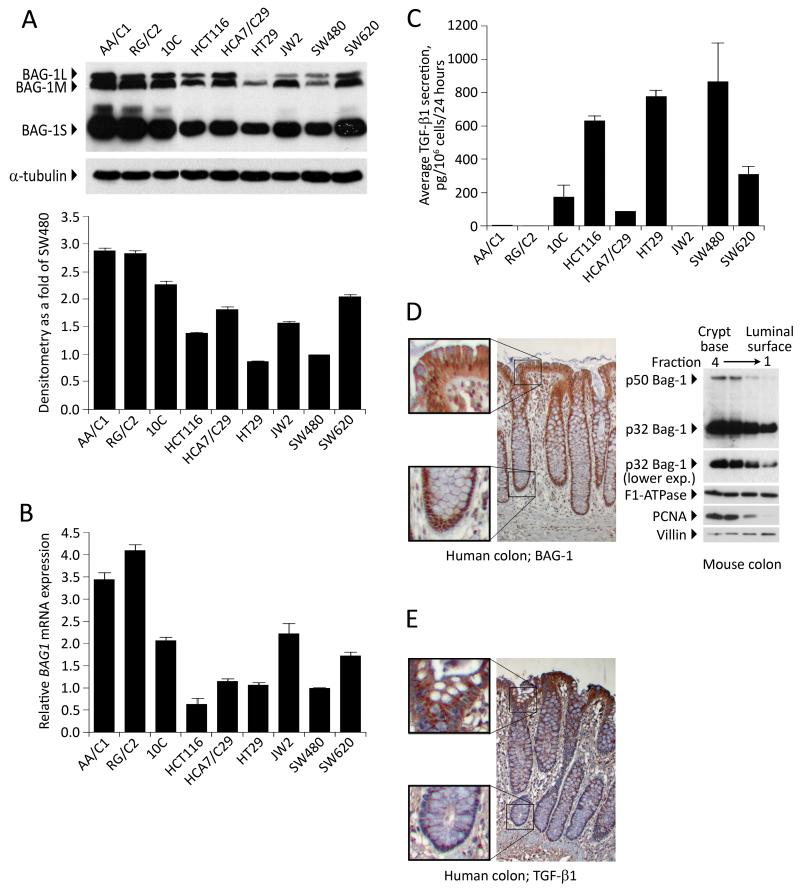
Figure 3. BAG-1 and TGF-β1 protein levels are negatively correlated in vitro and in vivo.
(A) BAG-1 protein levels were assessed in a panel of colorectal adenoma (AA/C1, RG/C2), transformed adenoma (10C) and carcinoma-derived cell lines (HCT116, HCA7/C29, HT29, JW2, SW480, SW620) by Western blot, using α-tubulin to indicate equal loading. Quantification of total BAG-1L expression levels was carried out using Image J Software, and depicted as fold change relative to basal BAG-1 levels in the SW480 cells. Quantification is the mean of 3 independent measurements of a representative result ±SD. Similar results were obtained in three independent experiments. (B) BAG1 mRNA levels were also quantified by Q-RT-PCR. Data show the mean from three independent experiments ±SD bars. (C) TGF-β1 production by these cell lines was also measured by ELISA and is presented as the mean from three independent experiments ± SD. (D) BAG-1 expression levels were assessed in normal human colonic epithelium by immunohistochemistry. Formalin-fixed, paraffin-embedded tissue sections were stained with the TB3 BAG-1 antibody (brown staining) and counterstained with Gill’s haematoxylin (blue); objective ×10, insets show higher magnification at base of crypt and luminal surface. Western blot of Weiser fractions of the mouse colonic epithelium in which Fraction 1 is enriched for cells from the luminal surface of the crypt and Fraction 4 is enriched for cells from the crypt base. This approach demonstrated a gradient of mBAG-1 expression (as shown by the two murine isoforms) along the crypt axis. Protein loading was assessed by F1-ATPase, fractionation confirmed using PCNA and Villin expression. (E) TGF-β1 expression levels were assessed in normal human colonic epithelium by immunohistochemistry. Formalin-fixed, paraffin-embedded tissue sections were stained with a TGF-β1 antibody (brown staining) and counterstained with Gill’s haematoxylin (blue); objective ×10, insets show higher magnification at base of crypt and luminal surface.

