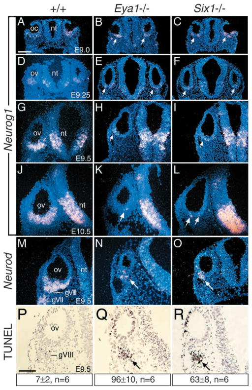
Fig. 1.
Eya1 and Six1 are required for the maintenance of normal neurogenesis during inner ear morphogenesis. Section in situ hybridization on transverse sections through the otic region. (A–C) Neurog1 is expressed in the ventral otic cup (oc) of E9.0 wild-type embryos (A), and its expression in Eya1−/− (B), and Six1−/− (C) embryos is indistinguishable from wild-type embryos. (D–L) Neurog1 expression became stronger from E9.25 to 10.5 after the vesicle (ov) is closed up (D,G,J); however, its expression is markedly reduced in Eya1−/− (arrows in E,H) or Six1−/− (arrows in F,I) from E9.25, and by E10.5 only a few Neurog1-positive cells were seen in the Eya1−/− (arrow in H) or Six1−/− (arrows in L) otic vesicle. Note that Neurog1 expression in the hindbrain neural tube (nt) is relatively normal in Eya1−/− or Six1−/− embryos compared to wild-type embryos. (M–O) Neurod is expressed in the differentiating neuroblast precursors within the otic vesicle (ov) and the cells migrating to form the VIIIth ganglion (gVIII) as well as within the VIIth ganglion (gVII) from E9.5 (M) in wild-type embryos. In Eya1−/− (N) or Six1−/− (O) embryos, Neurod expression in the cells within the otic vesicle and the cells migrating away from the otic vesicle was observed (arrow in N,O). (P–R) Transverse sections through E9.5 wild-type (P), Eya1−/− (Q) and Six1−/− (R) embryos labeled with the TUNEL method for detection of apoptotic cells. TUNEL-positive cells from 6 developing VIIIth ganglia (three embryos) for each genotype were counted using an image analysis system, and the number provided represents an average number per ganglion for each genotype. Apoptosis was markedly induced in the mutants (arrow). Scale bar: 100 μm.

