Abstract
Background: Botulinum neurotoxin (BoNT) complexes consist of neurotoxin and neurotoxin-associated proteins. Hemagglutinin-33 (HA-33) is a member of BoNT type A (BoNT/A) complex. Considering the protective role of HA-33 in preservation of BoNT/A in gastrointestinal harsh conditions and also its adjuvant role, recombinant production of this protein is favorable. Thus in this study, HA-33 was expressed and purified, and subsequently its antigenicity in mice was studied. Methods: Initially, ha-33 gene sequence of Clostridium botulinum serotype A was adopted from GenBank. The gene sequence was optimized and synthesized in pET28a (+) vector. E. coli BL21 (DE3) strain was transformed by the recombinant vector and the expression of HA-33 was optimized at 37°C and 5 h induction time. Results: The recombinant protein was purified by nickel nitrilotriacetic acid agarose affinity chromatography and confirmed by immunoblotting. Enzyme Linked Immunoassay showed a high titer antibody production in mice. Conclusion: The results indicated a highly expressed and purified recombinant protein, which is able to evoke high antibody titers in mice.
Key Words: Botulinum neurotoxin, Expression, Purification
INTRODUCTION
Botulinum neurotoxins (BoNT) are complexes which are produced by Gram-positive, anaerobic, and soporiferous Clostridium botulinum [1-3]. Respiratory tract, gastrointestinal tract, and wounds are the main entrances for BoNT [1, 4]. Based on antigenic properties, target sites, differences in structures, and toxicity, these neurotoxins are classified into seven serotypes: A, B, C, D, E, F, and G [4]. Serotypes A, B, E, and rarely F cause illness in humans, whereas serotypes C and D cause illness in animals [5]. Botulism syndrome is caused by one of the seven serotypes of C. botulinum [6]. BoNT are ~150-kDa single-chain polypeptides, which are cleaved by proteases into an N-terminal ~50-kDa light chain and a C-terminal ~100-kDa heavy chain connecting together by a disulfide bond [1, 7]. Heavy chain is composed of two functional domains: an N-terminal translocating domain and a C-terminal binding domain, whereas light chain has only a single domain named catalytic domain [7]. After intestinal absorption, blood circulation carries BoNT to neuromuscular synapses, where the toxins bind to nerve cells via their binding domains. The BoNT-catalytic domain has the ability of hydrolyzing the soluble N-ethylmaleimide-sensitive fusion protein attachment protein receptor proteins: vesicle-associated membrane protein/synaptobrevin, synaptosomal-associated protein, or syntaxin. The cleavage of mentioned proteins inhibits acetylcholine releasing at neuromuscular junctions and eventually causes flaccid paralysis [8-10].
To overcome the harsh conditions of high acidic pH in stomach and enzymatic digestion, BoNT are in complex of several nontoxic proteins, which assist BoNT to reach to blood circulation without any significant changes in their structure. The alkaline pH of intestine separates BoNT from associated proteins. The naked BoNT is then absorbed via intestinal epithelial or M cells [11].
The gene cluster of BoNT/A encodes seven proteins including neurotoxin-associated proteins (NAP) [12]. Most NAP are known as hemagglutinin proteins. Kouguchi et al. [13] indicated that a combination of HA-33, HA-17, and HA-55 are needed for hema-gglutination activity. In a study by Mutoh et al. [14] in 2005, HA-33 molecules showed hema-gglutination and erythrocyte-binding activity [14]. HA-33, a 33-kDa protein, is a subcomponent of NAP which is strongly resistant to proteases [15]. HA assists BoNT to be absorbed by intestinal epithelial cells [16], and they also increases endopeptidase activity of BoNT/A and E [17].
The role of HA-33 of BoNT/B complexes as an adjuvant in animal models has been proved [18]. In comparison to naked BoNT/B, the complex of BoNT/B and HA-33 increases IL-6 production in mouse spleen cells as well as B cell antigen presentation. These results indicated that HA-33 of BoNT/B complex has an adjuvant role [18]. Since HA-33 of BoNT/A has a great similarity to HA-33 of BoNT/B, it is expected that this protein could have an adjuvant role as well. In 2009, Kukreja et al. [19] also confirmed the adjuvant role of NAP especially HA-33. Thus, to study the HA-33 role as an adjuvant and also to study its protective role in oral botulism vaccines, expression of this protein is valuable. In this study, for the first time, the expression, purification, and antigenicity of recombinant BoNT/A HA-33 was evaluated.
MATERIALS AND METHODS
Bacteria, chemicals and media. All molecular biology grade chemicals and bacterial culture media were purchased from Merck (Germany). Chemical agents for nickel nitrilotriacetic acid agarose (Ni-NTA) resin were obtained from Qiagen (USA). Luria Bertani powder was obtained from Difco (Sparkes, MD, USA). Anti-BoNT/A complex antibodies were purchased from Medp (Moscow, Russia).
Gene expression and optimization. The BoNT/A ha-33 gene sequence was adopted from GenBank (GenBank ID: X87850). The gene length was 882 bp. The ha-33 gene sequence was optimized to achieve high expression of the recombinant protein in E. coli. The optimized sequence was then synthesized by Shinegene (China) in pET-28a (+) expression vector. NdeI and XhoI restriction sites were located at the 5’ end and 3’ end of the gene, respectively. By using the calcium chloride method, E. coli BL21 (DE3) competent cells were prepared at 4°C. Competent cells were then transformed by recombinant plasmids by heat-shock method. Bacterial colonies were screened by culturing in Luria Bertani agar containing 80 µg/ml kanamycin. Then recombinant plasmids were extracted. Transformant bacteria were chosen by restriction analysis (by NdeI and XhoI) [20]. The transforming bacteria were grown in Luria Bertani broth containing 40 μg/ml kanamycin at 37°C overnight. The cultures were then grown at 37°C with shaking at 150 rpm until the OD595 reached to 0.5. At this point, 1 mM IPTG was added to induce HA-33 expression, and incubation continued for additional 5-6 h. The cells were then harvested by centrifugation at 2500 ×g at 4°C for 10 min. The harvested cells were divided into two groups, one group was suspended in buffer A (10 mM imidazol, 50 mM NaH2PO4 and 300 mM NaCl, pH 8.0) and the other group was suspended in buffer B (100 mM NaH2PO4, 10 mM Tris-HCl, Urea 8 M, pH 8.0). The samples were kept for 2 h at room temperature. The cells were then lysed by sonication (4 times, for 10 s, with 30 s intervals and amplitude 75) and subsequently centrifuged at 18,000 ×g at 4°C for 20 min. The insoluble cell debris was discarded and the supernatant was stored at 4°C for purification [21]. The expression was analyzed by SDS-PAGE. Gene expression was evaluated in different conditions including induction temperature and induction time. The different induction times of 3, 5 and 12 and different induction temperatures of 25, 30 and 37°C were studied.
Recombinant protein purification . According to the manufacturer’s protocol, purification was performed by using of 50% Ni-NTA [21]. Initially, the Ni-NTA column was washed by MES buffer (20 mM 2-(N-morpholino) ethane sulfonic acid, pH 6.2)and then by 20 mM imidazol. Then the expressed proteins were added to the column and finally were eluted by 40 mM imidazol, 250 mM imidazol and MES buffer. Proteins were analyzed by SDS-PAGE. A ~33-kDa band corresponding to HA-33 recombinant protein was observed. Protein concentration measurement was carried out according to the Bradford's method [22].
Western-blot analysis . For immunoblotting assay, the protein was electrophoresed on a SDS- PAGE. The proteins were then transferred to a nitrocellulose membrane using Bio-Rad Mini Trans-Blot (Bio-Rad, USA). The transfer buffer contained 15 mM glycine, 20 mM Tris-base, and 20% methanol. Blocking process was performed at 4°C overnight by incubating the membrane in blocking buffer (5% skimmed milk). After washing by PBST (8 g NaCl, 0.2 g KCl, 2.9 g Na2HPO4, and 0.2 g K2HPO4 dH2O up to 1 lit + 0.5 ml Tween 20, pH 7.3), the membrane was incubated in a 1:3,000 dilution of horse anti-BoNT/A complex for 1 h. Then the membrane was washed three times with PBST (10 min each), and then it was incubated with a 1:10,000 dilution of the standard polyclonal anti-horse HRP conjugate for 1 h. After washing step, the presence of the HA-33 protein was evaluated by exposing the membrane to HRP staining solution (3,3’-diaminobenzidine tetrahydrochloride [DAB] buffer containing 0.3 mg/ml DAB, 50 mM Tris-HCI, pH 8, and 0.01% H202) for 15-30 min.
Vaccination of Mice. The purified HA-33 protein was mixed with an equal volume of complete Freund's adjuvant for first injection and with incomplete Freund's adjuvant for subsequent injections. Then, these compounds were injected intraperitoneally to the mice. The protein (15 µg) on 0, 2, 4 weeks were injected into each mouse. Negative controls were injected with adjuvant only. Animals were bled one week after the second and third injection, and the sera were stored for ELISA experiments at 4°C.
ELISA titration . An optimal concentration of purified HA-33 protein in coating buffer (15 mM Na2CO3 and 36 mM NaHCO3, pH 9.8) was coated in ELISA plates (3.5 µg of the protein for each well) and allowed to be adsorbed at 4°C overnight. Washing was then carried out four times with 400 µl PBST and then the reaction was blocked by adding 100 µl per well of skimmed milk (50 mg/ml) and placing the plate at 4°C overnight. After that, two-fold serial dilutions of mice serum in PBST, starting at 1:1,000 were added (100 µl per well), and the plate was incubated at 37°C for 30 min. After washing with PBST, the plate was incubated with anti-mouse immunoglobulin G-horseradish peroxidase conjugate (1:2,000, 100 µl per well) in binding buffer at 37 °C for 30 min. Afterward, the plate was washed four times with 400 µl PBST and incubated with 100 µl per well of orthophenyl-enediamine (Sigma) and with H2O2 as substrate. The reaction was then stopped by addition of 100 µg/well of 2.5 M H2SO4, and finally the absorbance at 490 nm was read by ELISA plate reader MRX (DYNEX).
RESULTS
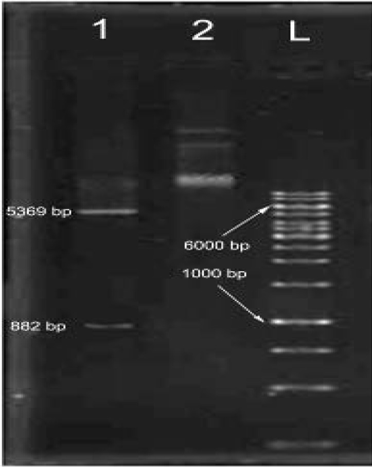
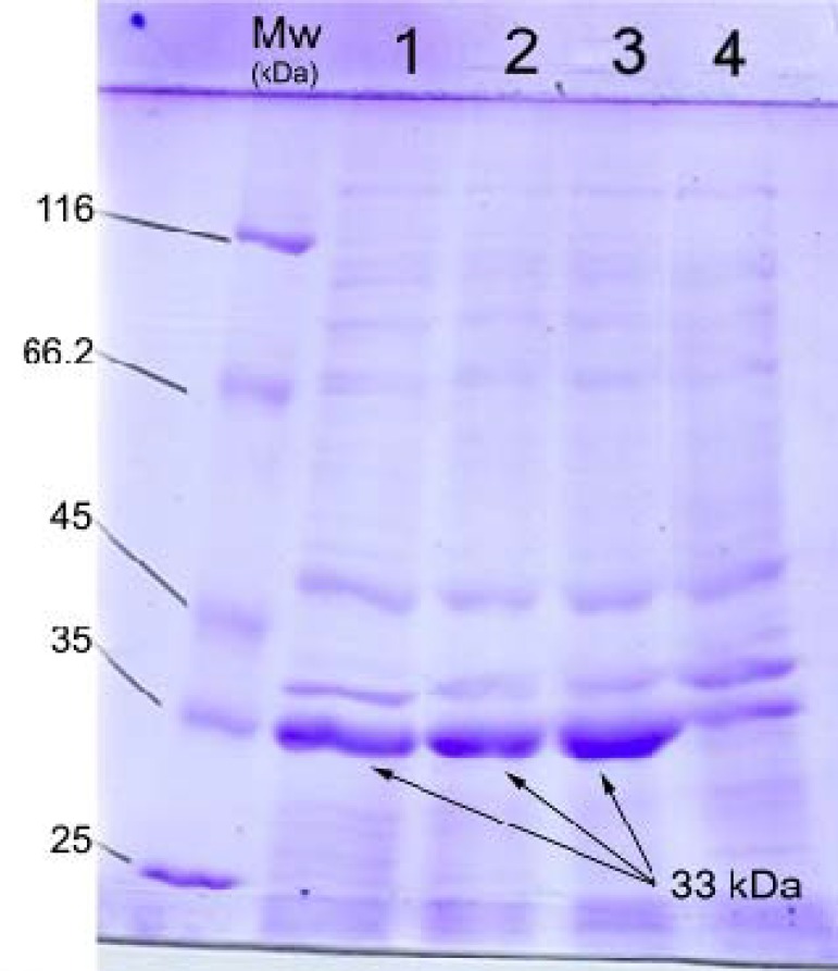
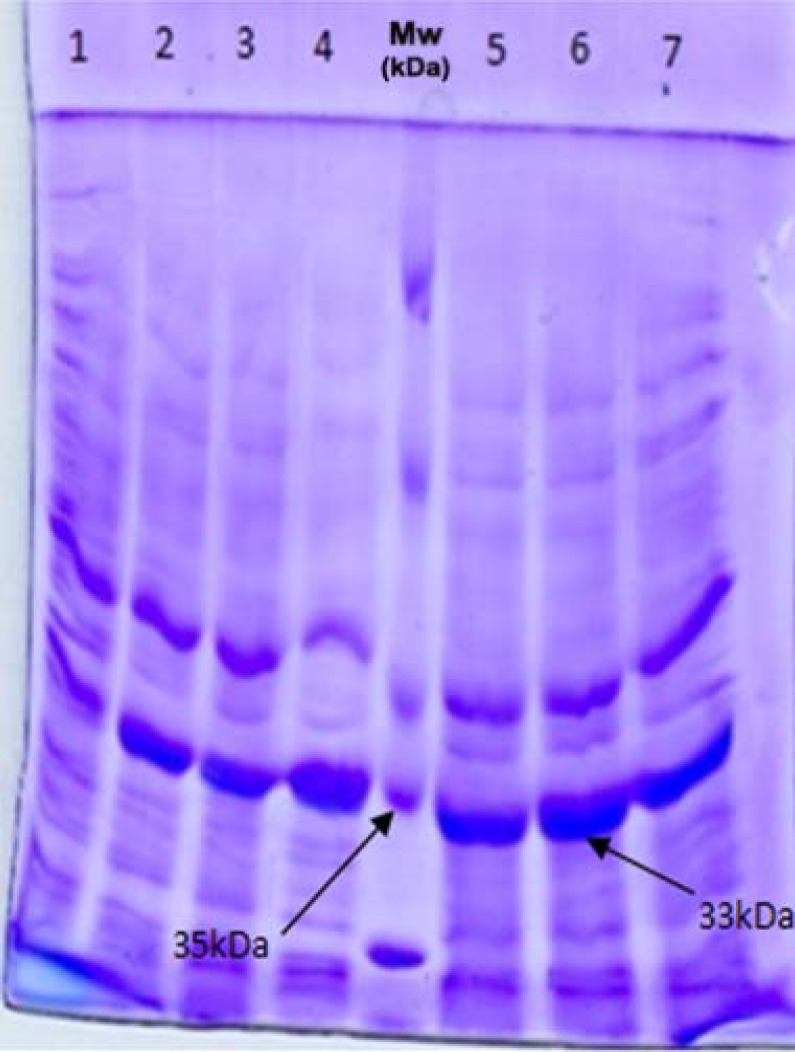
Expression evaluation and optimization. To achieve the high level of HA-33 expression, we used the expression vector pET-28a (+) with T7 promoter. After transformation and screening, the enzymatic digestion of the synthetic gene with Nde1 and Xho1 verified the presence of ha-33 gene in recombinant plasmids (Fig. 1). The expression of recombinant HA-33 protein was optimized as follows: 1 mM IPTG, incubation temperature of 37°C, shaking in 150 rpm for 5 h. The expression of the protein was evaluated by 12% SDS-PAGE (Fig. 2). Observation of a ~33-kDa band corresponding to HA-33 confirmed recombinant expression accuracy and sufficiency. In order to obtain the best condition of HA-33 expression, different values of time and temperature were applied. As Figure 3 shows, the best expression is seen after 5-h induction at 37°C.
Fig. 1.
Confirmation of ha-33 gene presence in recombinant plasmid. Enzymatic digestion of pET-28a-ha-33 with NdeI and XhoI was used to verify the presence of ha-33 gene. Lane 1, NdeI and XhoI digested pET-28a-ha33 producing two distinct bands (~5369 bp and ~882 bp); lane 2, pET-28a containing ha-33 gene; lane L, DNA ladder
Fig. 2.
SDS-PAGE analysis of HA-33 protein expression in E. coli BL21(DE3). Lanes 1-3, cell lysate of E. coli BL21(DE3) from three bacterial colonies containing pET28a-ha-33 gene after induction with IPTG; lane 4, cell lysate of E. coli BL21(DE3) containing pET28a-ha-33 gene before induction with IPTG; lane Mw, protein molecular weight markers. The expressed recombinant protein was observed at approximately 33 kDa
Fig. 3.
Optimization of HA-33 expression. Lane 1, cell lysate of non-induced bacteria; Lanes 2-4, E. coli lysate after induction with IPTG at 25, 30, and 37°C, respectively; lanes 5-7, IPTG-induced E. coli lysate after 3-, 5-, and 12-h induction at 37°C, respectively; Lane Mw, protein molecular weight markers. The best expressed recombinant protein was observed at 37°C and 5-h induction
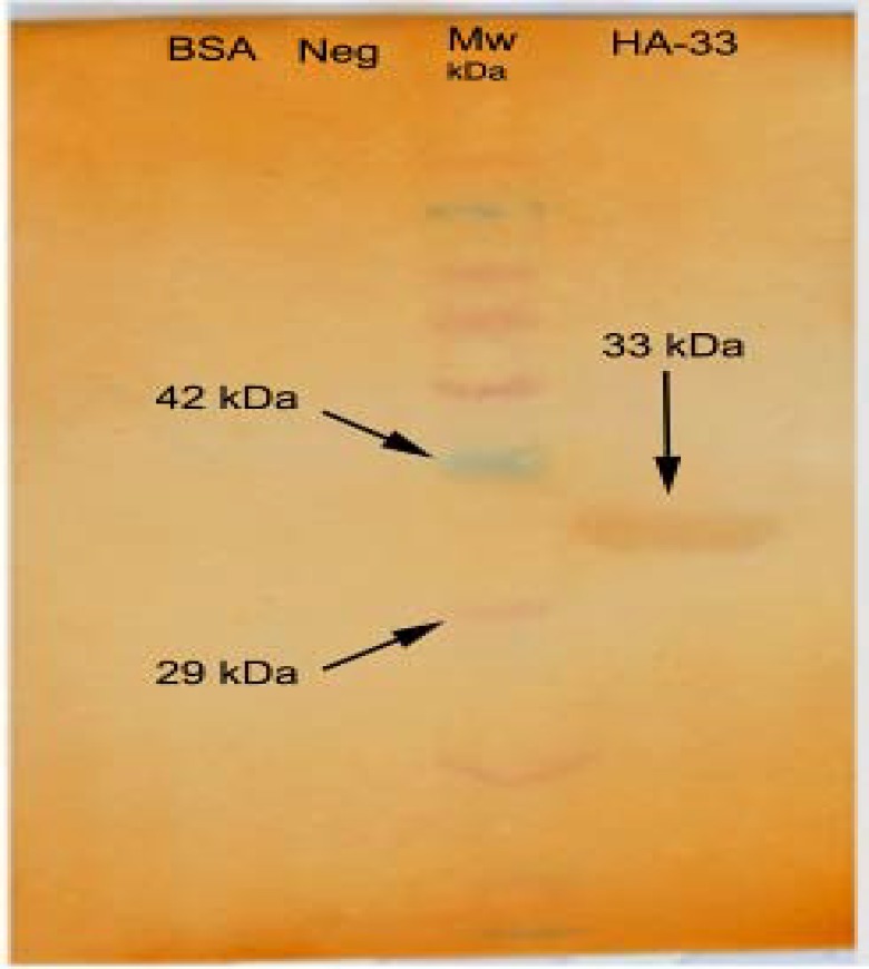
Western blotting. A ~33-kDa protein of interest was confirmed by western blotting. The result showed that the recombinant HA-33 is recognized by horse anti-BoNT/A serum, while no reaction was observed between these antibodies and non-induced bacteria or BSA, a nonspecific protein (Fig. 4).
Fig. 4.
Western-blot analysis of the recombinant HA-33 with antibody raised against BoNT/A complex. A single band (~33 kDa) was observed on HA-33 lane, showing HA-33 recognition “anti-BoNT/A complex” antibodies. There were no visible bands for non-induced bacteria with IPTG (Neg.) and BSA as controls
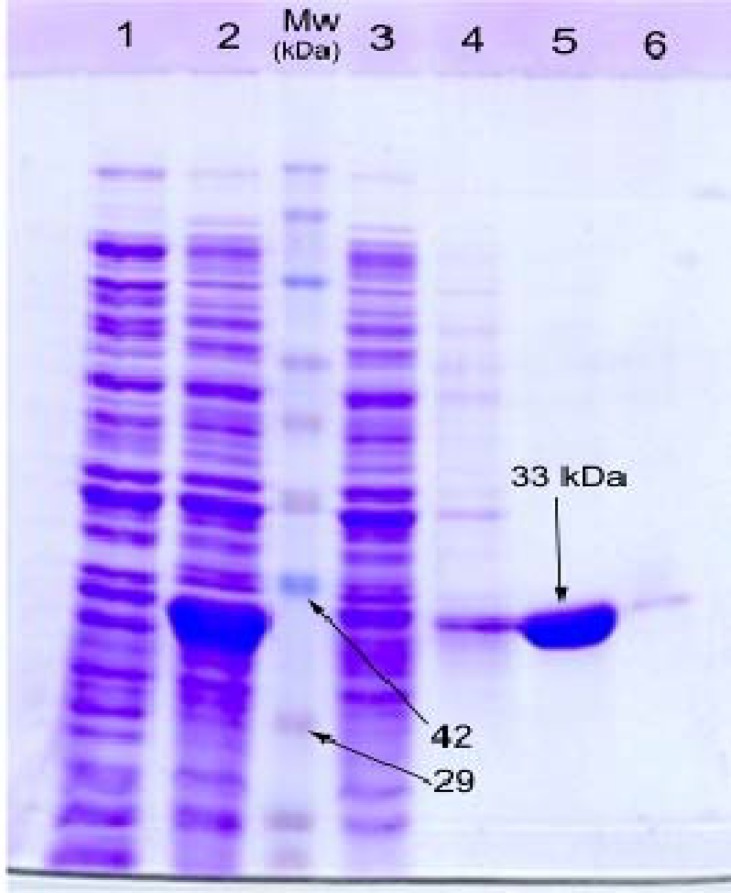
Purification of HA-33 protein. Since the recombinant HA-33 is a 6-His-tagged protein, a Ni-NTA column was used to purify the protein. After eluting the proteins by elution buffers and analyzing by 12% SDS-PAGE, a 33 kDa band corresponding to HA-33 was observed after the addition of 250 mM imidazol and MES buffer (Fig. 5). Concentration determination by Bradford's technique [22] showed 10 mg/L of the expressed protein in culture suspension.
Fig. 5.
Purification of HA-33 on a nickel nitrilotriacetic acid agarose affinity column. The separated protein fractions of purification process were run on 12% SDS-PAGE gel and stained with Coomassie blue stain. Lanes 1 and 2, bacterial lysate without and with induction with IPTG, respectively; lane 3, unbounded fraction of bacterial suspension; lanes 4-6, eluted fractions after adding 40 mM imidazol, 250 mM imidazol, and MES buffer, respectively; lane Mw, protein marker
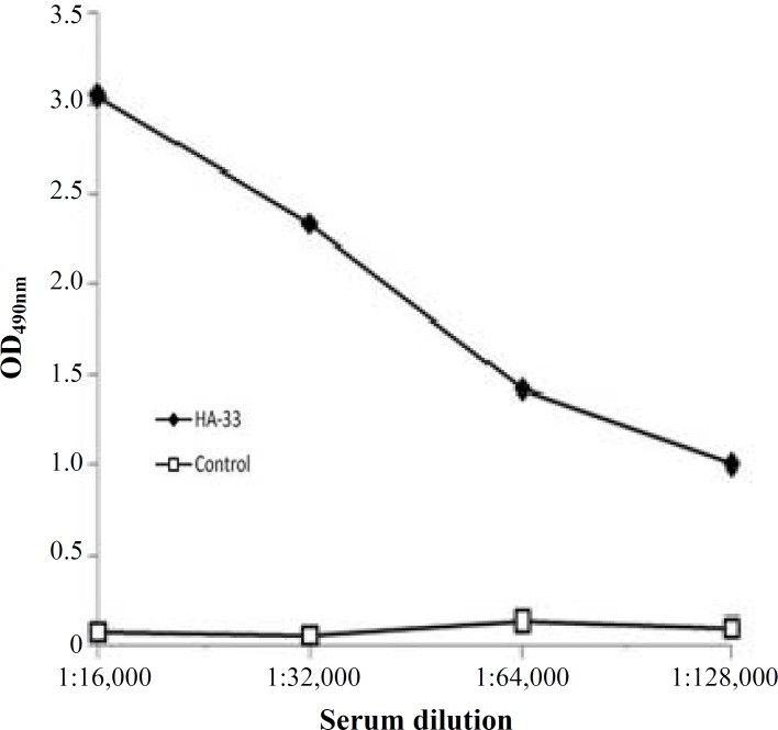
Antigenicity of recombinant HA-33. In order to study the antigenicity of HA-33 protein in mice, ELISA was performed. Sera of mice were taken two weeks after last vaccination and used in ELISA. Figure 6 shows the antibody titers of the vaccinated mice. Up to 1:128,000 dilution of the vaccinated mouse sera could recognize HA-33 properly.
Fig. 6.
ELISA results of mouse sera antibody titration against HA-33. Sera from mice bind to HA-33 up to 1:28,000. Serum from non-immunized mice did not react with HA-33 (control).
DISCUSSION
HA-33 is resistant to proteases and acidic pH, thus it increases the BoNT/A complex resistance to harsh condition of gastrointestinal tract [15]. This protein also has a role in the absorption of the toxin via intestinal epithelial cells and increases endopeptidase activity of the toxin [17]. In addition, it may serve as an adjuvant in vaccinology [18]. Therefore, researchers have a tendency to produce large amounts of HA-33.
Although HA-33 has been successfully purified and isolated from BoNT complex several times [13, 14, 18], some attempts were failed to isolate HA-33 with a high purity [22, 23]. One of the unsuccessful attempts were carried out by Tsuzuki et al. [23], when they tried to isolate HA components from BoNT/C progenitor toxin but only obtained partially purified HA-55/33. Another attempt was applied by Somers and DasGupta [24] to purify HA from BoNT/A progenitor toxin by using electroelution from SDS/PAGE gels. Due to protein denaturation during the SDS/PAGE, they could not isolate HA components properly [14]. In 2001, Kouguchi et al. [13] applied gel filtration and ion-exchange chromatography and succeeded to isolate different HA components (HA-33, HA-55, and HA-70) from BoNT complexes. The purification process was composed of several steps of precipitation by ammonium sulfate and chromatography (using mild denaturants such as guanidine hydrochloride and urea/lithium chloride) and finally Hiload Superdex gel filtration column. Although HA-33, HA-55, and HA-70 were isolated, they failed to separate HA-17 due to irreversible precipitation [13]. The attempts to obtain high purity HA components via isolation from BoNT complexes were continued [14], but there are still several defects. These results indicate that isolation of HA components from BoNT complexes is a complicated process that needs precision manipulation. Furthermore HA isolation needs large cultures of C. botulinum which increase dealing with hazardous bacteria. Due to these incompetents, recombinant production of HA components, which leads to high yield and high purified proteins without dealing with biosafety risks, is valuable. Therefore, we tried here to produce high purified recombinant HA-33 using pET system.
In order to obtain high yield of recombinant HA-33, the process should be optimized. Since, C. botulinum serotype A is a Gram-positive and AT rich, and E. coli, is a Gram-negative but not AT rich, the presence of rare codons, the GC content, and the Codon Adaptation Index of the gene was studied and codon optimization was done using DNAsis software and optimum genetic algorithm. The optimization process results in removal of rare codon from the sequence. Then the gene was synthesized in pET-28a (+) and expressed in optimized temperature and time (as mentioned before), which led to high expression and purification of the protein.
The adjuvant role of HA-33 of BoNT/B complex has been reported [18, 19], but there is no information about HA-33 of BoNT/A complex immunogenicity. Here as the first step of immunology studies, we tried to evaluate the antibody titer against recombinant HA-33. Because of the protective role of HA-33-associated protein [18] and also the role in absorption of the toxin via epithelial cell [25], it is suggested to analyze the effectiveness of the oral vaccination of mice by combining the neurotoxin and HA-33.
Since study on hemagglutinin-33 due to its function in BoNT complex resistance and epithelial absorption is important, recombinant production of HA-33 is highly valuable. Therefore in this study, we expressed and purified HA-33 protein using pET system. Using this system, we were able to obtain high yield and high purified HA-33. The protein was confirmed by western blotting. Then as the first step toward immunological studies, evaluation of antibody titer against HA-33 was conducted and showed that high titers of antibody are evoked in mice. Therefore, subsequent immunological studies are needed to perform in this field.
ACKNOWLEDGMENTS
We acknowledge the Biological Science Center of Imam Hussein University of Basic Science, Tehran, Iran for all supports and especially extend our vote of gratitude to Dr. Jamil Zargan and other researchers for all helpful ideas.
References
- 1.Simpson LL. Botulinum and tetanus neurotoxins. San Diego: Academic Press; 1989. [Google Scholar]
- 2.Hatheway CL. Toxigenic clostridia. Clin Microbiol Rev. 1990 Jan;3(1):66–98. doi: 10.1128/cmr.3.1.66. [DOI] [PMC free article] [PubMed] [Google Scholar]
- 3.Jawetz E, Melnick JL, Adelberg EA. Medical Microbiology. Stamford: Appleton and Longe press; 1995. [Google Scholar]
- 4.Simpson LL. The origin, structure, and pharmacological activity of botulinum toxin. Pharmacol Rev. 1981 Sep;33(3):155–88. [PubMed] [Google Scholar]
- 5.Santos-Buelga JA, Collins DM, East AK. Charac-terization of the genes encoding the botulinum neurotoxin complex in a strain of Clostridium botulinum producing type B and F neurotoxins. Curr Microbiol. 1998 Nov;37(5):312–8. doi: 10.1007/s002849900384. [DOI] [PubMed] [Google Scholar]
- 6.Dembek ZF, Smith LA, Rusnak JM. Botulism: cause, effects, diagnosis, clinical and laboratory identification, and treatment modalities. Disaster Med Public Health Prep. 2007 Nov;1(2):122–34. doi: 10.1097/DMP.0b013e318158c5fd. [DOI] [PubMed] [Google Scholar]
- 7.Schiavo G, Montecucco C. Tetanus and botulism neurotoxins: isolation and assay. Methods Enzymol. 1995;248:643–52. doi: 10.1016/0076-6879(95)48041-2. [DOI] [PubMed] [Google Scholar]
- 8.Dong M, Richards DA, Goodnough MC, Tepp WH, Johnson EA, Chapman ER. Synaptotagmins I and II mediate entry of botulinum neurotoxin B into cells. J Cell Biol. 2003 Sep;162(7):1293–303. doi: 10.1083/jcb.200305098. [DOI] [PMC free article] [PubMed] [Google Scholar]
- 9.Dong M, Liu H, Tepp WH, Johnson EA, Janz R, Chapman ER. Glycosylated SV2 A and SV2 B Mediate the Entry of Botulinum Neurotoxin E into Neurons. Mol Biol Cell. 2008 Dec;19(12):5226–37. doi: 10.1091/mbc.E08-07-0765. [DOI] [PMC free article] [PubMed] [Google Scholar]
- 10.Schiavo G, Santucci A, Dasgupta BR, Mehta PP, Jontes J, Benfenati F, et al. Botulinum neurotoxins serotypes A and E cleave SNAP-25 at distinct COOH-terminal peptide bonds. FEBS Lett. 1993 Nov;335(1):99–103. doi: 10.1016/0014-5793(93)80448-4. [DOI] [PubMed] [Google Scholar]
- 11.Chen F, Kuziemko GM, Stevens RC. Biophysical characterization of the stability of the 150-kilodalton botulinum toxin, the nontoxic component, and the 900-kilodalton botulinum toxin complex species. Infect Immun. 1998 Jun;66(6):2420–5. doi: 10.1128/iai.66.6.2420-2425.1998. [DOI] [PMC free article] [PubMed] [Google Scholar]
- 12.Dineen SS, Bradshaw M, Johnson EA. Neurotoxin gene clusters in Clostridium botulinum type A strains: sequence comparison and evolutionary implications. Curr Microbiol. 2003 May;46(5):345–52. doi: 10.1007/s00284-002-3851-1. [DOI] [PubMed] [Google Scholar]
- 13.Kouguchi H, Watanabe T, Sagane Y, Ohyama T. Characterization and reconstitution of functional hemagglutinin of the Clostridium botulinum type C progenitor toxin. Eur J Biochem. 2001 Jul;268(14):4019–26. doi: 10.1046/j.1432-1327.2001.02317.x. [DOI] [PubMed] [Google Scholar]
- 14.Mutoh S, Suzuki T, Hasegawa K, Nakazawa Y, Kouguchi H, Sagane Y, et al. Four molecules of the 33 kDa haemagglutinin component of the Clostridium botulinum serotype C and D toxin complexes are required to aggregate erythrocytes. Microbiology. 2005 Dec;151(Pt 12):3847–58. doi: 10.1099/mic.0.28323-0. [DOI] [PubMed] [Google Scholar]
- 15.Niwa K, Koyama K, Inoue S, Suzuki T, Hasegawa K, Ikeda T, et al. Role of nontoxic components of serotype D botulinum toxin complex in permeation through a Caco-2 cell monolayer, a model for intestinal epithelium. FEMS Immunol Med Microbiol. 2007 Apr;49(3):346–52. doi: 10.1111/j.1574-695X.2006.00205.x. [DOI] [PubMed] [Google Scholar]
- 16.Ito H, Sagane Y, Miyata K, Inui K, Matsuo T, Horiuchi R, et al. HA-33 facilitates transport of the serotype D botulinum toxin across a rat intestinal epithelial cell monolayer. FEMS Immunol Med Microbiol. 2011 Apr;61(3):323–31. doi: 10.1111/j.1574-695X.2011.00779.x. [DOI] [PubMed] [Google Scholar]
- 17.Sharma SK, Singh BR. Enhancement of the endopeptidase activity of purified botulinum neurotoxins A and E by an isolated component of the native neurotoxin associated proteins. Biochemistry. 2004 Apr;43(16):4791–8. doi: 10.1021/bi0355544. [DOI] [PubMed] [Google Scholar]
- 18.Lee JC, Yokota K, Arimitsu H, Hwang HJ, Sakaguchi Y, Cui J, et al. Production of anti-neurotoxin antibody is enhanced by two subcomponents, HA1 and HA3b, of Clostridium botulinum type B 16S toxin-haema-gglutinin. Microbiology. 2005 Nov;151(Pt 11):3739–47. doi: 10.1099/mic.0.28421-0. [DOI] [PubMed] [Google Scholar]
- 19.Kukreja R, Chang TW, Cai S, Lindo P, Riding S, Zhou Y, et al. Immunological characterization of the subunits of type A botulinum neurotoxin and different components of its associated proteins. Toxicon. 2009 May;53(6):616–24. doi: 10.1016/j.toxicon.2009.01.017. [DOI] [PubMed] [Google Scholar]
- 20.Sambrook J , Mac Callum P, Russell D. Molecular Cloning A laboratory Manual. New York: CSHL Press; 2001. [Google Scholar]
- 21.Zyskind JW, Bernstein SI. Recombinant DNA laboratory manual. New Delhi: Academic Press; 1992. [Google Scholar]
- 22.Bradford MM. A rapid and sensitive method for the quantitation of microgram quantities of protein utilizing the principle of protein dye binding. Anal Biochem. 1976 May;72:248–54. doi: 10.1016/0003-2697(76)90527-3. [DOI] [PubMed] [Google Scholar]
- 23.Tsuzuki K, Kimura K, Fujii N, Yokosawa N, Indoh T, Murakami T, et al. Cloning and complete nucleotide sequence of the gene for the main component of hemagglutinin produced by Clostridium botulinum type C. Infect Immun. 1990 Oct;58(10):3173–7. doi: 10.1128/iai.58.10.3173-3177.1990. [DOI] [PMC free article] [PubMed] [Google Scholar]
- 24.Somers E, DasGupta BR. Clostridium botulinum types A, B, C1, and E produce proteins with or without hemagglutinating activity: do they share common amino acid sequences and genes? J Protein Chem. 1991 Aug;10(4):415–25. doi: 10.1007/BF01025256. [DOI] [PubMed] [Google Scholar]
- 25.Fujinaga Y. Interaction of Botulinum Toxin with the Epithelial Barrier. J Biomed Biotechnol. 2010 Feb;2010:974943. doi: 10.1155/2010/974943. [DOI] [PMC free article] [PubMed] [Google Scholar]


