
Figure 9.
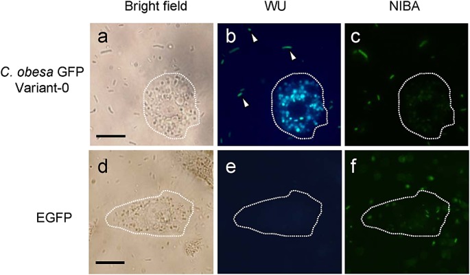
Bright-field (a,d) and fluorescence (b,c,e,f) microscopy of macrophages (RAW264.7) feeding on E. coli that expressed variant-0 CoGFP and EGFP. Fluorescence images were captured by the WU and NIBA mirror units. The dotted line in each image outlines a RAW264.7 cell, and arrowheads indicate E. coli cells. Scale bars, 20 µm.