Abstract
Follicular unit extraction (FUE) is a surgical procedure, which can be used to transplant follicular units into vitiliginous areas. Such follicular unit transplant has been recently used to repigment stable vitiligo patches. FUE was done for a 12-year-old female with a stable vitiligo patch with leukotrichia on the eyebrow. Repigmentation was noted in 6 weeks and complete pigmentation seen at 12 weeks. Leukotrichia resolved over a period of 6 months. No recurrence was noted at the end of 6 months follow-up with excellent colour match. This case is presented to highlight the simplicity, safety and effectiveness of FUE in stable vitiligo patches with leukotrichia.
KEY WORDS: Follicular unit extraction, leukotrichia, vitiligo
INTRODUCTION
Vitiligo is an acquired, idiopathic disorder characterised by circumscribed depigmented macules and patches and can have a major impact on personality. The psychological impact can be profound in deeply pigmented races.[1]
Clinically, with any treatment for vitiligo, the repigmentation usually begins at the orifice of hair follicles, which enlarges and coalesces to cover whole vitiliginous areas; meanwhile, on the palms and soles, mucosal surfaces, where hair follicles are absent, vitiliginous macules are very difficult to treat. A vitiliginous area with leukotrichia is considered to be a sign of poor prognosis. These observations indicate that the repigmentation of vitiligo is closely related to hair follicles. This forms the principle of follicular unit transplant.
The concept of follicular unit transplantation is based on the fact that hair, in general, does not grow singly, but with the exception of hairline, emerges from the scalp in groups called follicular units, histologically these units are comprised of 1-4 terminal and 1-2 vellus hairs that form a distinct group bounded by a circumferential band of adventitial collagen, the perifolliculum.[2] The existence of undifferentiated stem cells in the hair follicle in these units, which can serve as a source of melanocytes for repigmentation is the rationale behind follicular unit transplantation.
CASE REPORT
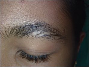
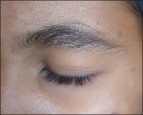
A 12-year-old female patient presented with depigmented macule on the right eyebrow of 8 years duration [Figure 1a]. She took various topical and oral medicines for the same without any relief. No new lesions were seen elsewhere in the body and size of the lesions remained stable for the past 2 years. On examination, she had single depigmented macule of the size 2 cm Χ 1.5 cm. Leukotrichia was found to be present. A diagnosis of focal type of vitiligo was made based on the clinical findings. Since the patient did not respond to the medical management, surgical correction with follicular unit transplant using the follicular unit extraction (FUE) technique was suggested. Written informed consent was taken. Donor hairs were harvested from the post auricular region using 1 mm skin biopsy punches. Donor area dressing was done using paraffin gauge dressing. The follicular units thus obtained were transplanted using an 18-G needle in the depigmented macules with 3 mm gap between the follicles. Paraffin gauze dressing was done for the recipient area and the dressing was removed after 5 days. Patient was followed-up every month. Patient was started on topical tacrolimus 2 weeks after the procedure. No post-operative complication was encountered. Repigmentation of the vitiligo patch was seen at the end of 6 weeks and complete pigmentation was seen at 12 weeks, with resolution of leukotrichia at the end of 6 months [Figure 1b]. Colour match was excellent. There was no recurrence after 6 months of follow-up.
Figure 1a.

Initial lesion of vitiligo with leukotrichia
Figure 1b.

Six months after treatment using follicular unit extraction
DISCUSSION
In the late 1950s, Staricco showed amelanotic melanocytes in the external root sheath of hair follicles that suggested immature pigment cells.[3] Further a melanocyte reservoir was proposed in the hair follicle by Ortonne et al., in 1980 who described the presence of DOPA negative, non-dendritic pigment cells along the external root sheath that migrated towards the basal cell layer to become functionally active in patients treated with Psoralen and UVA therapy for vitiligo it has been found that only active (DOPA positive) melanocytes existed in the epidermis of normal skin. There are some inactive melanocytes in the outer root sheath (ORS) of hair follicles, which form the melanocyte reservoir in the human skin.[4] Arrunátegui et al. suggested that melanocytes from the implanted lower third of the hair follicle can act as a reservoir and are able to migrate and repigment achromic areas in vitiligo.[5] Cui et al. studied the different stages of repigmentation of vitiligo and confirmed the existence of a melanocyte reservoir in the ORS of hair follicles.[6]
Vitiligo is a process in which only active (melanin-producing) melanocytes are destroyed and the inactive melanocytes in the ORS are preserved and serve as the only source for repigmentation.[3] Recovery of vitiligo is initiated by the proliferation of these inactive melanocytes, followed by the upward migration to the nearby epidermis to form perifollicular pigment islands and the downward migration to the hair matrices to produce melanin.[6] A retrograde movement of melanocyte to the depigmented hair follicles in vitiligo lesions has also been proposed to explain the repigmentation of leukotrichia seen after surgical treatment of vitiligo. After surgical repigmentation of vitiligo, the epidermis has a high concentration of melanotic melanocytes. Migration of these melanocytes from the area of high concentration in the epidermis to the hair follicle in the dermis, where the pigmented melanocytes are deficient, can explain the phenomenon of pigmentation of leukotrichia.[7]
Follicular unit transplant was first introduced to repigment vitiligo patches in 1998.[8] The existence of undifferentiated stem cells in the hair follicle in these units, which can serve as a source of melanocytes for repigmentation is the rationale behind follicular unit transplantation.
This procedure for vitiligo has been reported earlier, in one study by Na et al., perifollicular repigmentation around the grafted hair was observed in (71% patients) within 2-8 weeks. In cases of localised/segmental vitiligo, perifollicular pigmentation was seen in 82% patients and the study concluded follicular unit grafting to be an effective method for treating localised/segmental vitiligo, especially on hairy parts of the skin, including the eyelids and eyebrows and for small areas of vitiligo.[8] Malakar and Dhar in 1999 conducted a study of three patients with five vitiliginous patches.[9] They have reported a success in all five lesions with excellent colour matching. Single follicular unit transplant conducted by Kumaresan 2011 showed excellent repigmentation after 4-8 weeks with no recurrences in a case of vitiligo.[10]
All these studies have used the traditional follicular unit transplantation technique where in a strip was taken from post auricular region of the scalp, follicles dissected and transplanted. In our case, we have used FUE technique; the follicles were not dissected and transplanted as a whole unit in hopes of maintaining their natural milieu and promoting better proliferation of the cells, thus helping to improve the results. Apart from this, FUE technique is much simpler, suture less with less patient downtime and less complications (e.g., scarring).
Thus, FUE appears to be an effective method for treating localised/segmental vitiligo, especially on hairy parts of the skin, including the eyelids and eyebrows and for small areas of vitiligo. Though labour intensive, it was found to be associated with quicker patient recovery time, less morbidity and good colour match.
Footnotes
Source of Support: Nil.
Conflict of Interest: None declared.
REFERENCES
- 1.Pichaimuthu R, Ramaswamy P, Bikash K, Joseph R. A measurement of the stigma among vitiligo and psoriasis patients in India. Indian J Dermatol Venereol Leprol. 2011;77:300–6. doi: 10.4103/0378-6323.79699. [DOI] [PubMed] [Google Scholar]
- 2.Bernstein RM, Rassman WR, Szaniawski W, Halperin AJ. Follicular transplantation. Int J Aesthet Restor Surg. 1995;3:119–32. [Google Scholar]
- 3.Staricco RG. Amelanotic melanocytes in the outer sheath of the human hair follicle. J Invest Dermatol. 1959;33:295–7. doi: 10.1038/jid.1959.154. [DOI] [PubMed] [Google Scholar]
- 4.Ortonne JP, Schmitt D, Thivolet J. PUVA-induced repigmentation of vitiligo: Scanning electron microscopy of hair follicles. J Invest Dermatol. 1980;74:40–2. doi: 10.1111/1523-1747.ep12514597. [DOI] [PubMed] [Google Scholar]
- 5.Arrunátegui A, Arroyo C, Garcia L, Covelli C, Escobar C, Carrascal E, et al. Melanocyte reservoir in vitiligo. Int J Dermatol. 1994;33:484–7. doi: 10.1111/j.1365-4362.1994.tb02860.x. [DOI] [PubMed] [Google Scholar]
- 6.Cui J, Shen LY, Wang GC. Role of hair follicles in the repigmentation of vitiligo. J Invest Dermatol. 1991;97:410–6. doi: 10.1111/1523-1747.ep12480997. [DOI] [PubMed] [Google Scholar]
- 7.Agrawal K, Agrawal A. Vitiligo: Surgical repigmentation of leukotrichia. Dermatol Surg. 1995;21:711–5. doi: 10.1111/j.1524-4725.1995.tb00275.x. [DOI] [PubMed] [Google Scholar]
- 8.Na GY, Seo SK, Choi SK. Single hair grafting for the treatment of vitiligo. J Am Acad Dermatol. 1998;38:580–4. doi: 10.1016/s0190-9622(98)70121-5. [DOI] [PubMed] [Google Scholar]
- 9.Malakar S, Dhar S. Repigmentation of vitiligo patches by transplantation of hair follicles. Int J Dermatol. 1999;38:237–8. [PubMed] [Google Scholar]
- 10.Kumaresan M. Single-hair follicular unit transplant for stable vitiligo. J Cutan Aesthet Surg. 2011;4:41–3. doi: 10.4103/0974-2077.79191. [DOI] [PMC free article] [PubMed] [Google Scholar]


