Abstract
Context: In the last two decades, a color based concept of disease activity in peritoneal endometriosis has been in use in the clinical context, with red lesions being considered active and black or white lesions being interpreted as less active or dormant. Objective: Our aim was to analyze 4 main color categories of peritoneal endometriosis (black, white, red and brown) in one single patient group using histomorphological and immunohistochemical methods. Design: 65 endometriosis lesions (30 black, 17 white, 11 brown, 7 red) were resected from 47 premenopausal, nulliparous women which had not received exogenous hormones for at least six months prior to the operation. Specimen workup, histomorphological analysis and immunohistochemical analysis were performed in a standardized manner. Results: The color categories showed a broad overlap in proliferative activity and hormone receptor expression. Differences were found in lesion morphology. Adjacent stromal reaction in particular showed a marked increase from red through brown and black to white lesions. Differences were also seen in gland pattern and gland content. Conclusions: Lesion colors in peritoneal endometriosis seem to be determined by gland content and a varying adjacent stromal reaction and more likely reflect an aging process than different levels of disease activity.
Keywords: Endometriosis, colors, proliferation, hormone receptors, age
Introduction
Endometriosis affects between 4% and 30% of women of reproductive age, making this condition one of the most frequent benign gynecological diseases [1]. The disease is defined as the occurrence of patches of endometrial glands and/or stroma outside of the uterine cavity [2]. It was first described in 1860 by Rokitansky, who documented “new growth of uterine glands” in patients with “uterine and ovarian sarcomas” [3].
The etiology of endometriosis has not been fully explained yet despite a high clinical and pathological interest. The most widely accepted theory was proposed by Sampson who postulated in 1927 that endometriosis is caused by the retrograde flow of menstrual blood through the fallopian tubes with subsequent dissemination and implantation of endometrial cells in the peritoneum [4].
Standardized research on endometriosis has been hampered by the highly heterogeneous morphology of the disease. The spectrum of colors is quite broad, ranging from black through brown to red and white, transparent foci [5-7], rendering standardized description difficult. Sampson himself described “blueberry blue” and “raspberry red” lesions [7]. Clinical observation of the variable morphology of endometriosis lesions gave rise to the question of whether different colors in the lesions also reflect differing histology and biology in endometriosis. Several study groups addressed this issue with different methods. Redwine [8] and Goldstein et al. [9] reported, on the basis of laparoscopic observations, that red lesions precede other lesion colors. In 1991, Köhler and Lorenz were the first to present a color-related description of the histology of endometriosis. In a schematic overview, they described a decline in endometrial glands, endometrioid stroma, and hormone receptor expression along with a simultaneous increase in collagenous fibers from colorless through red to blue-black lesions. Unfortunately, the underlying data and methodology were not presented in the study [6]. Nisolle et al. investigated vascularization and proliferation in red, black, and white lesions, finding the strongest vascularization and highest level of proliferative activity in red lesions and the lowest level of proliferative activity in white lesions [10]. Donnez et al. reported a higher level of vascular endothelial growth factor (VEGF) content in red lesions in comparison with black lesions [11]. On the basis of this data, Nisolle et al. [12] suspected - like Köhler and Lorenz [6] and Brosens [13] - that red lesions represent “fresh” implants, while white foci correspond to dormant lesions.
Based on these observations, it was postulated that the different lesion colors also reflect differing disease activity [14,15]. However, up to now - to the best of our knowledge - the main color categories have not been comprehensively investigated in a single group of patients.
The aim of the present study was to investigate histology, proliferative activity and hormone receptor expression in black, white, brown and red endometriosis lesions obtained from a single group of patients and to compare the results with previously collected data.
Materials and methods
Patients
The study collective comprised 47 premenopausal, nulliparous women aged 19-52 years which had not received exogenous hormones for at least six months prior to the operation. Endometriosis was suspected clinically in all of the patients and a diagnostic laparoscopy with tissue resection was performed in all of the cases. All patients provided written informed consent to the histopathological workup of their endometriosis lesions. The local ethics committee approved the study. Any prior abdominal surgery was an exclusion criterion. Patients were recruited from September 2011 to November 2012.
Sampling and tissue processing
The patients’ pelvic and abdominal peritoneum was examined laparoscopically. Endometriosis lesions were photographed intraoperatively and their location, size, and color were documented. Lesions that could be clearly classified as “black”, “white”, “brown” or “red” were sharply resected without coagulation. All in all, 30 black, 17 white, 11 brown and 7 red endometriosis lesions were excised (total 65). Eutopic endometrium was also obtained in 27 patients (26 x streak curettage, 1 x hysterectomy).
After 24 hours of fixation, the peritoneal specimens were inked on the resection margin, laminated at right angles to the peritoneal surface and embedded in paraffin, oriented so that the entire width of the specimen was demonstrated on the cut surface. 3 μm sections were taken from all of the paraffin blocks and stained with hematoxylin-eosin (HE). On the basis of HE morphology, the block with the largest proportion of endometriosis lesions was selected for specialized histochemical staining (Berlin Blue (BB), Elastica van Gieson (EvG)) and immunohistochemistry.
Immunohistochemical staining
Immunohistochemical stains were performed on 1 μm sections using the fully automatic BenchMark ULTRA staining machine (Ventana Medical Systems, Inc., Tucson, Arizona, USA) in accordance with the manufacturer’s specifications. The immunohistochemical panel included: estrogen receptor (ER; CONFIRM anti-ER [SP1], alpha chain, ready-to-use; Ventana); progesterone receptor, (PR; lyophilized monoclonal mouse progesterone receptor [NCL-PGR-312], 1:200; Novocastra Laboratories Ltd., Newcastle upon Tyne, United Kingdom); Moleculare Immunologie Bortsel1 (MIB1; monoclonal mouse anti-human Ki-67 antigen [clone MIB-1], 1:100, Dako Ltd., Glostrup, Denmark); cluster of differentiation 10 (CD10; lyophilized monoclonal mouse antibody CD10 [NCL-CD10-270], 1:20, Novocastra); actin (monoclonal mouse anti-human smooth muscle actin [clone 1A4], 1:400, Dako); desmin (monoclonal mouse anti-human desmin [clone D33], 1:50, DakoCytomation); and caldesmon (monoclonal mouse anti-human caldesmon [clone h-CD], 1:100, Dako).
Histological and immunohistochemical analysis
Histological and immunohistochemical analyses were carried out by two pathologists (J.D.S. and D.L.W.) who worked independently and in ignorance of the clinically documented lesion colors. Divergent results were discussed until consensus was reached.
Hematoxylin-eosin analysis
Gland pattern, gland content and grade of endometrioid stroma were assessed. In addition, the presence or absence of neural structures and inflammatory infiltrates in the area of the endometriosis lesions were noted.
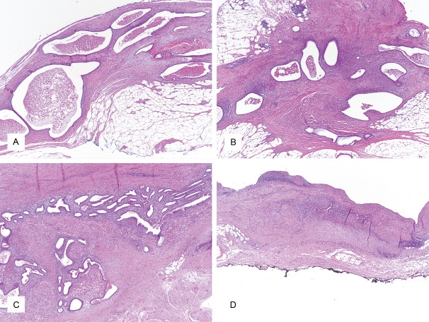
The gland pattern categories were defined as “large”, “medium”, “small” and “canalicular/collapsed” (Figure 1). “Large” endometrioid glandular structures characteristically were strongly dilated and showed irregular contours, whereas medium-sized and smaller glandular structures possessed round, regular shapes. Canalicular/collapsed glands had narrow, slit-like to completely obliterated glandular lumens.
Figure 1.
Gland patterns in peritoneal endometriosis; Hematoxylin-Eosin; 40x magnification. A: Large gland pattern, B: Medium gland pattern, C: Small gland pattern, D: Canalicular/collapsed gland pattern.
The glandular content was defined as “bloody” or “serous/empty”, with the category “bloody” including both fresh erythrocytes and also old-blood liquid and/or pigment-storing macrophages.
The amount of endometrioid stroma was graded as “sparse” (+), “moderate” (++), and “abundant” (+++). “Sparse” represented only focal patches of endometrioid stroma incompletely surrounding the endometrioid glands. A wider rim of endometrioid stroma at least partly surrounding the endometrioid glands was described as “moderate”. The category “abundant” referred to a broad rim of endometrioid stroma completely enveloping the endometrioid glands.
Berlin blue stain
In the BB stain, the endometriosis lesions were examined for hemosiderin deposits. The quantity and distribution pattern of intracellular and extracellular hemosiderin pigment within the endometriosis lesions were documented.
Elastic van gieson stain
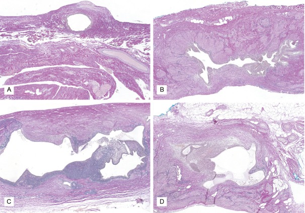
The adjacent stromal reaction in the endometriosis lesions was analyzed using the EvG stain, with the individual components “collagenous fibers”, “elastic fibers”, and “smooth-muscle metaplasia” being evaluated separately. In EvG stains collagen fibers are strongly pink, elastic fibers stain black, and smooth muscle appears yellowish to light brown (Figure 2). The grading of these three components of the stromal reaction was differentiated into the categories of “sparse” (+), “moderate” (++), and “abundant” (+++), as in the scoring system for endometrioid stroma detailed above. The grading of smooth muscle metaplasia was later refined with immunohistochemistry of smooth muscle markers.
Figure 2.
Components of the stromal reaction adjacent to endometriosis lesions; Elastica von Giesson; 40x magnification. A: Mostly collagen fibers (pink stain), B: Mostly smooth muscle metaplasia (yellowish stain), C: Mostly elastic fibers (grey-black stain), D: Mixed lesion.
Immunohistochemical analysis
Smooth-muscle metaplasia
Immunohistochemical staining for Actin, Desmin, and Caldesmon was carried out to confirm the smooth-muscle metaplasia previously identified using EvG staining. Fragmented, irregular courses of the smooth-muscle fibers were classified as evidence of smooth-muscle metaplasia. By contrast, a regular fascicular arrangement suggested localized smooth muscle.
Estrogen receptor/progesterone receptor
ER and PR expression were quantified separately in the endometrioid epithelium and stroma using the Immunoreactive Score (IRS). Hormone receptor expression was evaluated as “+” (low, scores 1-4), “++” (moderate, scores 5-8) or “+++” (high, scores 9-12). The entire endometriosis lesion was taken into account for quantification of hormone receptor expression.
Proliferative activity
Proliferative activity in the endometrioid epithelium and stroma was evaluated using MIB1 staining. Counting was performed in the areas of highest proliferation activity as identified in low magnification (25x). To determine proliferation activity in the endometrioid epithelium, the mean number of MIB1-positive cell nuclei in three areas of 100 adjoining epithelial cells was calculated. (“Number of MIB1-positive cells per 100 epithelial cells”). The proliferation activity in the endometrioid stroma was given as the mean number of MIB1-positive cell nuclei in three high power fields (hpf, at 400x) (“number of MIB1-positive cell nuclei per high-powered field with field number 25 (hpf/FN 25)”).
Data processing and statistical analysis
In addition to the 4 color categories, three menstrual cycle categories were defined: “proliferative phase” (day 1-14), “secretory phase” (day 15-28) and “irregular/absent cycle”. This categorization was based on the clinical history of the patients and the cycle day given by the patients. In 27 of the 47 cases, eutopic endometrium was available for examination and the cycle phase given by the patient could be corroborated histologically.
Histomorphological and immunohistochemical characteristics were analyzed in regard to color categories and menstrual cycle categories.
For proliferative activity and hormone receptor expression, means and standard deviation were calculated in the various categories. The color categories and menstrual cycle categories were assessed for significant differences using the Kruskal-Wallis test.
A P-value of 0.05 was considered significant. In case of significant results, the Levene test was used to check the equality of variances in the groups. In case of homogeneous variance, the Bonferroni test was carried out as a post-hoc test; if there was inhomogeneous variance, the Games-Howell test was used.
Results
Table 1 provides an overview of all of the results.
Table 1.
Overview of all of the results
| Subcategories | Black lesions (n=30) | White lesions (n=16) | Brown lesions (n=11) | Red lesions (n=7) | |
|---|---|---|---|---|---|
| Gland size | Large | 16/30 | 5/16 | 2/11 | 2/6 |
| Medium | 8/30 | 6/16 | 5/11 | 3/6 | |
| Small | 2/30 | 3/16 | 1/11 | 0/6 | |
| Collapsed | 4/30 | 2/16 | 3/11 | 1/6 | |
| Gland content | Serous/empty | 4/30 | 9/16 | 5/11 | 6/6 |
| Bloody | 26/30 | 7/16 | 6/11 | 0/6 | |
| Proliferation endometrioid epithelium | MIB + epithelial cells/100 epithelial cells | 21.2 ± 19.3 Range: 1-55 | 14.2 ± 10.8 Range: 0-35 | 15.0 ± 16.1 Range: 0-51 | 17.9 ± 15.9 Range: 10-28 |
| Proliferation endometrioid stroma | MIB + stromal cells/hpf | 13.2 ± 11.8 Range: 0-45 | 3.8 ± 4.2 Range: 0-11 | 11.3 ± 10.6 Range: 0-32 | 9.5 ± 7.4 Range: 1-20 |
| ER expression endometrioid epithelium | (+) | 1/30 | 0/16 | 0/11 | 0/7 |
| (++) | 2/30 | 1/16 | 1/11 | 1/7 | |
| (+++) | 27/30 | 15/16 | 10/11 | 6/7 | |
| ER expression endometrioid stroma | (+) | 0/30 | 0/16 | 0/11 | 0/6 |
| (++) | 2/30 | 1/16 | 0/11 | 1/7 | |
| (+++) | 28/30 | 15/16 | 11/11 | 6/7 | |
| PR expression endometrioid epithelium | (+) | 4/30 | 2/16 | 4/11 | 0/7 |
| (++) | 9/30 | 2/16 | 3/11 | 1/7 | |
| (+++) | 16/30 | 12/16 | 4/11 | 6/7 | |
| PR expression endometrioid stroma | (+) | 1/30 | 1/16 | 1/11 | 0/7 |
| (++) | 5/30 | 2/16 | 2/11 | 1/7 | |
| (+++) | 24/30 | 12/16 | 8/11 | 6/7 | |
| Collagen fibers | (+) | 3/30 | 1/16 | 2/11 | 1/7 |
| (++) | 16/30 | 5/16 | 7/11 | 5/7 | |
| (+++) | 11/30 | 10/16 | 2/11 | 1/7 | |
| Elastic fibers | (+) | 12/30 | 3/16 | 5/11 | 5/7 |
| (++) | 12/30 | 6/16 | 3/11 | 1/7 | |
| (+++) | 6/30 | 7/16 | 3/11 | 1/7 | |
| Smooth muscle metaplasia | (+) | 11/30 | 4/16 | 7/11 | 5/7 |
| (++) | 10/30 | 4/16 | 3/11 | 1/7 | |
| (+++) | 9/30 | 8/16 | 1/11 | 1/7 | |
| Endometriotic stroma | (+) | 11/30 | 10/16 | 3/11 | 1/7 |
| (++) | 15/30 | 5/16 | 7/11 | 6/7 | |
| (+++) | 4/26 | 1/16 | 1/11 | 0/7 | |
| Nerves | present | 4/30 | 2/16 | 2/11 | 1/7 |
| Not present | 26/30 | 14/16 | 9/11 | 6/7 | |
| Inflammatory infiltrates | present | 15/30 | 9/16 | 4/11 | 2/7 |
| Not present | 15/30 | 7/16 | 7/11 | 5/7 |
Histology
Clear trends were evident for the histological criteria of “glandular growth pattern” and “glandular content” in black and red lesions. Black lesions frequently showed large glandular structures (16/30), and the glandular content was usually bloody (26/30). By contrast, red lesions with identifiable glandular lumina had serous content in all cases (6/6). White and brown lesions showed variable results in relation to both categories.
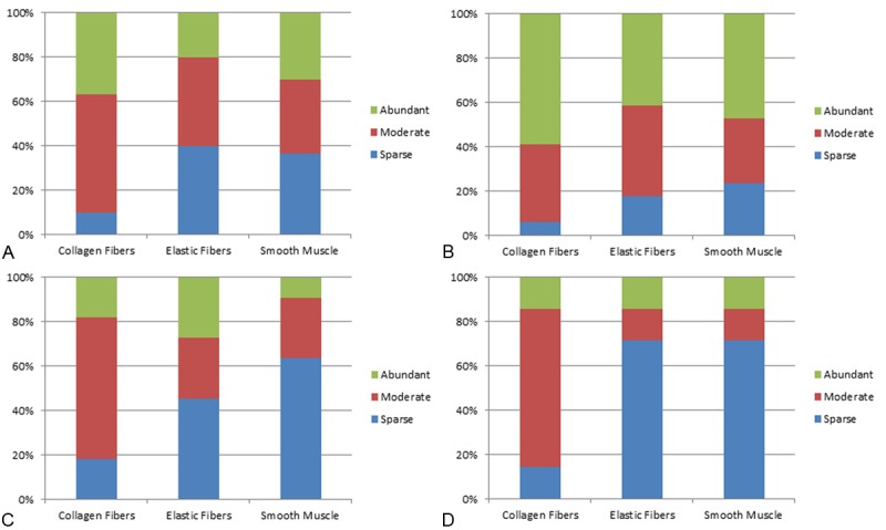
Trends were also evident in relation to the adjacent stromal reaction (Figure 3). Red lesions showed mainly sparse elastic fibers (5/7) and smooth-muscle metaplasia (5/7), with moderate collagenous fiber content (5/7). Many white lesions, in contrast, displayed an abundant amount of collagenous fibers (10/16), elastic fibers (7/16) and smooth-muscle metaplasia (8/16). Black lesions were in an intermediate position, with mainly moderate (16/30) to abundant (11/30) collagen content, variable smooth-muscle metaplasia, and low (12/30) to moderate (12/30) elastic fiber content. The brown lesions were also in an intermediate position. Overall, an increase in the degree of stromal reaction was evident from red lesions through black and brown lesions to white lesions.
Figure 3.

Stromal reaction adjacent to endometriosis lesions with regard to collagen fibers, elastic fibers and smooth muscle. A: Black lesions, B: White lesions, C: Brown lesions, D: Red lesions.
Most red lesions showed a moderate degree (6/7) of endometrioid stroma. Black and brown lesions contained mainly moderate (15/30 and 7/11, respectively) or sparse (11/30 and 3/11, respectively) amounts of endometrioid stroma. In contrast, white lesions showed mostly sparse endometrioid stroma (10/17). A slight declining trend from the red lesions through black and brown lesions to white lesions thus became apparent in this category.
Inflammatory infiltrates were absent in the majority of cases in red and brown lesions (5/7 and 7/11, respectively). However, they were present in half of the black lesions (15/30) and in more than half (10/17) of the white lesions.
By contrast, no trends were observed in relation to the detection of nerves inside the endometriosis lesions, with the presence of nerval structures being rare in all color categories.
Immunohistochemistry
Hormone receptors
In the immunohistochemical analysis of ER expression, no major differences were observed between the color categories either with regard to endometrioid epithelium or stroma. ER expression was uniformly high in all color categories (endometrioid epithelium: 27/30, 15/16, 11/11, and 6/7, respectively; endometrioid stroma: 28/30, 15/16, 11/11, and 6/7, respectively). With regard to the menstrual cycle categories, a declining trend was observed from the proliferative to the secretory phase in white, black, and red lesions. However, this trend was only slight, as all lesions had at least moderate ER expression.
PR expression showed a more variable picture. In the red lesions, epithelial PR expression was mainly strong (6/7). The proportion of cases with strong epithelial PR expression was lower in black and white lesions (16/30 and 12/16, respectively). Moderate epithelial PR expression was found in 9/30 cases in black lesions and 2/16 cases in white lesions. Brown lesions showed a more varied epithelial PR expression, with more than half of the cases showing a low (4/11) or moderate (3/11) PR expression. Stromal PR expression was high in the majority of cases in all of the colors (black, 24/30; white, 12/16; brown, 8/11; red, 6/7). Low to moderate stromal PR expression occurred in occasional cases in all color categories. When PR expression was analyzed with regard to the menstrual cycle categories, a slight declining trend in PR expression from the proliferative phase to the secretory phase was observed in red, black and brown lesions. There was no case with complete ER or PR negativity in the collective of 65 endometriosis lesions.
Proliferative activity
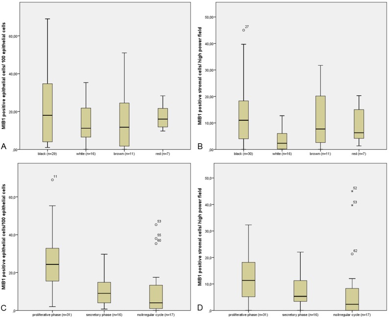
No significant differences were observed with regard to epithelial proliferative activity in the four color categories. Stromal proliferative activity was significantly higher in black than in white lesions (P=0.019). No other significant differences were identified in this context (Table 2, Figure 4A, 4B).
Table 2.
Proliferative activity of endometrioid epithelium
| Proliferation of endometrioid epithelium: MIB1 positive nuclei/100 epithelial cells | Black lesions | White lesions | Brown lesions | Red lesions |
|
| ||||
| All cases | 21.2 ± 19.3 (n=29) | 14.2 ± 10.8 (n=16) | 15.0 ± 16.1 (n=11) | 17.3 ± 7.2 (n=7) |
| Proliferative phase | 29.8 ± 18.7 (n=16)* | 20.1 ± 7.8 (n=5) | 26.1 ± 17.2 (n=5) | 16.2 ± 6.4 (n=4) |
| Secretory phase | 6.1 ± 6.7 (n=5)* | 12.8 ± 9.8 (n=5) | 5.5 ± 5.6 (n=3) | 18.8 ± 9.3 (n=3) |
| No/irregular cycle | 13.5 ± 17.8 (n=8) | 10.5 ± 13.1 (n=6) | 6.0 ± 9.9 (n=3) | - |
|
| ||||
| Proliferation of endometrioid stroma: MIB1 positive nuclei/hpf | Black lesions | White lesions | Brown lesions | Red lesions |
|
| ||||
| All cases | 13.23 ± 11.8 (n=30)** | 3.8 ± 4.2 (n=16)** | 11.3 ± 10.6 (n=11) | 9.5 ± 7.4 (n=7) |
| Proliferative phase | 14.1 ± 9.6 (n=5) | 5.4 ± 6.1 (n=5) | 15.0 ± 10.6 (n=5) | 11.2 ± 9.4 (n=3) |
| Secretory phase | 9.1 ± 7.2 (n=5) | 5.1 ± 3.6 (n=5) | 9.1 ± 11.2 (n=3) | 7.2 ± 4.4 (n=4) |
| No/irregular cycle | 14.0 ± 18.0 (n=8) | 1.4 ± 1.5 (n=6) | 7.3 ± 12.2 (n=3) | - |
Significant difference;
Significant difference.
Figure 4.

A, B: Proliferation activity in endometriosis lesions with regard to the color categories. A: Proliferation activity in endometrioid epithelium (No significant difference between the different color categories), B: Proliferation activity in endometrioid stroma (Significant difference between the categories “black” and “white”). C, D: Proliferation activity in endometriosis lesions with regard to menstrual cycle. C: Proliferation activity in endometrioid epithelium (Significant difference between the categories “proliferative phase” and “secretory phase” as well as between the categories “proliferative phase” and “no/irregular cycle”), D: Proliferative activity in endometrioid stroma (No significant differences between the cycle categories).
The menstrual cycle categories were regarded separately within the individual lesion colors. A statistically significant difference (P=0.037) was found between the proliferative and the secretory phase in the black category in relation to epithelial proliferation. No further significant differences were observed in the individual color categories regarding the menstrual cycle categories (Table 2).
Proliferative activity was also analyzed with in the menstrual cycle categories independently of lesion color (Figure 4C, 4D). A significant difference (P=0.002) was noted in epithelial proliferative activity between samples from the proliferative and the secretory phase and between samples from the proliferative phase and the group of absent/irregular cycle (P=0.002). There were no significant differences between the menstrual cycle categories with regard to stromal proliferation.
Discussion
Histology
Our analysis showed considerable overlapping of results in all of the color categories with regard to both histological and immunohistochemical characteristics. However, clear trends were also evident. Thus the black coloring of endometriosis lesions is well explained by the presence of large endometriosis glands with bloody content. However, on BB staining, only sparse hemosiderin deposits were found. Furthermore, no glandular or stromal fragments were evident in the glandular lumina. This contradicts the commonly held view that the blood seen in endometriosis lesions is caused by a process analogous to that of menstrual withdrawal bleeding [12] and points in the direction of simple lesional bleeding.
Regarding adjacent stromal reaction, there is an increase in the quantity of collagenous fibers, elastic fibers and smooth-muscle metaplasia from red lesions through brown and black to white lesions. Also, there is a slight declining trend in endometrioid stroma from red lesions through brown and black to white lesions. A strong adjacent stromal reaction is probably the reason why some endometriosis lesions appear macroscopically white, even though they contain endometrioid glandular structures that are partly filled with blood. Previously, it has been postulated that white lesions are almost completely scarified lesions which contain only sparse endometriosis foci [16,17]. The white lesions in our collective did indeed show high-grade adjacent stromal reaction. However, easily identifiable patches of endometriosis were found in all cases. These findings are more compatible with endometriosis being “walled in” by a strong stromal reaction than with “burnt-out” endometriosis.
All in all, our histomorphological findings seem to reflect an “aging process” from red through brown and black to white lesions. The fact that inflammatory infiltrates are found comparatively more often in black and white lesions may be regarded as further evidence of a greater “age” of black and white endometriosis. The concept that lesion color reflects on lesion age was already put forward by Köhler and Lorenz in 1991 [6]. More recently, the aging of endometriosis has been demonstrated in an animal model with baboons [18]. Furthermore, Sohler et al. [19] have shown through molecular studies that tissue remodeling generally plays an important role in endometriosis lesions. How ever no distinctions were made between lesion colors in this publication.
Immunohistochemistry
Hormone receptors
Epithelial and stromal ER expression and stromal PR expression were generally high in all color categories. Only epithelial PR staining showed some variability, with moderate to low epithelial PR expression being demonstrated in over half of brown and black lesions. This is in contrast to the color concept of Köhler et al who postulated a decline in hormone receptor expression from non-pigmented to pigmented lesions [6].
With relation to menstrual cycle categories, we found only a slight decrease of ER and PR expression from the proliferative to the secretory phase. This finding contrasts somewhat with the results reported by Nisolle et al. [12], who observed significantly higher levels of ER and PR expression in the proliferative phase in comparison with the secretory phase in the endometrioid epithelium of black and red lesions and in the endometrioid stroma of red lesions. The persistence of hormone receptor expression throughout the menstrual cycle documented in the present study may be regarded as further evidence of a certain degree of endocrine autonomy within endometriosis lesions.
Proliferative activity
No significant differences between the color categories were observed with regard to epithelial proliferation activity. However, a significant difference was found between black and white lesions regarding stromal proliferation activity. Our results thus partly corroborate and partly contradict those of Nisolle et al., who documented significantly lower proliferation activity in white lesions and significantly higher proliferation activity in red lesions compared to black lesions [10,12]. When endometriosis lesions were regarded irrespective of lesion color, there was a significant difference between the proliferative phase and the secretory phase and also between the proliferative phase and the group of irregular/absent menstruation. When analysis by menstrual cycle categories was performed on individual colors, significant differences were seen for epithelial proliferation between the proliferative and the secretory phase in black lesions. The present data therefore provide some evidence of a cycle-related variability of proliferation activity in endometriosis lesions.
Study design
In the present study, the assignment of the menstrual cycle phase was based on clinical history and the cycle day given by the patient. It is possible that imprecise information may have been provided in some cases. In 27 of 47 cases, eutopic endometrium was available for histomorphologic study. All of the samples of eutopic endometrium from patients with a regular cycle corresponded histologically to the cycle phase given by the patients. However, additional clinical testing with analysis of hormone levels would have rendered the menstrual cycle categories more robust.
Conclusions
In the literature the impression is often given that the histology and the biology of peritoneal endometriosis lesions have been comprehensively elucidated. However, our knowledge of endometriosis lesions is still incomplete and based on a small number of publications. The results of our study contradict the concept that the different colors of endometriosis lesions reflect different degrees of biologic activity as defined by proliferation activity and hormone receptor expression. Our data suggest that lesion color in endometriosis is associated with lesion age, gland pattern and gland content rather than with biologic activity. Regarding the menstrual cycle, we documented consistently high levels of hormone receptor expression in endometriosis lesions in all three cycle categories, a finding which may be indicative of endocrine autonomy. However, we also saw significant differences in proliferation activity between the three cycle categories, suggesting that endometriosis lesions are, to a certain degree, responsive to the menstrual cycle.
To date, there has only been one study providing evidence of a connection between proliferation activity and hormone receptor expression and the symptoms of peritoneal endometriosis [14]. Further studies are needed in order to determine whether the severity of symptoms in endometriosis does indeed correlate with a particular morphological, histological or immunohistochemical characteristic of the endometriosis lesion.
Acknowledgements
We acknowledge support by Deutsche Forschungsgemeinschaft and Friedrich-Alexander-Universität Erlangen-Nürnberg within the funding programme Open Access Publishing.
Disclosure of conflict of interest
None.
References
- 1.Renner SP, Lermann J, Hackl J, Burghaus S, Oppelt P, Binder H. Chronische Erkrankung. Endometriose. Geburtsh Frauenheilk. 2012;72:914–919. [Google Scholar]
- 2.Droegemueller W. Comprehensive Gynecology. Philadelphia: Mosby; 2001. [Google Scholar]
- 3.Rokitansky C. Ueber Uterusdruesen-Neubildung in Uterus und Ovarialsarcomen. Z Ges Aerzte Wien. 1860;16:577–581. [Google Scholar]
- 4.Sampson JA. Peritoneal endometriosis due to menstrual dissemination of endometrial tissue into the peritoneal cavity. Am J Obstet Gynecol. 1927;14:422. [Google Scholar]
- 5.Donnez J, Squifflet J, Casanas-Roux F, Pirard C, Jadoul P, Van Langendonckt A. Typical and subtle atypical presentations of endometriosis. Obstet Gynecol Clin North Am. 2003;30:83–93. viii. doi: 10.1016/s0889-8545(02)00054-2. [DOI] [PubMed] [Google Scholar]
- 6.Köhler G, Lorenz G. Zur Korrelation von endoskopischem und histologischem Bild der Endometriose. Endometriose. 1991;4:56–60. [Google Scholar]
- 7.Sampson JA. Benign and malignant endometrial implants in the peritoneal cavity and their relationship to certain ovarian tumors. Surg Gynecol Obstet. 1924;38:287–311. [Google Scholar]
- 8.Redwine DB. Age-related evolution in color appearance of endometriosis. Fertil Steril. 1987;48:1062–1063. [PubMed] [Google Scholar]
- 9.Goldstein DP, deCholnoky C, Emans SJ, Leventhal JM. Laparoscopy in the diagnosis and management of pelvic pain in adolescents. J Reprod Med. 1980;24:251–256. [PubMed] [Google Scholar]
- 10.Nisolle M, Casanas-Roux F, Anaf V, Mine JM, Donnez J. Morphometric study of the stromal vascularization in peritoneal endometriosis. Fertil Steril. 1993;59:681–684. [PubMed] [Google Scholar]
- 11.Donnez J, Smoes P, Gillerot S, Casanas-Roux F, Nisolle M. Vascular endothelial growth factor (VEGF) in endometriosis. Hum Reprod. 1998;13:1686–1690. doi: 10.1093/humrep/13.6.1686. [DOI] [PubMed] [Google Scholar]
- 12.Nisolle M, Casanas-Roux F, Donnez J. Immunohistochemical analysis of proliferative activity and steroid receptor expression in peritoneal and ovarian endometriosis. Fertil Steril. 1997;68:912–919. doi: 10.1016/s0015-0282(97)00341-5. [DOI] [PubMed] [Google Scholar]
- 13.Brosens IA. Is mild endometriosis a progressive disease? Hum Reprod. 1994;9:2209–2211. doi: 10.1093/oxfordjournals.humrep.a138422. [DOI] [PubMed] [Google Scholar]
- 14.Schweppe KW. Aktive und inaktive Endometriose-eine prognose- und therapierelevante Differentialdiagnose. Zentralbl Gynakol. 1999;121:330–5. [PubMed] [Google Scholar]
- 15.Viscomi FA, Dias R, De Luca L, Franco MF, Ihlenfeld MF. [Correlation between laparoscopic aspects and glandular hystological findings of peritoneal endometriotic lesions] . Rev Assoc Med Bras. 2004;50:344–348. doi: 10.1590/s0104-42302004000300046. [DOI] [PubMed] [Google Scholar]
- 16.Martin D. Color Atlas. Memphis: Resurge Press; 1990. Laparoscopic Appearance of Endometriosis. [Google Scholar]
- 17.Nisolle M, Donnez J. Peritoneal endometriosis, ovarian endometriosis, and adenomyotic nodules of the rectovaginal septum are three different entities. Fertil Steril. 1997;68:585–596. doi: 10.1016/s0015-0282(97)00191-x. [DOI] [PubMed] [Google Scholar]
- 18.Harirchian P, Gashaw I, Lipskind ST, Braundmeier AG, Hastings JM, Olson MR, Fazleabas AT. Lesion kinetics in a non-human primate model of endometriosis. Hum Reprod. 2012;27:2341–2351. doi: 10.1093/humrep/des196. [DOI] [PMC free article] [PubMed] [Google Scholar]
- 19.Sohler F, Sommer A, Wachter DL, Agaimy A, Fischer OM, Renner SP, Burghaus S, Fasching PA, Beckmann MW, Fuhrmann U, Strick R, Strissel PL. Tissue remodeling and nonendometrium-like menstrual cycling are hallmarks of peritoneal endometriosis lesions. Reprod Sci. 2013;20:85–102. doi: 10.1177/1933719112451147. [DOI] [PubMed] [Google Scholar]


