Introduction
Sarcoidosis is a systemic disease of unknown etiology (1). Pathologically it is characterized by the presence of non-caseating epithelioid-cell granulomas in the lungs, intrathoracic lymph nodes and other affected organs (2). The clinical course of sarcoidosis is widely variable, ranging from asymptomatic to bilateral hilar lymphadenopathy, pulmonary infiltration, skin and ocular lesions. Presentation of sarcoidosis with submental lymphadenopathy and renal failure with normal chest x-ray is quite rare. Here we describe this patient.
Case
A 68 -year-old woman referred to the hospital with various complaints, consisting of decreased appetite, early satiety, nausea and intense constipation. Patient also had polydipsia and nocturia and a history of 10 kg weight lose. Symptoms were started from 4 months before admission. There was not fever or chills. Except for a history of hysterectomy 10 years ago, no history of any disease in patient or her family was existed. On physical examination the blood pressure was 180/110 mmHg. In head and neck, no thyromegaly or lymphadenopathy in anterior and posterior cervical chains was found. There were three lymph nodes closed together with 1×1 cm2 sized in submental zone. There was not lymphadenopathy in axillae, bilateral supraclavicular, epitrochlear and inguinal zones. Heart and lung were normal in examination. In abdomen the edge of spleen was palpable. The neurologic examination and muscular forces were normal. One month before admission, patient was evaluated with the impression of a probable malignancy mostly from gastrointestinal (GI) origin in another hospital of neighbor province. The results were as follows; Upper GI series: only small hiatal hernia, upper GI endoscopy; deodenitis and small hiatal hernia, abdominal sonography; mild splenomegaly (span: 15 cm). Laboratory data were as follows; Hb: 12.3 g/dL, Hct: 34%, Platelets; 356x103 µL. Urine analysis was normal. Urine Bence Jones in 3 times was negative too. Stool exam for occult blood in three times was negative. Serologic tests were; BUN: 40 mg/dL, serum creatinine: 2.9 mg/dL, FBS: 113 mg/dL, Na: 143 meq/L, K: 4 meq/L, SGOT: 10 IU/L, ALP: 185 IU/L (100-200 IU/L), Ca: 12.5 mg/dL (8.2-10.6 mg/dL), P: 3.06 mg/dL (2.5-5 mg/dL), T4: 110 nmol/L (52-145 nmol/L), T3: 1.4 nmol/L (1.3-3.2 nmol/L), TSH: 1.4 Iu/mL (0.3-4 Iu/mL), Intact PTH: 54 pg/mL (13-54 pg/mL).
According to hypercalcemia and normal GI findings, we decided to reevaluate the patient for malignancy in other sites of body.
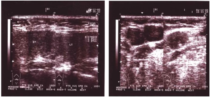
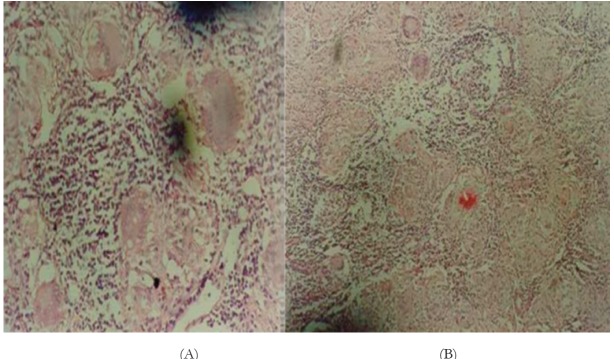
New paraclinical data included: Chest radiography was normal. Sonography of parathyroid glands were normal. Echocardiography was revealed a mild left ventricular hypertrophy. In kidneys sonography, right and left kidneys size were 93 and 98 cm respectively, otherwise was normal. Cervical CT scanning for evaluating of parathyroid glands or a probable mass was normal. Parathyroid radioisotopic scan with MIBI was normal. Bone scan and mamography were normal. Sub mental sonography, revealed three masses with 10-15 mm size (figure 1). Intact PTH was 26 pg/mL (9-55 pg/mL). Serum CRP was negative. ESR was 38 mm/h and serum albumin was 4.5 g/dL (3.5-5 g/dL). Serum proteins electrophoresis and peripheral blood smear were normal. Since, there was not an obvious reason for hypercalcemia, we referred patient for submental lymphoadenectomy. The result of tissue study was multiple foci of noncaseating granulomatous reaction with multinucleated giant cells with some Asteroid body, in favor of Sarcoidosis (figure 2A, B).
Figure 1.

Submental sonography, revealed three masses with 10-15 mm size.
Figure 2.

Multiple foci of noncaseating granulomatous reaction with multinucleated giant cells with some Asteroid body.
Angiotensin converting level was 62 Iu/L (8-52 Iu/L), confirmed the diagnosis of sarcoidosis. Then treatment with 40 mg/day prednisolone accompanied by a short course of Furosemid as a loop diuretic to treat hypercalcemia was began.
Serologic tests were after 8 months of treatment was as follows:
Ca: 9.3 mg/dL (8.1-10.4 mg/dL), P: 2.4 mg/dL (2.5-5 mg/dL), creatinine: 1.2 mg/dL (0.7-1.4 mg/dL), Urea: 42 mg/dL (15-45 mg/dL).
Oral prednisolon consumption was gradually reduced and after one year of treatment, discontinued. Patient’s symptom was less during annual referring for five years. Also, laboratory tests mentioned above remained unchanged for five years of evaluation.
Discussion
The clinical expression of sarcoidosis is protean, particularly as regards the number and sites of involved organs. The lung and the lymphatic system are predominantly affected, but virtually every organ may be affected. Overall, sarcoidosis is mostly revealed in the following circumstances: respiratory symptoms, extrathoracic localizations, mainly peripheral lymph nodes, eyes or skin, and constitutional symptoms such as fatigue (27%), weight loss (28%), fever (10–17%) or night sweats and also erythema nodosum (3–44%) (3). In this patient, chest radiography was normal and patient was presented with lymphodenopathy limited to the submental region accompanied by renal failure. The only confirmatory test is biopsy showing classic noncaseating granulomas (4,5). Renal involvement may occur as granulomatous interstitial nephritis as a consequence of calcium metabolism disorders, however renal insufficiency is uncommon and renal sarcoidosis is rare and may lead to renal failure in less than 3% of patients (6).
Authors’ contributions
ARM prepared the primary draft. HN, FGA and MDM wrote some parts of the manuscript. MRK and MBG edited the manuscript. HN prepared the final manuscript.
Conflict of interest
The author declared no competing interests.
Funding/Support
None declared.
Acknowledgments
The authors would like to thank all staffs of the Internal Medicine Department of Shahrekord University of Medical Sciences.
Implication for health policy/practice/research/medical education:
Sarcoidosis is an unknown etiology systemic disease. Pathologically, it is characterized by the presence of noncaseating epithelioid-cell granulomas in various organs. The clinical course of sarcoidosis is widely variable. In this report we describe a case of unusual sarcoidosis, presented with submental lymphadenopathy and renal failure with normal chest x-ray.
Please cite this paper as: Maghsoudi AR , Baradaran-Ghahfarokhi M, Ghaed-Amini F, Nasri H, Dehghani Mobarakeh M,Rafieian-Kopaei M. Renal failure and sub mental lymphadenopathy in a 68 years old woman. J Nephropathology. 2012; 1(3): 198-201. DOI:10.5812/nephropathol.8295
References
- 1. Dastoori M, Fedele S, Leao JC, Porter SR. Sarcoidosis - a clinically orientated review. J Oral Pathol Med 2012;. [DOI] [PubMed]
- 2.Porzezińska M, Słomiński JM. Sarcoidosis--clinical features, diagnosis and treatment. Przegl Lek . 2004;61(9):972–7. [PubMed] [Google Scholar]
- 3.Nunes H, Bouvry D, Soler P, Valeyre D. Sarcoidosis. Orphanet J Rare Dis . 2007;2:46. doi: 10.1186/1750-1172-2-46. [DOI] [PMC free article] [PubMed] [Google Scholar]
- 4.Bonfioli AA, Orefice F. Sarcoidosis. Semin Ophthalmol . 2005;20(3):177–82. doi: 10.1080/08820530500231938. [DOI] [PubMed] [Google Scholar]
- 5.Wu JJ, Schiff KR. Sarcoidosis. Am Fam Physician . 2004;70(2):312–22. [PubMed] [Google Scholar]
- 6. Unsal A, Basturk T, Koc Y, Sakacı T, Ahbap E, Ozagarı A, et al. Renal sarcoidosis with normal serum vitamin D and refractory hypercalcemia. Int Urol Nephrol 2012;. [DOI] [PubMed]
- 7.Sadek BH, Sqalli Z, Al Hamany Z, Benamar L, Bayahia R, Ouzeddoun N. Renal failure in sarcoidosis. Rev Pneumol Clin . 2011;67(6):342–6. doi: 10.1016/j.pneumo.2011.01.004. [DOI] [PubMed] [Google Scholar]


