

Abstract
Atomic Force Microscopy (AFM) has been extensively used in studies of biological interactions. Particularly, AFM based force spectroscopy and recognition imaging can sense biomolecules on a single molecule level, having great potential to become a tool for molecular diagnostics in clinics. These techniques, however, require affinity molecules to be attached to AFM tips in order to specifically detect their targets. The attachment chemistry currently used on silicon tips involves multiple steps of reactions and moisture sensitive chemicals, such as (3-aminopropyl)triethoxysilane (APTES) and N-hydroxysuccinimide (NHS) ester, making the process difficult to operate in aqueous solutions. In the present study, we have developed a user-friendly protocol to functionalize the AFM tips with affinity molecules. A key feature of it is that all reactions are carried out in aqueous solutions. In summary, we first synthesized a molecular anchor composed of cyclooctyne and silatrane for introduction of a chemically reactive function to AFM tips and a bi-functional polyethylene glycol linker that harnesses two orthogonal click reactions, copper free alkyne-azide cycloaddition and thiol-vinylsulfone Michael addition, for attaching affinity molecules to AFM tips. The attachment chemistry was then validated by attaching anti-thrombin DNA aptamers and cyclo-RGD peptides to silicon nitride (SiN) tips respectively, and measuring forces of unbinding these affinity molecules from their protein cognates human α-thrombin and human α5β1-integrin immobilized on mica surfaces. In turn, we used the same attachment chemistry to functionalize silicon tips with the same affinity molecules for AFM based recognition imaging, showing that the disease-relevant biomarkers such as α-thrombin and α5β1-integrin can be detected with high sensitivity and specificity by the single molecule technique. These studies demonstrate the feasibility of our attachment chemistry for the use in functionalization of AFM tips with affinity molecules.
Introduction
The human proteome consists of millions of proteins, many of which occur in minute concentrations below limits of detection (LOD) of current technologies such as ELISA, mass spectrometry and protein microarrays.1, 2 Therefore, there is a long felt need of a molecular tool capable of directly detecting those disease relevant protein biomarkers present in low abundance without any additional manipulation such as post-assay signal amplification. AFM has been envisioned as a mean of nanodiagnostics due to its single molecule sensitivity.3 It has been demonstrated that in combination with irreversible binding, AFM can reach a concentration sensitivity limit of 10−17 M.4 While AFM has been exploited in the analysis of DNA, proteins and cells, its chemical sensibility has grown tremendously as well. As illustrated in Figure 1, AFM is capable of “seeing and counting” target molecules when its tip is equipped with an affinity molecule. The interactions between antibody and antigen, ligand and receptor, DNA probe and target etc. can be determined and characterized at a single molecule level by AFM force measurements, termed as Molecular Recognition Force Spectroscopy (MRFS).5–11 Also, AFM has been enabled to scan individual proteins immobilized on a surface with an affinity molecule tethered to its tip, known as Recognition Imaging (RI).12–16 It is conceivable to employ both MRFS and RI for identification and detection of protein biomarkers in a clinic setting. This requires that these techniques are robust, supported with well-designed chemistry and bioassays. Recent advances in automated AFM-based force spectroscopy should facilitate the instrument operation.17 One of our efforts has been directed towards developing simple attachment chemistry that works in aqueous solutions without any of organic solvents involved so that it can easily be adapted in biological laboratories and clinics.
Figure 1.

Illustration of an AFM tip with an affinity molecule tethered at its apex to specifically recognize its protein cognates immobilized on a substrate (A). Using contact mode, a force of the affinity molecule unbinding from its cognate can be determined by retracting the tip along the Z direction (B). By tapping the functionalized tip on the surface along the X to Y direction, topographic and recognition images can be generated (C, D). In general, the affinity molecule can be a ligand, an antibody, a nucleic acid aptamer, and so on
A molecular linker is often employed to attach affinity molecules to AFM tips, which provides an advantage in distinguishing between specific and nonspecific interactions.18 The heterobifunctional poly[ethylene glycol] (PEG) has become a commonly used linker.19, 20 In general, the attachment is a three-step process that begins with functionalizing an AFM tip with a chemically reactive group, tethers the PEG linker to the AFM tip, and then reacts with an affinity reagent. (3-Aminopropyl)triethoxysilane (APTES) is a choice reagent for amination of silicon tips,21, 22 but it is notoriously problematic for forming uniform monolayers, especially when the reaction is carried out in a liquid phase.23, 24 APTES should be freshly redistilled before use in order to achieve reproducible results. Chemical vapor deposition of APTES has been developed to improve the outcome,21 but the process is tedious, requiring a thorough purge of the deposition chamber with argon to remove trace of moisture. Without developing an automated apparatus, it is difficult to be scaled up. The reaction of amine with NHS (N-Hydroxysuccinimide) ester has been one of the most commonly used methods for tethering carboxylated PEG linkers to AFM tips.25–30 The NHS ester is sensitive to moisture, and prone to rapid hydrolysis with increase in pH (a half-life time of 4–5 hours at pH 7 and one hour at pH 8).31, 32 On the other hand, the amine exists in an aminium form at the neutral pH, requiring a basic condition to be deprotonated for its nucleophilic activity. These caveats make it difficult to handle the NHS ester reaction in aqueous solutions and one has to fine tune pH, reaction time in order to achieve optimal outcomes.
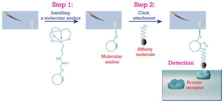
Here, we report on a new scheme of attaching affinity molecules to AFM tips based on click chemistry (Figure 2). Chen et al have employed a copper-catalyzed alkyne-azide reaction to attach antibodies to a gold coated AFM tip through an azido-PEG-thiol linker.33 To take it further, we implement two orthogonal catalyst-free click reactions for the attachment of affinity molecules to silicon tips. First, we have synthesized a molecular anchor composed of cyclooctyne and silatrane for the introduction of an alkyne function to the silicon tip. In aqueous solution, the silatrane moiety reacts with silanol on silicon surfaces to form a monolayer. It has been known that silatrane is less reactive than alkoxysilanes and extremely resistant to polymerization at a neutral pH.34 Gruber and Lyubchenko et al have employed 1-(3-aminopropyl)silatrane (APS) as a substitute of APTES in functionalization of AFM tips and mica surfaces.35–38 Thus, we expected that the new anchoring molecule would form a uniform cyclooctyne monolayer on the silicon tips. The ring strained cyclooctyne promotes the alkyne-azide reaction without the copper catalyst.39 In addition, we have synthesized a new class of molecular linkers, azido-PEG-vinyl sulfone with defined lengths, for connection of affinity molecules to AFM tips. In the present study, we have focused on attaching thiolated oligonucleotide aptamers and affinity peptides to AFM tips because they are rapidly growing areas in molecular diagnostics.40 The reaction of vinyl sulfone with thiol in aqueous solution comprises another category of click chemistry in bioconjugation,41 being used for the labeling of proteins42 and proteomes.43 We adapt it as a first click reaction to connect thiolated affinity molecules to the PEG linker as illustrated in Figure 2. The second click (azide to alkyne) finishes the process of the attachment. These two click reactions are orthogonal so that there are no cross talks between each other.
Figure 2.

Illustration of a new chemical approach for attaching affinity molecules to an AFM tip
Results and Discussion
Synthesis
The molecular anchor (3) was synthesized simply by reacting APS (1)36 with 2-(cyclooct-2-yn-1-yloxy)acetic acid (2)39 in the presence of 1-ethyl-3-(3-dimethylaminopropyl)carbodiimide (EDC, Scheme 1-a). The desired product was separated as a white solid by silica gel chromatography with a yield of 60% (see Supporting Information for details). The molecular linker for RI (6a, Scheme 1-b) was synthesized starting from hexaethylene glycol. First, azido-(CH2CH2O)6-Ts (4, Ts = tosyl) was synthesized in a multi-gram scale following a method reported in literature.44, 45 The azido-(CH2CH2O)12-H (5) was prepared in a 71% yield by reacting 4 with sodium hexaethylene glycoxide (3 times excess) that was generated in situ by treating hexaethylene glycol with sodium hydride. In presence of potassium t-butoxide, 5 reacted with divinyl sulfone to furnish the desired product 6a in a fairly good yield (64%). In the same manner, the linker azido-(CH2CH2O)36-vinyl sulfone (6b) was synthesized by reacting azido-dPEG®36-alcohol with divinyl sulfone in a yield close to that of 6a (Scheme 1-c). These two products were characterized with FTIR, NMR, and mass spectroscopy (see supporting information). Although it has been reported that vinyl sulfones react with azides in presence of CuSO4 and sodium ascorbate,46 we found by NMR monitoring that 6a and 6b were stable both in its pure form and in chloroform at room temperature at least for two days. They have been stored at −78°C already for one year and no degradation has been observed. Maleimide is another widely used reactive group that functions similarly to vinyl sulfone in bioconjugation,47 but it may not be amenable to coexisting with azide because a [3 + 2] cycloaddition could spontaneously take place between these two functions in some circumstances.48 In addition, maleimide can undergo the thiol exchanges and ring hydrolysis (above pH 8),49, 50 which would complicate outcomes of the conjugating reaction. It is also a reason why we chose vinyl sulfone as a Michael addition receptor of thiols in our attachment chemistry.42
Scheme 1.

Schematic diagram of the chemical syntheses
Click 1: tethering molecular linkers to affinity molecules
Two affinity molecules, thrombin-binding DNA aptamer (TBA)51 and cyclic RGDfC peptide containing a RGD motif that binds to integrin receptors such as α5β1,52 were chosen to study the attachment chemistry. First, the disulfide at the 3′-end of the DNA aptamer from custom synthesis was reduced to thiol using tris(2-carboxyethyl)phosphine (TCEP), which then reacted with linker 6a and 6b at pH 8.0 in phosphate buffered aqueous solutions, respectively. Through the Michael addition of thiol to vinyl sulfone (Scheme 2-a), the DNA aptamer was converted to azido-PEGylated products D-1a with a 95% yield and D-1b with a 89% yield, based on HPLC analysis (see Supporting Information). The disulfide DNA was used as a negative control and it did not react with 6a and 6b, implying that the vinyl sulfone is specific to thiol under the current conditions. Also, we observed that the reaction between the vinyl sulfone and the thiolated DNA at pH 7–7.5 was very slow and did not complete even after one day. The thiol reaction is driven by the thiolate that is a much stronger nucleophile than its conjugate acid thiol. Since the alkylthiol is fairly acidic with pKa of about 10 to 11, the increase of pH surely increases existence of the thiolate anion, resulting in an increased reaction rate. This is consistent with what has been reported in literature.53 Under the similar conditions, the thiolated RGDfC was converted to products P-1a and P-1b quantitatively (Scheme 2-b). We did not observe any side products by MALDI mass spectrometer and reverse phase HPLC analysis. In addition, no apparent time differences between reacting with 6a and 6b were observed (see Supporting Information for details). In sum, all these reactions were completed within three hours when starting with DNA or peptides in a range of millimolar concentrations.
Scheme 2.

Schematic description of tethering the molecular linkers to affinity molecules through the Michael addition of thiol to vinyl sulfone
It should be noted that the vinyl sulfone also reacts with alkyl amines under basic conditions.54 In our case, the amine functionalized aptamer and the cyclic RGDfK reacted with both 6a and 6b in phosphate buffered solutions at pH 8.8, but the reactions were very slow and not completed even after ten hours. This shows that the vinyl sulfone can specifically react with thiol in the presence of amino function with well-tuned pH.
Click 2: Attaching affinity molecules to AFM tips
At present, there is no effective way to directly monitor chemical reactions and characterize their products on AFM tips. To have insights into our new attachment chemistry, we first carried out a pilot study on planar thermally oxidized silicon substrates, presumably the surface of which has a chemical reactivity similar to that of silicon AFM tips. We found that compound 3 formed a monolayer with its physical properties close to those of the monolayer generated by reacting cyclooctynyloxy-acetic acid 2 with the APTES functionalized silicon substrate. As illustrated in Scheme 3, when APTES was deposited on a silicon oxide surface by chemical vapor deposition (route i-a), it changed the contact angle of water on the surface from 0° to ~ 46° (Table 1), a value that is consistent to data reported in literature.55 The measured thickness of the organic layer was about 7.3 Å, slightly larger than the calculated distance from nitrogen to oxygen of APTES (see S-2 in Scheme 3), indicating formation of a monolayer (See Supporting Information for details about contact angle and thickness measurement). Treating the APTES monolayer with compound 2 in the presence of EDC increased the contact angle to ~76° and thickness to ~15.9 Å, close to the expected value (see S-3 in Scheme 3). When the same silicon substrates were treated directly with an aqueous solution of compound 3 (route ii in Scheme 3), the measured contact angle and thickness were ~78° and 15.3 Å, respectively, well matching with those data just mentioned above. This indicates that compound 3 may form a monolayer with a structure as suggested in S-3. In turn, we treated the cyclooctyne surface with a solution of fluorescent TBA containing an azide at its 3′-end and it became highly fluorescent after one hour incubation, whereas the same surface treated with the fluorescent aptamer containing disulfide at the 3′-end (negative control) had negligible fluorescence (see Figure S1 in Supporting Information). Note that we had confirmed that the azide functionalized TBA and cyclo-peptides reacted with the cyclooctyne effectively in the liquid phase (monitored by MALDI mass) before applying them to silicon substrates or tips.
Scheme 3.
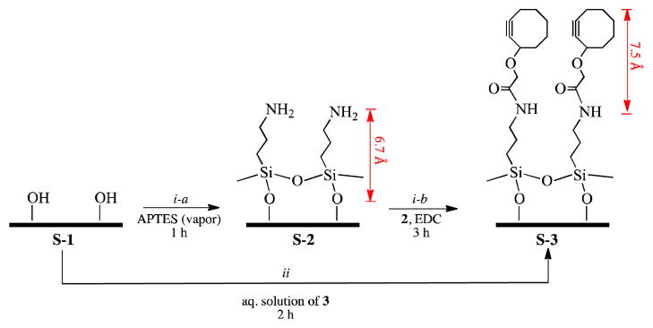
Functionalization of the silicon oxide surface: (i-a) chemical vapor deposition of APTES; (i-b) coupling of compound 2 to the APTES surface in DCM; (ii) reacting with compound 3 in an aqueous solution. The numbers in red are estimated molecular lengths from ChemDraw 3D modeling.
Table 1.
Physical properties of surfaces derivatized with chemical functions
| Contact Angle (°) | Thickness (Å) | |
|---|---|---|
| S-2 (Route i-a) | 45.8±0.9 | 7.3±0.3 |
| S-3 (Route i-b) | 75.8±0.8 | 15.9±0.5 |
| S-3 (Route ii) | 77.9±1.2 | 15.3±0.3 |
Based on the study above-mentioned, we have developed a two-step protocol for the attachment. As illustrated in Figure 3, a bare AFM tip is first functionalized with the molecular anchor 3 in aqueous solution, followed by reacting with the azide functionalized affinity molecules under physiological conditions (see Experimental Section). It is worth noting that all the reactions were carried out in aqueous solutions without using any of organic solvents. The reaction between cyclooctyne and azide may yield two regioisomeric triazoles,56 but it will be challenging for us to confirm their existence on the AFM tip with currently available analytical tools. However, we have not observed any apparent regioisomeric effects on the following AFM measurements. The attachment chemistry has worked well on two different AFM tip materials: SiN tipped probes (from Olympus and Bruker) and silicon probes (from NanoWorld). Before the chemical functionalization, these tips were cleaned sequentially with UV-ozone and oxygen plasma to increase the silanol density on the silicon surface for the silanization reaction.
Figure 3.

Process of functionalizing AFM tips with affinity molecules: (a) coupling cyclooctyne to an AFM tip through silanization; (b) attaching affinity molecules to an AFM tip through alkyne-azide click reaction (only one regioisomeric product is drawn).
Force Measurement
The attachment chemistry was validated by measuring forces of affinity molecules tethered to SiN tips unbinding from their protein cognates. The protein samples wers immobilized on APS-modified mica substrates using glutaraldehyde as a crosslinker according to a procedure reported in literature.57 Initially, we collected about 1000 force-distance curves from each of measurement experiments with either D-1b against thrombin or P-1b against integrin α5β1. The blue solid lines in Panel A and D of Figure 4 show typical retracting force-distance curves we used for data analysis, which accounts for more than one forth of the collections. The selection was based on an assumption that a rupture directly related to unbinding of an affinity molecule from its protein cognate is likely to take place around the distance corresponding to the stretched length of a PEG linker (~ 13.5 nm in our case). A distance histogram was created from each data set (Panel B and E of Figure 4). They show that the unbinding events were mainly distributed in the regions of 2–7 nm and 13–16 nm. Ratios of the rupture events between these two regions were 1 : 1.2 for the TBA tip against the thrombin and 1 : 1.3 for the RGD tip against the integrin respectively. After finishing each of the initial measurements, a thrombin or integrin solution was injected to the flow cell accordingly, and then another set of force curves were collected to determine the specificity of unbinding.58 We expected disappearance of the specific unbinding ruptures from the force-distance curves because the interactions of the affinity molecule tethered to the tip with its cognates on the substrate were blocked by protein from the solution. In fact, we obtained force-distance curves appearing like those red dotted lines in Panel A and D of Figure 4. Overall, the ruptures around the longer distances were reduced to a great extent and those around the shorter distances remained (Panel C and F of Figure 4) in comparison with those prior to blocking. The rupture ratios between these two regions were changed to 12.3 : 1 for the TBA tip against the thrombin modified surface and 12 : 1 for the RGD tip against the integrin modified surface. To best interpret these results shown in Panel B and E of Figure 4, we assigned the rupture events occurring at the distance around ~ 13.5 nm as specific unbinding of the affinity molecule from its protein cognate and those events in the range of 2 to 7 nm are a consequence of multi-molecular interactions and unraveling that do not involve stretching of the PEG linker between the surface and the tip.
Figure 4.

(A) Blue solid line: a force-distance curve of a TBA functionalized tip retracting from a thrombin immobilized mica surface; red dotted line: a force-distance curve from repeating the measurement after blocking the TBA tip with thrombin; (B) A distance histogram of the ruptures taking place with the TBA functionalized tip retracting from the surface; (C) A distance histogram of the ruptures taking place after blocking the TBA tip with thrombin; (D) Blue solid line: a force-distance curve of a RGDfC functionalized tip retracting from a α5β1 integrin immobilized mica surface; red dotted line: a force-distance curve from repeating the measurement after blocking the RGDfC tip with integrin; (E) A distance histogram of the ruptures taking place with the RGD functionalized tip retracting from the surface; (F) A distance histogram of the ruptures taking place after blocking the RGD tip with integrin.
In total, there were 26.5 % of force-distance curves containing the specific ruptures of TBA unbinding from thrombin in the initial collection. They were plotted as a force histogram and fitted into a Gaussian function, yielding a curve with the peak at ~ 80 piconewton (pN) (Panel A of Figure 5). Similarly, 29.4 % of the initial force-distance curves showed the specific unbinding ruptures for the RGD-integrin interactions, which results in a Gaussian curve with the peak at ~ 48 pN (Panel B of Figure 5). These unbinding force data were comparable with those reported in literature.59, 60
Figure 5.

(A) A force histogram of TBA unbinding from thrombin immobilized on the mica surface; (B) A force histogram of RGD unbinding from integrin immobilized on the mica surface.
We further examined the non-specific interactions between functionalized AFM tips with both bare and bovine serum albumin (BSA) immobilized mica substrates. The results are given in Table 2. In summary, the functionalized AFM tips generally formed featureless force-distance curves on these surfaces. Only 6.1% of collected curves show unbinding ruptures from the nonspecific TBA-BSA interaction (median force 14.4 pN) and 5.6% from the nonspecific RGD-BSA interaction (median force 15.3 pN) around the expected distance, respectively. The functionalized tips interacted with the bare mica surfaces with even lower statistics and smaller unbinding forces. All of non-specific unbinding forces we measured were significantly smaller than those specific ones. These data demonstrate that our attachment chemistry has effectively tethered affinity molecules to AFM tips as well as maintained their specificity.
Table 2.
Statistical data of functionalized AFM tips interacting with varied surfaces based on force-distance curves
| On the tip | On the substrate | Unbinding events (%)* | Unbinding force (pN) |
|---|---|---|---|
| TBA | thrombin | 26.5 | 80.2±34.5 |
| TBA (blocked) | thrombin | 7.0 | 15.6±12.9 |
| TBA | BSA | 6.1 | 14.4±9.4 |
| TBA | bare mica | 3.0 | 6.9±3.8 |
| RGD | integrin | 29.4 | 48.0±27.8 |
| RGD (blocked) | integrin | 6.7 | 11.1±10.7 |
| RGD | BSA | 5.6 | 15.3±14.4 |
| RGD | bare mica | 2.5 | 6.7±4.0 |
The percentage of ruptures taking place around the specific unbinding distance over total collected force curves.
Recognition Imaging (RI)
The AFM based recognition imaging has great potential to be an effective tool for clinical diagnostics. It is important for us to confirm that our chemistry works with the RI technique. Unexpectedly, the recognition imaging of clinically relevant proteins thrombin and integrin has not been reported. It has been demonstrated that a PEG linker with 12 ethyleneoxy (CH2CH2O) units can effectively produce quality recognition images.13 Linker 6a was tailored for RI. Its conjugate D-1a or P-1a was attached to Ni-coated MacMode tips (from Nanoworld) following the same protocol above mentioned. The protein samples (thrombin or α5β1 integrin) were deposited on mica using the same glutaraldehyde chemistry. However, the optimal protein concentration (50 pg/μL in 1X PBS buffer, pH 7.4) for the RI was 20 times lower than that for the force measurements, which was pre-determined by imaging the surface with bare AFM tips in the air mode, ensuring that the protein molecules were well distributed in a predefined area. For one measurement, only 2–3 μL of protein sample is needed in the current setup. Thus, a few femtomoles of proteins can readily be detected by the AFM based recognition imaging. Figure 6 shows the images obtained from our RI experiments. In general, RI simultaneously produces both topographic and recognition images. Each bright round spot in the topographic image presumably represents a protein molecule (thrombin in Panel A and integrin in panel D of Figure 6). This can be verified by examining the recognition images (Panel B and E of Figure 6) where the dark spots represent recognition of those bright ones within the corresponding locations in the topographic image as expected protein molecules. We obtained about 77% recognition of thrombin and 84% recognition of integrin by comparison between their topography and recognition images. The recognition was further confirmed by the same blocking experiments as in the force measurements. After injecting a protein (thrombin or integrin) solution to the flow cell, most of the dark spots disappeared from the recognition images (Panel C and F of Figure 6). These experiments demonstrate that our attachment chemistry works effectively for RI as well.
Figure 6.

(A) Topographic image of thrombin proteins on mica; (B) Corresponding recognition image of A; (C) a recognition image from using a thrombin solution to block the TBA tip; (D) Topographic of integrin proteins on mica; (E) Corresponding recognition imaging of D; (F) a recognition image from using a integrin solution to block the RGD tip; (the circles in the images indicate those protein molecules that were recognized whereas the square indicates the protein that was not recognized)
Conclusions
We have developed a new scheme to attach affinity molecules to AFM tips for force spectroscopy and recognition imaging, based on two orthogonal click chemistries: catalyst free azide-alkyne cycloaddition and thiol-vinyl sulfone Michael addition. All the reactions can be carried out in aqueous solutions without the use of organic solvents. We synthesized two new reagents for this implementation. The first one is an APS derivative of cyclooctyne for introduction of a chemically reactive group to AFM tips. The silatrane chemistry allows for the formation of a uniform monolayer in aqueous solution, which is particularly useful when the chemical is not volatile and the vapor deposition would not work. The operation is more convenient compared to the vapor deposition technique and the resulting surface is highly reproducible. The second one is a class of heterobifunctional linkers with a form of “azido-PEG-vinyl sulfone”. Our data show that it works for both AFM based force measurement and recognition imaging. The attachment process is easy to follow since there are no special requirements for the chemical reactions. With an increasing number of affinity oligonucleotides and peptides, more and more proteins will be detected with these synthetic materials. Incorporating thiol to peptides and oligonucleotides has become a routine process in custom synthesis. Hence, our attachment method should be applicable to a broad range of affinity molecules.
Experimental Section
General procedure for attaching affinity molecules to AFM tips
AFM tips (a batch of four or five) were first immersed in ethanol for five minutes, dried with ultrapure argon, and then treated with ultraviolet-ozone in a Boekel UV cleaner for 15 minutes and oxygen plasma (medium power) in a Harrick Plasma Cleaner for 2 minutes. These tips were immediately placed in a petri dish, to which an aqueous solution of compound 3 (50 mM) was added. After one hour, the tips were taken out, rinsed with water thrice, and dried gently with nitrogen.
In a humid surrounding, the cyclooctyne functionalized tips were placed in a petri dish and a solution of D-1a (50 μM, 20 μl) in 1X PBS buffer (pH 7.4) was added to cover all the tips, incubated at room temperature for one hour, and then the tips were rinsed thrice with the same buffer and used immediately. Other conjugates including D-1b, P-1a, and P-1b were also attached to the AFM tips under the exactly same conditions.
Immobilization of proteins on mica substrates
Freshly cleaved mica sheets (1.5 × 2.0 cm2) were immersed in an aqueous solution of APS (50 mM). After one hour, the mica sheets were taken out and rinsed thoroughly with water thrice. In a humid surrounding, an aqueous solution of glutaraldehyde (1 mM, 200 μL) was added on the APS mica sheet. After 10 minutes, the mica substrates were rinsed with water thrice, and then a solution of protein in a 1X PBS buffer (3 μL) was placed on it, incubated for one hour, and rinsed with the 1X PBS buffer thrice. In general, protein concentrations were made around 10 ng/μL for force measurements and 0.05 ng/μL for recognition imaging. It should be noted that integrin we used was a lyophilized product from a solution containing: 0.26 mg/ml α5β1, 20 mM Tris-HCl pH 7.5, 150 mM NaCl, 2 mM MgCl2, 0.2% Triton X-100, which was reconstituted by dissolving it in 1X PBS buffer.
AFM experimental setup
An Agilent AFM 5500 (with inverted light microscope) system was used for the AFM experiments. Both force measurement and recognition imaging were carried out in 1X PBS buffer (pH 7.4). For force measurements, Veeco probes (Bruker, SiN tips) were used, having a force constant 0.05 N/m and a gold back coating, and the loading rate was 25000 pN/s.
For Recognition Imaging, magnetic cantilevers were used in AC (MAC) mode operation. Tips from NanoWorld were made of silicon and had a length of 125 μm, width 35 μm and thickness 800 nm with force constant value of 0.14 N/m. Backsides of these tips were coated with 1 nm Ti/40 nm Ni. They have a remarkably low spread in force constant (a few percentage) and give stable MacMode operation in even quite reactive solution. Also, Olympus tips (silicon nitride, a force constant 0.08 N/m) were functionalized and used for few recognition experiments. Each of images was taken by scanning a 1 × 1 μm2 area.
For a blocking experiment, a protein solution (50 μL, 0.01 ng/μL in 1X PBS buffer, pH 7.4) was added to the flow cell, and the surface was imaged again. In general, a 15–20 minute waiting time is needed to effectively block the tip. The blocking for force measurements proceeded in the same way.
Data analysis
All the topography images, recognition images and force spectra were recorded using Agilent PicoView software. The force-distance curves were analyzed in PicoView, and the corresponding unbinding forces were plotted in the form of histograms and fitted into the Gaussian function using MathWorks-MATLAB.
Supplementary Material
Acknowledgments
This research was supported by a grant (U54CA143862) from National Cancer Institute (NCI). We would like to thank Dr. Parminder Kaur for her suggestions on force measurement and recognition imaging, Natalya Zolotova for HRMS measurements, and Dr. Douglas Daniel for assisting in fluorescence imaging.
Footnotes
Detailed descriptions of synthesis and characterization of organic compounds, reactions of PEG likers with DNA aptamer and cyclo-peptide, chemical reactions on silicon substrates can be found in the supporting information. This material is available free of charge via the Internet at http://pubs.acs.org.
References
- 1.Anderson NL, Anderson NG. The Human Plasma Proteome: HISTORY, CHARACTER, AND DIAGNOSTIC PROSPECTS. Mol Cell Proteomics. 2002;1:845–867. doi: 10.1074/mcp.r200007-mcp200. [DOI] [PubMed] [Google Scholar]
- 2.Wilson B. Sensitivity and specificty: two goals of proteomics assays. Can they be combined? Expert Rev Proteomics. 2013;10:135–149. doi: 10.1586/epr.13.7. [DOI] [PubMed] [Google Scholar]
- 3.Archakov AI, Ivanov YD. Analytical nanobiotechnology for medicine diagnostics. Mol Biosyst. 2007;3:336–42. doi: 10.1039/b618285b. [DOI] [PubMed] [Google Scholar]
- 4.Archakov AI, Ivanov YD, Lisitsa AV, Zgoda VG. AFM fishing nanotechnology is the way to reverse the Avogadro number in proteomics. Proteomics. 2007;7:4–9. doi: 10.1002/pmic.200600467. [DOI] [PubMed] [Google Scholar]
- 5.Florin EL, Moy VT, Gaub HE. Adhesion Forces Between Individual Ligand-Receptor Pairs. Science. 1994;264:415–417. doi: 10.1126/science.8153628. [DOI] [PubMed] [Google Scholar]
- 6.Dammer U, Hegner M, Anselmetti D, Wagner P, Dreier M, Huber W, Guntherodt HJ. Specific Antigen/Antibody Interactions Measured by Force Microscopy. Biophys J. 1996;70:2437–2441. doi: 10.1016/S0006-3495(96)79814-4. [DOI] [PMC free article] [PubMed] [Google Scholar]
- 7.Avci R, Schweitzer M, Boyd RD, Wittmeyer J, Steele A, Toporski J, Beech I, Arce FT, Spangler B, Cole KM, McKay DS. Comparison of Antibody-Antigen Interactions on Collagen Measured by Conventional Immunological Techniques and Atomic Force Microscopy. Langmuir. 2004;20:11053–11063. doi: 10.1021/la036376i. [DOI] [PubMed] [Google Scholar]
- 8.Neuert G, Albrecht C, Pamir E, Gaub HE. Dynamic force spectroscopy of the digoxigenin-antibody complex. FEBS Lett. 2006;580:505–509. doi: 10.1016/j.febslet.2005.12.052. [DOI] [PubMed] [Google Scholar]
- 9.Carvalho FA, Connell S, Miltenberger-Miltenyi G, Pereira SV, Tavares A, Ariëns RAS, Santos NC. Atomic Force Microscopy-Based Molecular Recognition of a Fibrinogen Receptor on Human Erythrocytes. ACS Nano. 2010;4:4609–4620. doi: 10.1021/nn1009648. [DOI] [PubMed] [Google Scholar]
- 10.Meng J, Paetzell E, Bogorad A, Soboyejo WO. Adhesion between peptides/antibodies and breast cancer cells. J Appl Phys. 2010;107:114301. [Google Scholar]
- 11.Zapotoczny S, Biedron R, Marcinkiewicz J, Nowakowska M. Atomic force microscopy-based molecular studies on the recognition of immunogenic chlorinated ovalbumin by macrophage receptors. J Mol Recognit. 2012;25:82–88. doi: 10.1002/jmr.2160. [DOI] [PubMed] [Google Scholar]
- 12.Stroh C, Wang H, Bash R, Ashcroft B, Nelson J, Gruber H, Lohr D, Lindsay SM, Hinterdorfer P. Single-molecule recognition imaging microscopy. Proc Natl Acad Sci USA. 2004;101:12503–12507. doi: 10.1073/pnas.0403538101. [DOI] [PMC free article] [PubMed] [Google Scholar]
- 13.Lin L, Wang H, Liu Y, Yan H, Lindsay S. Recognition imaging with a DNA aptamer. Biophys J. 2006;90:4236–4238. doi: 10.1529/biophysj.105.079111. [DOI] [PMC free article] [PubMed] [Google Scholar]
- 14.Wang H, Dalal Y, Henikoff S, Lindsay S. Single-epitope recognition imaging of native chromatin. Epigenetics Chromatin. 2008;1:10. doi: 10.1186/1756-8935-1-10. [DOI] [PMC free article] [PubMed] [Google Scholar]
- 15.Chtcheglova LA, Hinterdorfer P. Simultaneous topography and recognition imaging on endothelial cells. J Mol Recognit. 2011;24:788–794. doi: 10.1002/jmr.1126. [DOI] [PubMed] [Google Scholar]
- 16.Wang B, Guo C, Zhang M, Park B, Xu B. High-Resolution Single-Molecule Recognition Imaging of the Molecular Details of Ricin-Aptamer Interaction. J Phys Chem B. 2012;116:5316–5322. doi: 10.1021/jp301765n. [DOI] [PubMed] [Google Scholar]
- 17.Allen S, Winkel A. Pharmaceutical Applications of Force Spectroscopy. Innovations in Pharmaceutical Technology. 2011;(39):28, 30, 32, 34. [Google Scholar]
- 18.Bizzarri AR, Cannistraro S. The application of atomic force spectroscopy to the study of biological complexes undergoing a biorecognition process. Chem Soc Rev. 2010;39:734–749. doi: 10.1039/b811426a. [DOI] [PubMed] [Google Scholar]
- 19.Hinterdorfer P, Kienberger F, Raab A, Gruber HJ, Baumgartner W, Kada G, Riener C, Wielert-Badt S, Borken C, Schindler H. Poly(Ethylene Glycol): An Ideal Spacer for Molecular Recognition Force Microscopy/Spectroscopy. Single Mol. 2002;1:99–103. [Google Scholar]
- 20.Ebner A, Wilding L, Zhu R, Rankl C, Haselgrübler T, Hinterdorfer P, Gruber HJ. Top Curr Chem. Vol. 285. Springler; 2008. Functionalization of Probe Tips and Supports for Single-Molecule Recognition Force Microscopy; pp. 29–76. [DOI] [PubMed] [Google Scholar]
- 21.Limansky AP, Shlyakhtenko LS, Schaus S, Henderson E, Lyubchenko YL. Aminomodified Probes for Atomic Force Microscopy. Probe Microscopy. 2002;2:227–234. [Google Scholar]
- 22.Ebner A, Hinterdorfer P, Gruber HJ. Comparison of different aminofunctionalization strategies for attachment of single antibodies to AFM cantilevers. Ultramicroscopy. 2007;107:922–927. doi: 10.1016/j.ultramic.2007.02.035. [DOI] [PubMed] [Google Scholar]
- 23.Vandenberg ET, Bertilsson L, Liedberg B, Uvdal K, Erlandsson R, Elwing H, Lundstrom I. Structure of 3-Aminopropyl Triethoxy Silane on Silicon Oxide. J Colloid and Interface Sci. 1991;147:103–118. [Google Scholar]
- 24.Thakurta SG, Subramanian A. Fabrication of dense, uniform aminosilane monolayers: A platform for protein or ligand immobilization. Colloids Surf, A. 2012;414:384–392. [Google Scholar]
- 25.Riener CK, Kienberger F, Hahn CD, Buchinger GM, Egwim IOC, Haselgrübler T, Ebner A, Romanin C, Klampfl C, Lackner B, Prinz H, Blaas D, Hinterdorfer P, Gruber HJ. Heterobifunctional crosslinkers for tethering single ligand molecules to scanning probes. Anal Chim Acta. 2003;497:101–114. [Google Scholar]
- 26.Ebner A, Wildling L, Kamruzzahan ASM, Rankl C, Wruss, Hahn CD, Hölzl M, Zhu R, Kienberger F, Blaas D, Hinterdorfer P, Gruber HJ. A New, Simple Method for Linking of Antibodies to Atomic Force Microscopy Tips. Bioconjugate Chem. 2007;18:1176–1184. doi: 10.1021/bc070030s. [DOI] [PubMed] [Google Scholar]
- 27.Kamruzzahan ASM, Ebner A, Wildling L, Kienberger F, Riener CK, Hahn CD, Pollheimer PD, Winklehner P, Hölzl M, Lackner B, Schörkl DM, Hinterdorfer P, Gruber HJ. Antibody Linking to Atomic Force Microscope Tips via Disulfide Bond Formation. Bioconjugate Chem. 2006;17:1473–1481. doi: 10.1021/bc060252a. [DOI] [PubMed] [Google Scholar]
- 28.Li G, Xi N, Wang DH. In situ Single Bio-Molecule Recognition by Atomic Force Microscopy Using Functionalized Tip. Proceedings of 2005 5th IEEE Conference on Nanotechnology. 2005;2:849–852. [Google Scholar]
- 29.Limanskii AP. Functionalization of amino-modified probes for atomic force microscopy. Biophysics. 2006;51:186–195. [PubMed] [Google Scholar]
- 30.Wildling L, Unterauer B, Zhu R, Rupprecht A, Haselgrubler T, Rankl C, Ebner A, Vater D, Pollheimer P, Pohl EE, Hinterdorfer P, Gruber HJ. Linking of sensor molecules with amino groups to amino-functionalized AFM tips. Bioconjug Chem. 2011;22:1239–48. doi: 10.1021/bc200099t. [DOI] [PMC free article] [PubMed] [Google Scholar]
- 31.Cuatrecasas P, Parikh I. Adsorbents for Affinity Chromatography. Use Of N-Hydroxysuccinimide Esters of Agarose. Biochemistry. 1972;11:2291–2299. doi: 10.1021/bi00762a013. [DOI] [PubMed] [Google Scholar]
- 32.Hermanson GT. Bopconjugate Techniques. 2. Elsevier; 2008. pp. 171–172. [Google Scholar]
- 33.Chen G, Ning X, Park B, Boons GJ, Xu B. Simple Clickable Protocol for Atomic Force Microscopy Tip Modification and Its Application for Trace Ricin Detection by Recognition Imaging. Langmuir. 2009;25:2860–2864. doi: 10.1021/la803523b. [DOI] [PMC free article] [PubMed] [Google Scholar]
- 34.Lyubchenko YL, Shlyakhtenko LS, Ando T. Imaging of nucleic acids with atomic force microscopy. Methods. 2011;54:274–283. doi: 10.1016/j.ymeth.2011.02.001. [DOI] [PMC free article] [PubMed] [Google Scholar]
- 35.Riener CK, Stroh CM, Ebner A, Klampfl C, Gall AA, Romanin C, Lyubchenko YL, Hinterdorfer P, Gruber HJ. Simple test system for single molecule recognition force microscopy. Anal Chim Acta. 2003;479:59–75. [Google Scholar]
- 36.Shlyakhtenko LS, Gall AA, Filonov A, Cerovac Z, Lushnikov A, Lyubchenko YL. Silatrane-based surface chemistry for immobilization of DNA, protein-DNA complexes and other biological materials. Ultramicroscopy. 2003;97:279–287. doi: 10.1016/S0304-3991(03)00053-6. [DOI] [PubMed] [Google Scholar]
- 37.Lyubchenko YL, Shlyakhtenko LS, Gall AA. Atomic Force Microscopy Imaging and Probing of DNA, Proteins, and Protein-DNA Complexes: Silatrane Surface Chemistry. Methods Mol Biol. 2008;543:337–351. doi: 10.1007/978-1-60327-015-1_21. [DOI] [PubMed] [Google Scholar]
- 38.Lyubchenko YL, Shlyakhtenko LS. AFM for analysis of structure and dynamics of DNA and protein-DNA complexes. Methods. 2009;47:206–213. doi: 10.1016/j.ymeth.2008.09.002. [DOI] [PMC free article] [PubMed] [Google Scholar]
- 39.Agard NJ, Baskin JM, Prescher JA, Lo A, Bertozzi CR. A Comparative Study of Bioorthogonal Reactions with Azides. ACS Chem Biol. 2006;1:644–648. doi: 10.1021/cb6003228. [DOI] [PubMed] [Google Scholar]
- 40.Mascini M, Palchetti I, Tombelli S. Nucleic acid and peptide aptamers: fundamentals and bioanalytical aspects. Angew Chem Int Ed. 2012;51:1316–1332. doi: 10.1002/anie.201006630. [DOI] [PubMed] [Google Scholar]
- 41.Nimmo CM, Shoichet MS. Regenerative biomaterials that “click”: simple, aqueous-based protocols for hydrogel synthesis, surface immobilization, and 3D patterning. Bioconjug Chem. 2011;22:2199–209. doi: 10.1021/bc200281k. [DOI] [PubMed] [Google Scholar]
- 42.Morales-Sanfrutos J, Lopez-Jaramillo J, Ortega-Munoz M, Megia-Fernandez A, Perez-Balderas F, Hernandez-Mateo F, Santoyo-Gonzalez F. Vinyl sulfone: a versatile function for simple bioconjugation and immobilization. Org Biomol Chem. 2010;8:667–675. doi: 10.1039/b920576d. [DOI] [PubMed] [Google Scholar]
- 43.Javier Lopez-Jaramillo F, Hernandez-Mateo F, Santoyo-Gonzalez F. Vinyl Sulfone: A Multi-Purpose Function in Proteomics. In: Man T-K, Flores RJ, editors. Integrative Proteomics. InTech; 2012. pp. 301–326. [Google Scholar]
- 44.Loiseau FA, Hii KKM, Hill AM. Multigram Synthesis of Well-Defined Extended Bifunctional Polyethylene Glycol (PEG) Chains. J Org Chem. 2004;69:639–647. doi: 10.1021/jo035042v. [DOI] [PubMed] [Google Scholar]
- 45.Svedhem S, Hollander C-Ak, Shi J, Konradsson P, Liedberg B, Svensson SCT. Synthesis of a Series of Oligo(ethylene glycol)-Terminated Alkanethiol Amides Designed to Address Structure and Stability of Biosensing Interfaces. J Org Chem. 2001;66:4494–4503. doi: 10.1021/jo0012290. [DOI] [PubMed] [Google Scholar]
- 46.Huang Z, Wang R, Sheng S, Zhou R, Cai M. Preparation of polystyrene-supported vinyl sulfone and its application in the solid-phase organic synthesis of 1-monosubstituted 1,2,3-triazoles. React Funct Polym. 2013;73:224–227. [Google Scholar]
- 47.Stenzel MH. Bioconjugation Using Thiols: Old Chemistry Rediscovered to Connect Polymers with Nature’s Building Blocks. ACS Macro Lett. 2013;2:14–18. doi: 10.1021/mz3005814. [DOI] [PubMed] [Google Scholar]
- 48.Sinclair AJ, del AV, Philp D. Structure-reactivity relationships in a recognition mediated [3+2] dipolar cycloaddition reaction. Org Biomol Chem. 2009;7:3308–3318. doi: 10.1039/b908072d. [DOI] [PubMed] [Google Scholar]
- 49.Alley SC, Benjamin DR, Jeffrey SC, Okeley NM, Meyer DL, Sanderson RJ, Senter PD. Contribution of Linker Stability to the Activities of Anticancer Immunoconjugates. Bioconjugate Chem. 2008;19:759–765. doi: 10.1021/bc7004329. [DOI] [PubMed] [Google Scholar]
- 50.Lin D, Saleh S, Liebler DC. Reversibility of Covalent Electrophile-Protein Adducts and Chemical Toxicity. Chem Res Toxicol. 2008;21:2361–2369. doi: 10.1021/tx800248x. [DOI] [PMC free article] [PubMed] [Google Scholar]
- 51.Bock LC, Griffin LC, Latham JA, Vermaas EH, Toole JJ. Selection of single-stranded DNA molecules that bind and inhibit human thrombin. Nature. 1992;355:564–566. doi: 10.1038/355564a0. [DOI] [PubMed] [Google Scholar]
- 52.Ruoslahti E. RGD and other recognition sequences for integrins. Annu Rev Cell Dev Biol. 1996;12:697–715. doi: 10.1146/annurev.cellbio.12.1.697. [DOI] [PubMed] [Google Scholar]
- 53.Boeckler PS, Frisch C, Schuber BF. Differential Reactivity of Maleimide and Bromoacetyl Functions with Thiols: Application to the Preparation of Liposomal Diepitope Constructs. Bioconjugate Chem. 2000;11:118–123. doi: 10.1021/bc990122k. [DOI] [PubMed] [Google Scholar]
- 54.Li L, Tsai SW, Anderson AL, Keire DA, Raubitschek AA, Shively JE. Vinyl Sulfone Bifunctional Derivatives of DOTA Allow Sulfhydrylor Amino-Directed Coupling to Antibodies. Conjugates Retain Immunoreactivity and Have Similar Biodistributions. Bioconjugate Chem. 2002;13:110–115. doi: 10.1021/bc015535b. [DOI] [PubMed] [Google Scholar]
- 55.Zeng X, Xu G, Gao Y, An Y. Surface Wettability of (3-Aminopropyl)triethoxysilane Self-Assembled Monolayers. J Phys Chem B. 2011;115:450–454. doi: 10.1021/jp109259b. [DOI] [PubMed] [Google Scholar]
- 56.Agard NJ, Prescher JA, Bertozzi CR. A Strain-Promoted [3 + 2] Azide-Alkyne Cycloaddition for Covalent Modification of Biomolecules in Living Systems. J Am Chem Soc. 2004;126:15046–15047. doi: 10.1021/ja044996f. [DOI] [PubMed] [Google Scholar]
- 57.Wang H, Bash R, Yodh JG, Hager GL, Lohr D, Lindsay SM. Glutaraldehyde Modified Mica: A New Surface for Atomic Force Microscopy of Chromatin. Biophys J. 2002;83:3619–3625. doi: 10.1016/S0006-3495(02)75362-9. [DOI] [PMC free article] [PubMed] [Google Scholar]
- 58.Lee CK, Wang YM, Huang LS, Lin S. Atomic force microscopy: determination of unbinding force, off rate and energy barrier for protein-ligand interaction. Micron. 2007;38:446–461. doi: 10.1016/j.micron.2006.06.014. [DOI] [PubMed] [Google Scholar]
- 59.Ge L, Jin G, Fang X. Investigation of the Interaction between a Bivalent Aptamer and Thrombin by AFM. Langmuir. 2012;28:707–713. doi: 10.1021/la203954x. [DOI] [PubMed] [Google Scholar]
- 60.Lehenkari PP, Horton MA. Single integrin molecule adhesion forces in intact cells measured by atomic force microscopy. Biochem Biophys Res Commun. 1999;259:645–650. doi: 10.1006/bbrc.1999.0827. [DOI] [PubMed] [Google Scholar]
Associated Data
This section collects any data citations, data availability statements, or supplementary materials included in this article.