Abstract
Purpose
To analyze the relationship between the cervical spine and global spinal-pelvic alignment in young patients with idiopathic scoliosis based on a morphological classification, and to postulate the hypothesis that cervical kyphosis is a part of cervico-thoracic kyphosis in them.
Methods
120 young patients with idiopathic scoliosis were recruited retrospectively between 2006 and 2011. The following values were measured and calculated: cervical angles (CA), cervico-thoracic angles (CTA), pelvic incidence (PI), pelvic tilt (PT), sacral slope (SS), spinal sacral angle (SSA), hip to C7/hip to sacrum, thoracic kyphosis (TK), lumbar lordosis (LL), Roussouly sagittal classification, Lenke Type Curve and Lumbar Modifier. The cervical curves were classified as lordosis, straight, sigmoid and kyphosis. They were categorized into four groups as cervical non-kyphosis group (CNK Group), cervical kyphosis group (CK Group), cervical-middle-thoracic kyphosis group (CMTK Group), and cervical-lower-thoracic kyphosis group (CLTK Group) according to their morphological characters of sagittal alignments. All parameters were compared and analyzed among groups.
Results
The incidence of cervical kyphosis was 40 % (48/120). The CA and the CTA were in significant correlation (r = 0.854, P = 0.00). The cervical spine alignments were revealed to be significantly different among groups (r = 85.04, P = 0.00). Significant differences among groups in CA, CTA and TK were also detected. A strong correlation between the group type and Lenke Lumbar Modifier was still seen (P < 0.05). Fisher’s exact test revealed that the individual vertebral body kyphosis and wedging were directly related to the overall cervical kyphosis (P = 0.00, respectively).
Conclusion
The cervical kyphosis is correlated with global sagittal alignment, and is a part of cervico-thoracic sagittal deformity in young patients with idiopathic scoliosis. Despite the deformity in cervical alignment, the global spine could still be well-balanced with spontaneous adjustment. The correlation between our grouping based on the morphological characteristics of the sagittal alignments and Lenke Lumbar Modifier suggests that the coupled motion principle be appropriate to explain the modifications both in coronal and sagittal planes.
Keywords: Young idiopathic scoliosis, Cervical kyphosis, Cervico-thoracic sagittal alignment groups, Global sagittal alignment
Introduction
The discussion about cervical kyphosis in idiopathic scoliosis has lasted for over three decades [1]. Hilibrand et al. [2] reported a result of 6° of kyphosis for cervical spine and a phenomenon that a hypo-kyphotic thoracic spine accompanies a kyphotic cervical spine before operation. Canavese et al. [3] found that 34.4 % of patients with adolescent idiopathic scoliosis (AIS) had an average of 11° cervical kyphosis, with their thoracic kyphosis <17°.
The previous researches provide us the fact that a hypo-kyphotic thoracic spine coexists with a kyphosis in cervical spine in idiopathic scoliosis [2, 3]. However, over-emphasis on the modification of isolated angles could render us neglect of their further association with the global spine alignment. For example, according to Cobb’s measurement, caudal segment of some cervical spine tilts from a cervical kyphosis into a thoracic curve, which becomes a consecutive deformity, and subsequently equilibrates by a hyper-lordosis of lumbar spine.
So far the incidence of this kind of cervical kyphosis occurring in young patients with idiopathic scoliosis is unknown. Despite the investigations in the relationship between the curves and pelvic parameters [4], little effort was given to explore the relationship between the cervical alignment and global spinal curvature, both in coronal and sagittal planes, which could be instructive if an association between them is found. Hence, we retrospectively observed the morphological characteristics of the spine in a sagittal plane in 120 young idiopathic scoliotic patients, and analyzed their relationship with pelvic parameters. We hypothesized that the cervical kyphosis in idiopathic scoliosis is correlated with the change of global sagittal curve.
Materials and methods
A cohort of 120 patients with idiopathic scoliosis who received operation at the Massues Center (Lyon, France) between January 2006 and March 2011 were recruited retrospectively for the study. Among them, 16 were males, and 104 were females. The average age was 15.9 ± 3.7 years (range 11–27 years).
All pre-operative full-length posterior–anterior and lateral X-rays of the spine were taken at the Massues Center. Patients stood in an erect position with their hands placed on supports (Fig. 1) and then two 30–90 cm exposures were taken from the base of the skull to the proximal femora in the posterior to anterior plane and left to right lateral plane. The distance from the radiographic source to the film was maintained at 230 cm for all exposures and the edges of the radiographic film were square in respect to the horizontal and vertical axes. The films were digitized with a commercially available optical scanner (VIDARVXR-8, Vidar Systems Inc., USA) [5]. In order to reduce any inaccuracy caused by head motion, patients were asked to stand in comfortable position and gaze horizontally. Digital images of the radiographs were stored in a computer database as either JPEG or bitmap format at a minimum of 75 dots per square inch, which is 8.7 dots per linear inch (i.e. pixel size 3 mm).
Fig. 1.
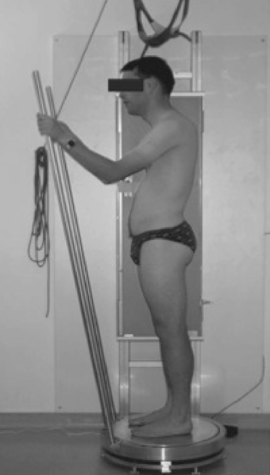
A lateral radiograph of the spine and pelvis is made with a patient in a controlled standing position. The hands are placed on rest, and the patient is asked to stand in a comfortable but erect posture
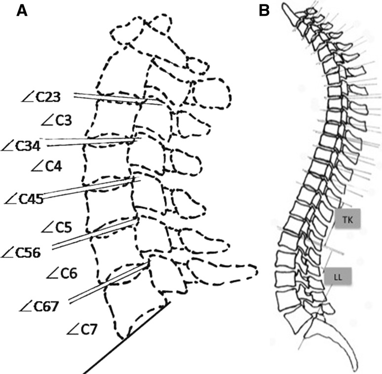
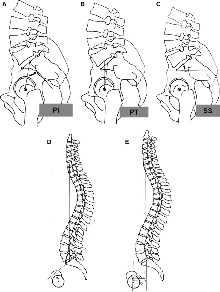
The relative inclinations of lines passing through the superior and inferior endplate of vertebral bodies from C2 to C7 were recorded, which provided a measurement of angles of endplates and discs (Fig. 2a). The cervical spine angles were calculated by the addition of each endplate angle from C3 to C7, and each inter-vertebral angle from C23 to C67. A wedging vertebral body or inter-vertebral kyphosis was considered if the angle was <0° and the angle open to ventral side was considered as positive. The cervico-thoracic angles (CTA) were the addition of both the cervical spine angles and the remaining angles of thoracic spine according to Cobb’s measurement (Fig. 2b). The results of thoracic kyphosis (TK) and lumbar lordosis (LL) were also measured as described in Fig. 2b based on Cobb’s measurement. Additional radiological parameters included pelvic incidence (PI) [6], sacral slope (SS) [6], pelvic tilt (PT) [6], spinal sacral angle (SSA) [5], ratio of hip to C7/hip to sacrum [5] (Fig. 3), Roussouly sagittal classifications [7], Lenke Curve Type, Lenke Lumbar Modifier [8]. Two trained specialists conducted the marking procedure separately and the average value of their calculation was adopted for analysis.
Fig. 2.
a The cervical spine angles were calculated by the addition of each endplate angle from C3 to C7, and each inter-vertebral angle from C23 to C67. b The cervico-thoracic angles were the addition of angles between endplates and intervertebral discs of cervical and thoracic spine according to Cobb’s measurement. The angles of thoracic kyphosis (TK) and lumbar lordosis (LL) were also measured between the lines of upper and lower end vertebral body according to Cobb’s method. The lower end vertebral body of lumbar lordosis was the upper endplate of sacrum
Fig. 3.
a Pelvic incidence (PI) is defined as the angle subtended by the line drawn from the hip axis (HA, center of the line connecting the center of each femoral heads) to the center of upper sacral endplate and the line perpendicular to upper sacral endplate, b Pelvic Tilt (PT) is defined as the angle subtended by the vertical line and the line drawn from HA to the center of upper sacral endplate. c Sacral slope (SS) is defined as the angle subtended by the horizontal line and upper sacral endplate. d Spinal sacral angles (SSA): sacral endplate and the line from the center of C7 vertebral body to the center of upper sacral endplate. e Hip to C7/hip to sacrum: horizontal distance from the center of upper sacral endplate to C7 plumb line (drawn from the center of C7 vertebral body) divided by horizontal distance from the center of upper sacral endplate to HA
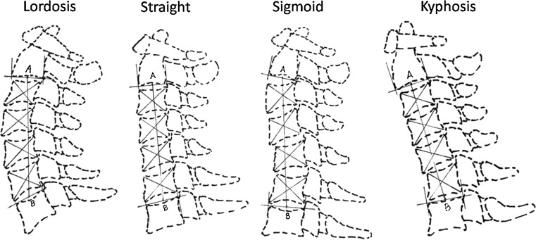
We categorized the cervical sagittal alignment into four types: lordosis, straight, sigmoid and kyphosis [9] (Fig. 4). Two diagonal lines were drawn after constructing four contour tangents for each body. Each line connected two corners of the vertebra, where adjacent contour tangents intersected. The intersection of these two lines was the vertebral centroid. Line AB, connecting midpoint A on the inferior surface of C2 and midpoint B on the superior surface of C7, was constructed. The alignment was then determined by the position of centroids relative to line AB. The classification of the vertebra types was based on the following description. Lordosis: all centroids were ventral to AB and the distance between them was more than 2 mm; straight: each centroid was ventral to or on line AB and the distance was <2 mm; sigmoid: centroids were ventral or dorsal to line AB and at least one centroid was more than 2 mm; kyphosis: all the centroids were dorsal to line AB and the distance between at least one centroid and the AB was 2 mm or more. To avoid the intra-observer bias, all of the radiographs were judged by two experienced surgeons separately. If they disagreed, a third one was invited to make a final decision.
Fig. 4.
Method of judgment of cervical alignment as lordosis, straight, sigmoid and kyphosis
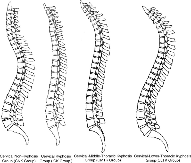
We finally divided the global sagittal alignment into four groups according to their morphological characteristics described as below (Fig. 5):
Fig. 5.
CNK Group: The cervical spine manifested in a lordotic or straight alignment. The transitional zone was usually located between T1 and T4. The thoracic and lumbar spines were well-aligned in physiological kyphosis and lordosis respectively. CK Group: The cervical spine presented a kyphotic deformity involving either C6 or C7 as the lower end vertebral body. Both the thoracic kyphosis and the lumbar lordosis were in small curves, occasionally forming a complete thoraco-lumbar lordosis. CMTK Group: A kyphotic cervical alignment extended to the middle thoracic spine. The transitional zone of lower thoracic curves from T9 to T12 was linked between an entity of kyphotic cervico-thoracic spine and lordotic lumbar spine. CLTK Group: The cervical and thoracic spines were in an overall kyphotic curve, without obvious transitional zone. Both cervico-thoracic kyphosis and lumbar lordosis were in large curves
Cervical non-kyphosis group (CNK Group): the cervical spine manifested a lordotic or straight alignment. The transitional zone was usually located between T1 and T4. The physiological kyphosis and lordosis of the thoracic and lumbar spines were well aligned.
Cervical kyphosis group (CK Group): the cervical spine presented a kyphotic deformity involving either C6 or C7 as the lower end vertebral body. Subsequently, both the thoracic kyphosis and the lumbar lordosis were in small curves, occasionally forming a complete thoraco-lumbar lordosis.
Cervical-middle-thoracic kyphosis group (CMTK Group): a kyphotic cervical alignment extended to the middle thoracic spine, such as T5, T6, T7 or T8. The transitional zone of lower thoracic curves from T9 to T12 was linked between an entity of kyphotic cervico-thoracic spine and lordotic lumbar spine, both of which were in small curves.
Cervical-lower-thoracic kyphosis group (Group CLTK): the cervical and thoracic spines were in an overall kyphotic curve, without obvious transitional zone. Both the cervico-thoracic kyphosis and lumbar lordosis were in large curves.
The values of CA, CTA, PI, SS, PT, SSA, hip to C7/hip to sacrum, TK and LL were compared among groups. The relationship between cervical alignments, Roussouly sagittal classification, Lenke Curve Type, Lenke Lumbar Modifier and Group classification were also tested, respectively. Moreover, the relationship between the cervical kyphosis and wedging vertebral body, intervertebral kyphosis were analyzed.
Data were analyzed with SPSS 15.0 software (SPSS Inc, Chicago, IL, USA). Statistical significance was set at 0.05. An adaptation of Kolmogorov–Smirnov test was applied to test for normally distributed data. Descriptive statistics in the form of mean and SD for all spine parameters were provided for all patients. The relationships were assessed using χ2 test. The one-way ANOVA test, Student t tests or u test for independent samples were also utilized to evaluate the parameters among different groups.
Results
The inter-observer correlation in classifying the cervical spine sagittal profile was 0.97.
The incidence of cervical kyphosis was 40 % (48/120). The values of CA and CTA were − 4.29 ± 15.70° and − 6.69 ± 20.40, respectively, with significant correlation (r = 0.854, P = 0.00). The distribution of the lower end vertebral bodies of cervico-thoracic curve generally conformed to our classification of four groups (Table 1). The cervical spine alignments were demonstrated to be significantly different among groups (Table 2, r = 85.04, P = 0.00).
Table 1.
Distribution of lower end vertebral body of cervico-thoracic curve
| C6 | C7 | T1 | T2 | T3 | T4 | T5 | T6 | T7 | T8 | T11 | T12 | L1 | L2 | |
|---|---|---|---|---|---|---|---|---|---|---|---|---|---|---|
| Group CNK | 14 | 24 | 28 | 7 | ||||||||||
| Group CK | 2 | 22 | ||||||||||||
| Group CMTK | 2 | 3 | 9 | 3 | ||||||||||
| Group CLTK | 2 | 1 | 2 | 1 |
Table 2.
Distribution of different cervical alignments in the four groups
| Cervical spine alignment | Total | ||||
|---|---|---|---|---|---|
| Kyphosis | Straight | Sigmoid | Lordosis | ||
| Group | |||||
| CNK | 6 (8.2 %) | 40 (54.8 %) | 8 (11.0 %) | 19 (26.0 %) | 73 |
| CK | 24 (100 %) | 0 | 0 | 0 | 24 |
| CMTK | 13 (76.5 %) | 4 (23.5 %) | 0 | 0 | 17 |
| CLTK | 5 (83.3 %) | 1 (16.7 %) | 0 | 0 | 6 |
| Total | 48 | 45 | 8 | 19 | 120 |
Fisher’s exact test = 85.04, P = 0.00
The distribution of the four groups in Roussouly sagittal classification (Table 3) revealed that 83.3 % (100/120) of the sagittal alignment belonged to Type 3 and 4. Significant differences in CA, CTA and TK were found between the curves of different groups (Table 4).
Table 3.
Distribution of the four groups in Roussouly sagittal classification
| Roussouly sagittal classification | |||||
|---|---|---|---|---|---|
| Type 1 | Type 2 | Type 3 | Type 4 | ||
| Group CNK | 2 (2.7 %) | 11 (15.1 %) | 34 (46.6 %) | 26 (35.6 %) | 73 |
| Group CK | 0 | 2 (8.3 %) | 9 (37.5 %) | 13 (54.2 %) | 24 |
| Group CMTK | 1 (5.9 %) | 4 (23.5 %) | 6 (35.3 %) | 6 (35.3 %) | 17 |
| Group CLTK | 0 | 0 | 2 (33.3 %) | 4 (66.7 %) | 6 |
| 3 | 17 | 51 | 49 | ||
Pearson χ 2 P = 0.59
Table 4.
Comparison of pre-operative global parameters among four Groups with one-way ANOVA test
| Group CNK | Group CK | Group CMTK | Group CLTK | P value | ||||||
|---|---|---|---|---|---|---|---|---|---|---|
| CNK and CK | CNK and CK | CNK and CLTK | CK and CMTK | CK and CLTK | CMTK and CLTK | |||||
| CA (°) | 4.2 ± 10.7 | −18.3 ± 6.7 | −18.4 ± 13.8 | −1.8 ± 10.4 | 0.00* | 0.00* | 0.03* | 1.0 | 0.13 | 0.17 |
| 0.00* | 0.00* | 0.00* | ||||||||
| CTA (°) | 6.5 ± 1 2.3 | −19.7 ± 6.9 | −33.8 ± 10.4 | −40.0 ± 11.2 | 0.00* | 0.00* | 0.00* | 0.01* | 0.00* | 0.61 |
| PI (°) | 52.7 ± 12.2 | 53.9 ± 15.3 | 53.5 ± 13.8 | 57.4 ± 12.3 | 1.00 | 0.94 | 0.87 | 0.97 | 0.91 | 0.99 |
| PT (°) | 11.1 ± 8.0 | 7.9 ± 9.0 | 13.8 ± 6.5 | 8.7 ± 5.8 | 0.36 | 0.66 | 0.92 | 0.13 | 0.99 | 0.62 |
| SS (°) | 41.7 ± 8.2 | 46.0 ± 10.4 | 39.7 ± 12.3 | 48.7 ± 8.6 | 0.48 | 1.00 | 0.39 | 0.59 | 0.89 | 0.42 |
| SSA (°) | 128.6 ± 7.7 | 134.2 ± 9.4 | 128.9 ± 9.9 | 136.9 ± 8.4 | 0.13 | 0.92 | 0.16 | 0.71 | 0.85 | 0.44 |
| Hip to C7/hip to sacrum | 11.0 (−10.4, 761.0) | −0.3 (−17.7, 6.4) | 0.4 (−0.6, 1.8) | 0.0 (−0.8, 1.9) | 0.50 | 0.58 | 0.71 | 0.97 | 0.99 | 0.99 |
| LL (°) | 55.0 ± 10.1 | 54.4 ± 12.8 | 46.7 ± 9.8 | 60.9 ± 11.7 | 0.98 | 0.13 | 0.64 | 0.40 | 0.56 | 0.10 |
| TK (°) | 36.0 ± 14.4 | 15.7 ± 13.9 | 3.3 ± 5.9 | 30.9 ± 6.9 | 0.00* | 0.00* | 0.85 | 0.02* | 0.12 | 0.00* |
In the post hoc Scheffe was selected in the one-way ANOVA test
*P < 0.05 was defined as statistically significant
In CNK Group, the values of CA and CTA were positive whereas they were negative in other groups. The values of hip to C7/hip to sacrum and TK ranked the first among the four groups. However, the values of PI and SSA were the smallest ones among the four groups.
In CK Group, the values of PT and hip to C7/hip to sacrum were the smallest among groups.
In CMTK Group, the values of CA in kyphosis and PT were the largest among groups. On the contrary, the values of SS, LL and TK were the smallest ones.
In CLTK Group, the value of CTA presented as the largest but the value of CA was the smallest in kyphosis among groups. Moreover, the values of PI, SS, SSA and LL were the largest ones.
A strong correlation between our grouping and Lenke Lumbar Modifier was revealed (P < 0.05) (Table 6). In CNK Group, the ratio of Modifier C was 56.2 % (41/73). In CK Group, the ratio of Modifier A was 50 % (12/24). In CMTK, the ratio of Modifier A was 58.8 % (10/17). In CLTK Group, Modifiers B and C occupied 50 % (3/6), respectively. No significant difference was found among Lenke’s Curve Types in the four groups (Table 5).
Table 6.
Distribution of four groups in Lenke Lumbar modifier
| Lenke Lumbar modifier | Total | |||
|---|---|---|---|---|
| A | B | C | ||
| Group | ||||
| CNK | 18 (24.7 %) | 14 (19.1 %) | 41 (56.2 %) | 73 |
| CK | 12 (50.0 %) | 6 (25.0 %) | 6 (25.0 %) | 24 |
| CMTK | 10 (58.8 %) | 4 (23.5 %) | 3 (17.7 %) | 17 |
| CLTK | 0 (0 %) | 3 (50 %) | 3 (50 %) | 6 |
| Total | 40 | 27 | 53 | 120 |
Fisher’s exact test P = 0.003
Table 5.
Distribution of four groups in Lenke curve type
| Lenke curve type | |||||||
|---|---|---|---|---|---|---|---|
| 1 | 2 | 3 | 4 | 5 | 6 | Total | |
| Group CNK | 32 (43.8 %) | 6 (8.2 %) | 5 (6.8 %) | 1 (1.4 %) | 9 (12.4 %) | 20 (27.4 %) | 73 |
| Group CK | 13 (54.2 %) | 5 (20.8 %) | 3 (12.5 %) | 0 (0 %) | 1 (4.2 %) | 2 (8.3 %) | 24 |
| Group CMTK | 9 (52.9 %) | 4 (23.6 %) | 0 (0 %) | 0 (0 %) | 1 (5.9 %) | 3 (17.6 %) | 17 |
| Group CLTK | 3 (50 %) | 1 (16.7 %) | 0 (0 %) | 0 (0 %) | 0 (0 %) | 2 (33.3 %) | 6 |
| 57 | 16 | 8 | 1 | 11 | 27 | 120 | |
Pearson χ 2 P = 0.57
Fisher’s exact test revealed that the individual vertebral body kyphosis and wedging was directly related to the overall cervical kyphosis (P = 0.00, Tables 7, 8).
Table 7.
Correlation between cervical kyphosis and wedging vertebral body
| No wedging vertebral body | Wedging vertebral body | ||
|---|---|---|---|
| No kyphosis | 59 | 13 | 72 |
| Kyphosis | 18 | 30 | 48 |
| 77 | 43 | 120 |
χ 2 test presents significant correlations. Fisher’s exact test P = 0.00
Table 8.
Correlation of preoperative cervical kyphosis and inter-vertebral kyphosis
| No intervertebral space kyphosis | Intervertebral space kyphosis | ||
|---|---|---|---|
| No kyphosis | 69 | 3 | 72 |
| Kyphosis | 24 | 24 | 48 |
| 93 | 27 | 120 |
Fisher’s exact test P = 0.00
Discussion
This study found that the cervical kyphosis in patients with idiopathic scoliosis correlates with its global sagittal alignment, as we saw significant difference in the alignment of cervical spine among the four groups based on global spinal curvature (Table 2).
Cervico-thoracic curves in different groups
The identification of cervical kyphosis is not as uncommon as one might expect. Cochran et al. [1] subjectively observed a cervical flattening or kyphosis in 49/95 patients without measurement. Hilibrand et al. [2] reported a straight (lordosis <5°) or kyphotic cervical alignment in 34/39 patients (89 %) and concluded that patients with idiopathic scoliosis developed lordosis within thoracic spine and compensatory kyphosis within the cervical and lumbar segments. The development of the cervical lordosis is believed to begin in the infantile period following a child raising its head and the subsequent lordosis correlates with the larger anterior height in the sagittal plane of the intervertebral disc.
In our cohort, the incidence of cervical kyphosis was 40 %, whereas it was 9 % in a previous radiographic survey of asymptomatic people [10], and the incidence of cervical kyphosis in Groups CK, CMTK and CLTK was all over 90 %. We thought the deformity correlates with their characteristics of global spine alignments both in sagittal and coronal planes. In this study, we defined cervical angle as the addition of the small angles, which could avoid possible inaccuracy of sagittal tangent method [9], which is to measure the angles between two lines designated parallel with posterior margins of C2 and C7. Though our calculation is far more complicated than the traditional measurement, it is likely to be more precise to reflect the authentic overall alignment of cervical spine than the conventional one. For example, a straight curve from C3 to C6 could be concealed by a positive result of measurement from C2 to C7. Moreover, the conventional way of measurement from C3 to C7 or T1 to T12 cannot accurately depict the curvature if a cervical kyphosis prolongs to thoracic spine. Some researchers also proposed the way of evaluations of the global thoracic kyphosis and lumbar lordosis is to measure angles between inflection points, which could provide better representation and understanding of the sagittal plane balance [11].
The distribution of lower end vertebral bodies (Table 1) supported our hypothesis in classification of different groups according to morphological characteristics. Despite the 40 % incidence of kyphosis in our cohort, the straight and lordotic alignments of cervical spine were seen in most of the cases, due to the flexible spine of young people and spontaneous adjusting mechanism of spinal-pelvic system. In Group CK, all the cervical alignments were categorized as kyphosis, and the deformities were judged to be mostly restricted to cervical spine. Nevertheless, the cervical kyphosis in both Groups CMTK and CLTK extended their curves to thoracic spine, which resulted in the coexistence of straight and kyphotic alignments. The sigmoid alignment seemed to be a short kyphosis. Anyway, it is obligatory to be classified as a specific type of curve if considering its isolated existence in cervical spine in Group CK and without any correlation to thoracic kyphosis.
Analysis of various spinal-pelvic parameters and Lenke classification between groups
We consider that the cervico-thoracic kyphosis is an extension of the cervical kyphosis, as we found significant correlation between the cervical angles and the CTA in the four groups. The difference of other parameters among groups explains the correlation between cervical and global spinal alignment and they could not be regarded separately (Table 4).
The parameters specific to each group are discussed individually as below.
Group CNK: the majority of the patients were well aligned in spite of occasional presence of cervical kyphosis. The average value of PI in this group was 52.7°, which was similar to that of asymptomatic people (51.99°) [7], and also close to that of people with AIS as 57.3° [4]. We thought the nearly normal value of PI was the main reason for spontaneous adjustment of spine in spite of deformity. The average value of PT was also close to that of asymptomatic people (11.99°) [7], but was higher than that in other researches [4]. This implies that patients in Group CNK had a well-balanced spinal-pelvic linkage and were more liable to equilibrate. The sacro-pelvic parameters are more influenced by the spinal deformity in the lumbar segment with a larger PT in the thoracic-right-lumbar-left type curves [12], which conforms to our results as a predominance of type 1 or 6 of Lenke curves (Table 5), with lumbar modifier C over 50 % (Table 6). In this group there were still 11 patients of Lenke Lumbar Modifier C besides the ones of Lenke curve types 4, 5 and 6. The distribution of such structure was presented as nine, one and one case of Lenke Curve Types 1, 2 and 3, respectively. In spite of the main thoracic curve, the patients could still have a large coronal curve in lumbar spine, and meanwhile a standard lumbar lordosis. We thought this could be related to the value of pelvic parameters, such as PI, that were the nearest to the normal.
The thoracic kyphosis consisted of similar segments as Group CK, but different average angles. This could be attributed to the fact that our grouping was based on sagittal curvature, but the coronal curvatures, represented by Lenke Curve Type and Lumbar Modifier, were different (Lenke Curve Type 1: 43.8 vs. 54.2 %; Lumbar Modifier C: 56.2 vs. 25.0 %) (Tables 5, 6). It was reported that the thoracic kyphosis angles were significantly lower for King II and III curves than for lumbar curves, which could be explained as the correlation between the shape and orientation of the discs and vertebrae that are distorted in AIS [4]. The coronal and sagittal curvatures always coexist with axial modifications in vertebrae and disc. Hence, the structural thoracic coronal curves influenced the sagittal plane, consequently the cervical alignment as what Hilibrand et al. [2] described.
Moreover, in spite of the largest value of hip to C7/hip to sacrum, most patients in this group still had a plumb line falling around sacrum, which indicated a balanced global alignment. There was only one case (761) that was very different from the others, which greatly affected the outcome of this group.
Group CK: the PI value similar to that of Group CNK was thought to be the reason of pelvic adjustment against deformity in Group CK and CMTK. Several patients in Group CK had no thoracic kyphosis but entire thoraco-lumbar lordosis, represented by a negative PT and PI <45°. Hilibrand et al. [2] postulated that in patients with a large cervical kyphosis, a compensatory thoracic lordosis occurred in order to maintain a forward gaze. Meanwhile, the hypo-kyphosis of thoracic spine is presumed to correlate with this coronal deformity, for most cases were classified as Lenke Curve Type 1 or 2, and 50 % as Lumbar Modifier A. The patients of Lenke curve type 1 or 2 did not have the Lumbar Modifier C, which was contrary to the similar patients in Group CNK. This might be attributed to the decrease of value of TK. A restricted cervical kyphosis and thoracic hypo-kyphosis reduces the possibility of large lumbar lordosis. Nonetheless, both the spinal sacral angles and sacral slope were larger in this group than in Group CNK and Group CMTK, implying that a pelvic anterior tilt contributed to an increased sacral slope to restore the lumbar lordosis. Possibly, such a phenomenon is a compensation of the values of an average negative hip to C7/hip to sacrum, which indicated that the C7 plumb line was located in the ventral side of hip axis while the sacrum in the dorsal, or vice versa. Mac-Thiong et al. [11] reported 14.2 % of patients with their C7 plumb line anterior to both hip axis and center of upper sacral endplate (which is assumed to be the anterior possible landmark in maintaining a non-pathological posture) remained asymptomatic. The incidence of such phenomenon in this group was 29.2 % (7/24). The segments of cervico-thoracic kyphosis were confined to cervical spine, which was distinguished from other kyphotic cervical spines. We thought the restriction of cervical kyphosis was attributed not only to the position of plumb line between hip axis and sacrum, but also to the augmentations of spinal sacral angles and sacral slope to compensate hypo-thoracic curves, which predispose to form a complete thoracic and lumbar lordosis.
Group CMTK: the cervical kyphosis constituted the majority of cervico-thoracic deformity, which was different from Group CLTK. The thoracic curves, the lower end vertebrae of CTA stopping at middle thoracic spine, were responsible for the smallest value, which could explain their corresponding size of lumbar curves. This is consistent with what was previously described as an association between lumbar lordosis and thoracic kyphosis, and also between lumbar lordosis and sacral slope [13], which could account for their sagittal difference. Besides, we observed that patients with Lenke Curve Type 1 or 2 often had Lumbar Modifier A or B, with similar distribution as Group CK. In addition, the spinal sacral angles in this group were similar to that of Group CNK, which can be deemed as C7 plumb line and hip axis that were almost superimposed. This conforms to the idea that a reduced spinal sacral angle will be observed in a spine with an increased pelvic tilt and reduced sacral slope when the pelvic incidence is greater than 45° [13]. We thus assume that lumbar hypo-lordosis is protective against vertebral rotation in flexible curves and a severe sagittal deformity has more influence on the pelvic parameters than a coronal one.
Group CLTK: the largest sacral slope and lumbar lordosis were the result of anterior version of pelvis, which was possibly a compensation for the largest cervico-thoracic kyphosis with the thoracic curves in majority. This compensation might also be responsible for the increase of spinal sacral angles while the C7 plumb line was migrating over the hip axis as in Group CMTK. The thoraco-lumbar zone, lower end vertebras of cervico-thoracic kyphosis, seems to be the inflexion point of the great kyphosis and lordosis with the largest value of pelvic incidence. The Lenke Lumbar Modifier C ranked the first, with the Curve Type 1 or 6 mostly. Whatever the scoliosis curve type, the coronal lumbar curve is probably more influenced by the sagittal alignment and pelvic factors. The largest PI correlated with a relevant lumbar lordosis, which is theoretically protective for the straight spine, because the sagittal curvatures tend to protect against the initiation of a coronal deformity by a buckling mechanism [14]. Consequently, in spite of cervico-thoracic deformity, a large lumbar coronal and sagittal curve, benefitting from the largest pelvic incidence, could interact with each other in order to maintain overall sagittal balance, so long as vertebral bodies are able to rotate in a coupled motion manner in scoliosis. That is why the lumbar curve is immune to the structural curve, but not the pelvic parameters.
The majority of sagittal classification presenting Roussouly Type 3 or 4 was deduced to be the fundamental factor in adjusting to maintain equilibrium.
As is well known to all, scoliosis is a three-dimensional deformity and there is a reciprocal relationship between the coronal and sagittal plane deformity during its progression. A rotation of the vertebral body in axial plane produces frontal and sagittal angles. And subsequently the alignment of cervical spine follows. As is postulated by Hilibrand et al. [2], lordosis in the thoracic spine could be a primary manifestation and cervical kyphosis might simply reflect a continuation of this sagittal malalignment.
The precaution that we have to consider is that a restoration of sagittal physiological alignment should be done when we correct coronal deformity, and lumbar and pelvic parameters provide us the surgical scheme.
Analysis of vertebral body wedging
Vertebral body growth is produced by endochondral ossification at the superior and inferior epiphyseal growth plates [15]. Our results demonstrated that the cervical kyphosis correlates with the incidence of wedge-shaped vertebras and inter-vertebral kyphosis, which is the morphological modification of this deformity.
The Hueter and Volkmann law indicates that an increased compression of epiphyseal growth plates prevents growth whereas distraction allows growth [15]. Whereas the Mechanostat theory with its Chondral Growth Force Response Curve predicts that the initial response to compression would be an accelerated growth response that was selective to anterior parts of the vertebral endplate to achieve spontaneous correction [16]. The former one might explain that the displacement of C7 plumb line over the hip and sacrum influenced the transmission of the gravity right down to the lower extremities, which prohibits the physiological growth of cervical vertebral bodies. And this was especially seen in Groups CK, CMTK and CLTK. But the growth of anterior column was not altered at all. And whether there would be a spontaneous correction needs our further study. And biomechanical and histological investigations are required to prove this.
It is not clear whether vertebrae or disc primarily causes the deformity of cervical spine. Nevertheless, the effect of vertebrae or disc on coronal deformity in scoliosis is certain [17]. However, without biomechanical and histological evidence, it is not convincing to conclude a causal relationship.
Limitation
In this study, we focused only on the morphological characteristics of global sagittal alignment in patients before surgery. We believe that the evaluation could have been more complete if post-surgery characteristics, such as the cervical alignment, thoracic and lumbar curves, pelvic parameters, were considered.
Conclusion
The cervical kyphosis in young patients with idiopathic scoliosis is correlated with global sagittal alignment. Despite the great curves in cervical alignment, the global spine could still be well-balanced by spontaneous adjustment. The correlation between our grouping based on the sagittal morphological characteristics and Lenke Curve Type/Lumbar Modifier suggests that the coupled motion principle be appropriate to explain the modifications both in coronal and sagittal planes. The morphology of cervical kyphosis can be manifested in wedging-shaped vertebrae and kyphotic inter-vertebral spaces.
Acknowledgments
I really appreciate the selfless and generous kindness from my British colleague, Dr. Kamran Hassan M.D., who corrected English language in this article. I will always remember the help and friendship beyond the countries and the enthusiastic discussion on the medical topics between us.
Conflict of interest
None.
Contributor Information
Miao Yu, Phone: +86-10-82267378, FAX: +86-10-82267368, Email: miltonyu1977@gmail.com.
Clement Silvestre, Phone: +33-04-72384855, FAX: +33-04-72384891, Email: clement.silvestre1@gmail.com.
Tanguy Mouton, Phone: +33-04-72384855, FAX: +33-04-72384891, Email: tanguy.mouton1@gmail.com.
Rami Rachkidi, Phone: +33-04-72384855, FAX: +33-04-72384891, Email: ramirachkidi1@gmail.com.
Lin Zeng, Phone: +86-10-82265732, FAX: +86-10-82265732, Email: zlwhy@163.com.
Pierre Roussouly, Phone: +33-04-72384855, FAX: +33-04-72384891, Email: chort@cmcr-massues.com.
References
- 1.Cochran T, Irstam L, Nachemson A. Long-term anatomic and functional changes in patients with adolescent idiopathic scoliosis treated by Harington rod fusion. Spine. 1983;8:576–583. doi: 10.1097/00007632-198309000-00003. [DOI] [PubMed] [Google Scholar]
- 2.Hilibrand A, Tannenbaum D, Graziano G, Loder R, Hensinger R, et al. The sagittal alignment of the cervical spine in adolescent idiopathic scoliosis. J Pediatr Orthop. 1995;15(5):627–632. doi: 10.1097/01241398-199509000-00015. [DOI] [PubMed] [Google Scholar]
- 3.Canavese F, Turcot K, De Rosa V, et al. Cervical spine sagittal alignment variations following posterior spinal fusion and instrumentation for adolescent idiopathic scoliosis. Eur Spine J. 2011;12(7):1141–1148. doi: 10.1007/s00586-011-1837-z. [DOI] [PMC free article] [PubMed] [Google Scholar]
- 4.Mac-Thiong JM, Labelle H, Charlebois M. Sagittal plane analysis of the spine and pelvis in adolescent idiopathic scoliosis according to the coronal curve type. Spine. 2003;28(13):1404–1409. doi: 10.1097/01.BRS.0000067118.60199.D1. [DOI] [PubMed] [Google Scholar]
- 5.Roussouly P, Gollogly S, Noseda O, et al. The vertical projection of the sum of the ground reactive forces of a standing patient is not the same as the C7 plumb line: a radiographic study of the sagittal alignment of 153 asymptomatic volunteers. Spine. 2006;31(11):E320–E325. doi: 10.1097/01.brs.0000218263.58642.ff. [DOI] [PubMed] [Google Scholar]
- 6.Legaye J, Duval-Beaupere G, Hecquet J, et al. Pelvic incidence: a fundamental parameter for three-dimensional regulation of spinal sagittal curves. Eur Spine J. 1998;7(2):99–103. doi: 10.1007/s005860050038. [DOI] [PMC free article] [PubMed] [Google Scholar]
- 7.Roussouly P, Gollogly S, Berthonnaud E, et al. Classification of the normal variation in the sagittal alignment of the human lumbar spine and pelvis in the standing position. Spine. 2005;30(3):346–353. doi: 10.1097/01.brs.0000152379.54463.65. [DOI] [PubMed] [Google Scholar]
- 8.Lenke GL, Betz R, Harms J, et al. Adolescent idiopathic scoliosis: a new classification to determine extent of spinal arthrodesis. J Bone Jt Surg. 2001;83(8):1169–1181. [PubMed] [Google Scholar]
- 9.Ohara A, Miyamoto K, Naganwa T, et al. Reliabilities of and correlations among five standard methods of assessing the sagittal alignment of the cervical spine. Spine. 2006;31(22):2585–2591. doi: 10.1097/01.brs.0000240656.79060.18. [DOI] [PubMed] [Google Scholar]
- 10.Gore DR, Sepic SB, Gardner GM. Roentgenographic findings of the cervical spine in asymptomatic people. Spine. 1986;11:512–514. doi: 10.1097/00007632-198607000-00003. [DOI] [PubMed] [Google Scholar]
- 11.Mac-Thiong JM, Roussouly P, Berthonnaud E, et al. Sagittal parameters of global spinal balance normative values from a prospective cohort of seven hundred nine Caucasian asymptomatic adult. Spine. 2010;35(22):E1193–E1198. doi: 10.1097/BRS.0b013e3181e50808. [DOI] [PubMed] [Google Scholar]
- 12.Pasha S, Sangole AP, Aubin CE, et al. Characterizing pelvis dynamics in adolescent with idiopathic scoliosis. Spine. 2010;35(17):E820–E826. doi: 10.1097/BRS.0b013e3181e6856d. [DOI] [PubMed] [Google Scholar]
- 13.Roussouly P, Nnadi C. Sagittal plane deformity: an overview of interpretation and management. Eur Spine J. 2010;19(11):1824–1836. doi: 10.1007/s00586-010-1476-9. [DOI] [PMC free article] [PubMed] [Google Scholar]
- 14.Stokes IAF. Three-dimensional terminology of spinal deformity: a report presented to the Scoliosis Research Society by the Scoliosis Research Society Working Group on 3-D terminology of the spinal deformity. Spine. 1994;19:236–248. doi: 10.1097/00007632-199401001-00020. [DOI] [PubMed] [Google Scholar]
- 15.Mehlman CT, Araghi A, Roy DR. Hyphenated history: the Hueter-Volkmann law. Am J Orthop. 1997;26:798–800. [PubMed] [Google Scholar]
- 16.Rajasekaran S, Natarajan RN, Babu JN, et al. Lumbar vertebral growth is governed by “chondral growth force response curve” rather than “Hueter-Volkmann law”. Spine. 2011;36(22):E1435–E1445. doi: 10.1097/BRS.0b013e3182041e3c. [DOI] [PubMed] [Google Scholar]
- 17.Will R, Stokes I, Qiu X, et al. Cobb angles progression in adolescent scoliosis begins at intervertebral disc. Spine. 2009;34(25):2782–2786. doi: 10.1097/BRS.0b013e3181c11853. [DOI] [PMC free article] [PubMed] [Google Scholar]


