Abstract
Heterocyclic aromatic hydrocarbons are, together with their un-substituted analogues, widely distributed throughout all environmental compartments. While fate and effects of homocyclic PAHs are well-understood, there are still data gaps concerning the ecotoxicology of heterocyclic PAHs: Only few publications are available investigating these substances using in vitro bioassays. Here, we present a study focusing on the identification and quantification of clastogenic and aneugenic effects in the micronucleus assay with the fish liver cell line RTL-W1 that was originally derived from rainbow trout (Oncorhynchus mykiss). Real concentrations of the test items after incubation without cells were determined to assess chemical losses due to, e.g., sorption or volatilization, by means of gas chromatography-mass spectrometry. We were able to show genotoxic effects for six compounds that have not been reported in vertebrate systems before. Out of the tested substances, 2,3-dimethylbenzofuran, benzothiophene, quinoline and 6-methylquinoline did not cause substantial induction of micronuclei in the cell line. Acridine caused the highest absolute induction. Carbazole, acridine and dibenzothiophene were the most potent substances compared with 4-nitroquinoline oxide, a well characterized genotoxicant with high potency used as standard. Dibenzofuran was positive in our investigation and tested negative before in a mammalian system. Chemical losses during incubation ranged from 29.3% (acridine) to 91.7% (benzofuran) and may be a confounding factor in studies without chemical analyses, leading to an underestimation of the real potency. The relative potency of the investigated substances was high compared with their un-substituted PAH analogues, only the latter being typically monitored as priority or indicator pollutants. Hetero-PAHs are widely distributed in the environment and even more mobile, e.g. in ground water, than homocyclic PAHs due to the higher water solubility. We conclude that this substance class poses a high risk to water quality and should be included in international monitoring programs.
Introduction
One of the key processes that built the foundation of the organic chemical industry in Germany and many other European countries in the 19th century was the distillation of coal tar-oil, by which common building-blocks for numerous syntheses were derived, e.g. for textile dyes [1]. However, tar-oil and coal tar can contain up to 85% polycyclic aromatic hydrocarbons (PAHs) and 5–13% heterocyclic aromatic hydrocarbons containing nitrogen, oxygen or sulphur (hetero-PAHs) [2], [3], while the latter can constitute 40% of the water-soluble fraction. In industrial areas, e.g. gas plants, coke manufacturing or wood preservation sites, long ground water plumes contaminated with hetero-PAHs have been detected [4], [5], potentially also endangering drinking water resources [6] and aquatic ecosystems.
While PAHs were already extensively investigated with regard to fate, biodegradation, toxicology and ecotoxicology [7], [8], [9], only limited knowledge exists for the hetero-PAHs. Many studies focused on few individual compounds and toxicological effects, e.g. mutagenicity of carbazoles [10] or chromosome aberrations induced by quinolines [11]. Researchers just recently began to conduct comparative studies investigating a range of hetero-PAHs using different mechanism-specific in vitro bioassays. The biological effects so far cover, e.g. toxicity to Daphnia and green algae, mutagenicity in the Ames assay, embryotoxicity to embryos of the zebrafish (Danio rerio) or effects mediated by the aryl hydrocarbon and retinoid receptor, respectively [12], [13], [14], [15], [16], [17], [18].
To further fill the data gap mentioned above, we report on the first comparative study in which the clastogenic and aneugenic effects of heterocyclic PAHs typically found at tar-oil contaminated sites were investigated using the micronucleus assay with the permanent rainbow trout liver cell line RTL-W1 [19]. It is one of the best characterized cell lines derived from fish for mechanism-specific biotests [20] and was extensively used in genotoxicity studies [21], [22], [23], [24]. The in vitro micronucleus assay was validated [25] and standardized by an international guideline [26] with the Chinese hamster lung fibroblast cell line V79. Furthermore, it is well-accepted that a substance’s potential to induce micronuclei represents an important toxicological effect with potential adverse effects on the population level [21], [27], [28]. The substances tested were chosen to match the set of compounds identified earlier by the project framework KORA (retention and degradation processes to reduce contaminations in groundwater and soil) [12], [29] and comprised indole, 1-benzothiophene, benzofuran, 2-methyl benzofuran, 2,3-dimethyl benzofuran, quinoline, 6-methyl quinoline, carbazole, dibenzothiophene, dibenzofuran, acridine, and xanthene. To account for the dissipation of the compounds from the test medium, changes in chemical concentration during incubation were measured by means of gas chromatography-mass spectrometry (GC-MS) analyses [13].
Materials and Methods
Chemicals
Stock solutions of the used heterocyclic PAHs were prepared in dimethyl sulfoxide (DMSO). Indole (>99%), quinoline (>98%), carbazole (approx. 95%), 6-methylquinoline (>98%), benzothiophene (>98%), dibenzothiophene (>98%) were supplied by abcr (Karlsruhe, Germany). Acridine (>98%) was purchased from Merck (Darmstadt, Germany). Xanthene (99%), benzofuran (>99%), 2-methylbenzofuran (≥96%), 2,3-dimethylbenzofuran (≥97%) and dibenzofuran (approx. 98%) were supplied by Sigma-Aldrich (Deisenhofen, Germany).
Micronucleus Assay with RTL-W1 Cells
The protocols recently published by Rocha et al. [30] for cell culture and the micronucleus assay with RTL-W1 cells were followed, with slight modifications. Cells were originally derived from rainbow trout liver [19] and generously provided by Drs Niels C Bols and Lucy Lee (University of Waterloo, Canada). Cells were cultured at 20°C in Leibovitz L15 medium with L-glutamine (Sigma–Aldrich) containing 9% fetal bovine serum (FBS, Biochrom, Berlin, Germany) and 1% (v/v) penicillin/streptomycin solution (Biochrom) according to Klee et al. [31]. Before use in the micronucleus assay, cells were rinsed twice with phosphate buffered saline (PBS, Sigma-Aldrich) and suspended following trypsinisation [32]. Cells of passage number 83 were used for the experiments.
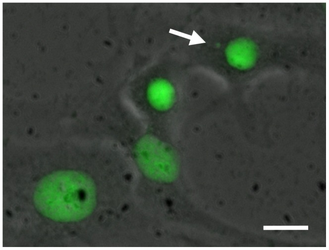
A volume of 2 ml of the cell suspension at a density of 5–6 104 cells/ml was seeded onto ethanol pre-cleaned microscopic glass cover slips in 6-well plates (TPP, Trassadingen, Switzerland) and incubated for 12 h at 20°C (resulting in approx. 6–7 103 cells/cm2) in a cooling incubator (Binder, Tuttlingen, Germany). Subsequently, the medium was aspired and completely exchanged with dilutions of the investigated hetero-PAHs and the plates incubated for 20 h at 20°C. For each substance, a serial dilution of each stock solution (1∶2) comprising five concentrations was tested in duplicate (i.e. on two different slides), while the highest concentration was equal to the NR80 of the substance, i.e. the concentration at which 80% viability of RTL-W1 cells was measured in the neutral red retention assay (Table 1). The maximum concentration of DMSO in the test was 1%. The exposure medium was aspired and completely exchanged with fresh L15 medium and the plates incubated for 72 h at 20°C to give cells enough time to divide at least once [33]. Subsequently, cells were fixed for 10 min in a PBS-diluted (1∶1 v/v) mixture of methanol and glacial acetic acid (4∶1 v/v). Fixation was repeated for 10 min in the undiluted mixture. After air-drying, the cover slips were mounted onto glass slides using DePeX (Serva, Heidelberg, Germany). Acridine orange was used for staining of the slides after fixation [34]. A total number of 2000 cells per slide were analyzed under an epifluorescence microscope (Nikon Instruments, Düsseldorf, Germany) with oil-immersion at 1000× magnification. The scoring criteria of the ISO guideline 21427-2 were used: (a) only cells with intact cellular structure were read, micronuclei shall have (b) the same staining intensity as and (c) a maximum size of about 30% of the main nucleus. Furthermore, cells must be (d) clearly separated from the nucleus [26]. A representative photomicrograph of a cell with micronucleus is shown in Figure 1.
Table 1. Corrected and uncorrected REPs relative to NQO, as well as respective corrected and uncorrected EC25 values in the micronucleus assay with RTL-W1 cells.
| Substance | Maximum concentration1 | Uncorrected EC25 | Uncorrected REP | Corrected EC25 | Corrected REP | Genotoxicity in mammalian models | LOQ | Loss |
| mg L−1 | mg L−1 | mg L−1 | µg L−1 | % | ||||
| 4-nitroquinoline oxide (STD) | 0.19 | 20.44 · 10−3 | 1 | |||||
| Benzofuran | 120.9 | 41.7 | 4.9 · 10−4 | 3.5 | 5.90 · 10−3 | + [51] | 0.2 | 91.7 |
| 2-Methylbenzofuran | 210.0 | 51.2 | 4.0 · 10−4 | 8.8 | 2.33 · 10−3 | n.a. | 0.2 | 82.9 |
| 2,3-Dimethylbenzofuran | 110.0 | n.d. | n.d. | n.d. | n.d. | n.a. | 0.1 | 51.8 |
| Dibenzofuran | 56.9 | 21.8 | 9.4 · 10−4 | 5.0 | 4.12 · 10−3 | − [52] | 0.1 | 77.2 |
| Benzothiophene | 136.1 | n.d. | n.d. | n.d. | n.d. | n.a. | 0.2 | 75.0 |
| Dibenzothiophene | 40.8 | 10.8 | 1.9 · 10−3 | 3.2 | 6.45 · 10−3 | n.a. | 0.3 | 70.6 |
| Acridine | 40.9 | 10.3 | 2.0 · 10−3 | 7.3 | 2.82 · 10−3 | + [60], [61] | 0.3 | 29.3 |
| Xanthene | 75.5 | 47.9 | 4.3 · 10−4 | 8.4 | 2.44 · 10−3 | n.a. | 0.1 | 82.5 |
| Carbazole | 25.6 | 7.5 | 2.7 · 10−3 | 3.5 | 5.81 · 10−3 | + [48] | 0.2 | 53.1 |
| Indole | 162.7 | 55.5 | 3.7 · 10−4 | 11.6 | 1.76 · 10−3 | n.a. | 0.1 | 79.1 |
| Quinoline | 226.0 | n.d. | n.d. | n.d. | n.d. | + [11], [38], [39], [40], [41], [42], [43] | 0.3 | 61.7 |
| 6-Methylquinoline | 528.0 | n.d. | n.d. | n.d. | n.d. | − [62] | 0.3 | 41.1 |
Chemical losses in the micronucleus assay used for calculation of corrected EC25 (i.e. by multiplying the residual compound fraction with the EC25) and REP values were derived from GC-MS measurements in 6-well microplates without cells.
n.d.: Inactive in assay system, i.e. substances which did not reach 25% induction of the NQO standard; n.a.: not available; STD: standard substance; LOQ: limit of quantification.
1 Maximun tested concentrations based on cytotoxicity data from Hinger & Brinkmann et al. [13].
Figure 1. Composite photomicrograph of a micronucleus in RTL-W1 cells directly after cytokinesis (arrow).

Nuclei and micronuclei were stained using Acridine Orange dye. The micrograph was captured at 1000× magnification and is a composite of bright-field and epifluorescence microscopy. Scale bar = 5 µm.
Calculation of EC and REP Values
Induction factors (fold-changes) relative to blanks, i.e. controls without treatment containing only the Leibovitz L15 medium, were calculated for each concentration by dividing the micronucleus rate of the respective concentration level by the mean of the blank replicates. Resulting concentration-response curves for individual substances and the well-characterized standard substance NQO (4-nitroquinoline oxide) were calculated with the software GraphPad Prism 5 (GraphPad, San Diego, CA, USA) using the four-parameter logistic equation model following log-transformation of the concentration values. Since a number of samples did not exceed 50% effect of the maximum NQO concentration, fixed-effect-level based REP (relative potency) values (Equation 1) were calculated according to Brack et al. [35]. Unlike in receptor-mediated assays, these REP values cannot be used for mass-balance analyses (i.e. to answer which portion of a measured effect is caused by which compound classes or single compounds) due to the different possible modes of action and are just intended for reference and comparison among samples.
| (1) |
Effect concentrations (EC25,NQO) refer to the concentration of the substance causing 25% of the maximum effect level of NQO. The arbitrary level of 25% effect was chosen since it includes more valid curves than using the EC50 but is still within the linear portion of the concentration-response curves. The EC25 value for NQO, EC25,NQO (NQO), was derived from the same test repetition of the micronucleus assay.
Chemical Analysis
To account for the effects of e.g. volatilization or sorption to plastic plates, real concentrations were compared to nominal concentrations comparable to the methodology recently published in Hinger & Brinkmann et al. [13] and Peddinghaus et al. [17]. Here, 6-well microplates were prepared in the same way as for the micronucleus assay without adding cells. Before addition to the plate, and after a 20 h incubation period in the microplate, solutions were stored in a glass vial with PTFE cap. The heterocyclic PAHs were extracted (liquid – liquid extraction) with methyl tert-butyl ether (MTBE). For this purpose 45 mL of the diluted sample were spiked with 10 µL internal standard solution in acetone (0.63 µg/µl toluene d8 and 0.66 µg/µl naphthalene d8, both obtained from Merck, Darmstadt, Germany) and extracted with 5 mL MTBE. Extraction time was 20 minutes. After phase separation, the extract was dried with sodium sulphate and subsequently analysed using gas chromatography (Agilent technologies GC 6890 N). The GC was equipped with an autosampler (Agilent technologies) and mass selective detector (MSD) (Agilent technologies MS 5973 Network) operated in SIM (Selective Ion Monitoring) mode. For Separation of the substances, a ZB-5 Inferno column (60 m×0.25 mm×0.25 µm) by Phenomenex was used. The concentrations of the internal standard in samples and external standards were equal. The limit of detection was 0.1 to 0.3 µg L−1 for the investigated substances. To correct the bioassay data for the measured chemical losses, EC25 values were multiplied with the remaining compound fraction and the REPs re-calculated with the corrected EC25.
Statistical Analysis
All spreadsheet calculations were performed using Microsoft Excel™ 2007. Statistical analyses were conducted with Sigma Stat 3.11 (Systat Software, Erkrath, Germany). All tested treatments and levels were tested for statistically significant differences from the blanks, i.e. controls without treatment, by use of one-way ANOVA (p≤0.001). Dunnett’s test was used as the multiple range test to identify significant differences between treatments and blanks. The probability of Type I error (α) was set to p≤0.05. Values are expressed as mean value ± standard deviation, unless indicated.
Results and Discussion
Clastogenic and Aneugenic Effects of Heterocyclic PAHs
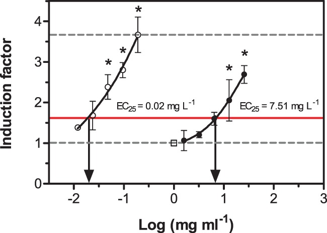
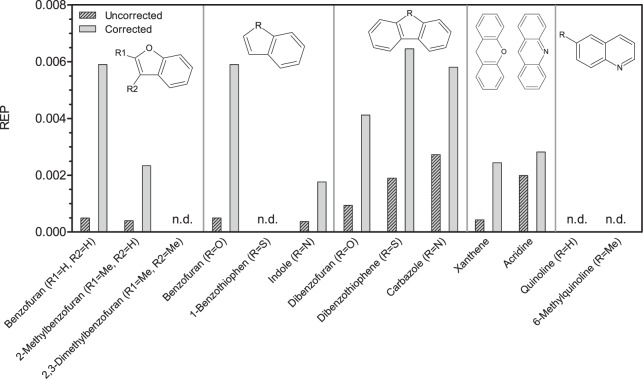
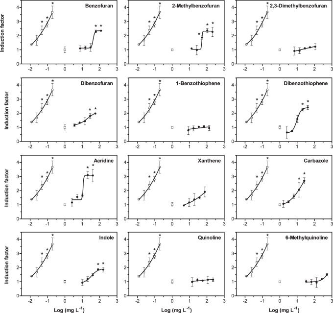
The substances 2,3-dimethylbenzofuran, benzothiophene, quinoline and 6-methylquinoline did not cause significant induction of micronuclei in the permanent fish liver cell line RTL-W1, i.e. did not reach 25% induction of the nitroquinoline oxide (NQO) standard (Table 1). Acridine caused the highest absolute induction (approx. 3-fold compared to blanks). Carbazole, acridine and dibenzothiophene were the most potent substances. To be able to better compare the investigated substances, relative potency (REP) values were calculated, comparing the potency of the reference compound NQO with the potency of the test item. Figure 2 illustrates this data analysis approach. REP values (Figure 3) ranged from 3.7 · 10−4 (indole) to 2.7 · 10−3 (carbazole). The three substances with the highest potency were only approx. 500-fold less potent than 4-nitroquinoline oxide. Full concentration-response curves are given for reference (Figure 4). While concentrations of hetero-PAHs in contaminated aquifers are typically in the µg L−1 range, values up to the mg L−1 range (at which effects in the micronucleus assay were observed) have occasionally been detected at highly contaminated sites [29]. However, care should be taken when comparing in vitro results with aqueous exposure concentrations, since substances with differing physicochemical properties can be absorbed and accumulated to a varying extent and through different tissues and organs [36].
Figure 2. Exemplary concentration-response curve (carbazole) measured in the micronucleus assay with RTL-W1 cells (closed circles).

The concentration-response curve for NQO (open circles) and the blanks, i.e. control cells without treatment (open square and lower dashed line), are given for reference. Induction factors are fold-changes relative to blanks. EC25 values relative to the maximum induction of NQO (upper dashed line) were calculated to derive fixed-effect-level-based REPs. Concentration values on the x-axis refer to nominal medium concentrations of the substances. Circles represent mean values measured in duplicate experiments, error bars the standard deviation. The red line depicts 25% of the maximum induction caused by the standard NQO. Asterisks denote statistically significant differences compared to blanks (one-way ANOVA with Dunnet’s test, p≤0.05).
Figure 3. Uncorrected and corrected relative potency (REP) values of hetero-PAHs.
Uncorrected values refer to nominal concentrations and corrected to measured concentrations in the exposure medium after incubation without cells. N.d.: inactive in assay system, i.e. substances which did not reach the stipulated level of 25% induction of the NQO standard.
Figure 4. Dose-response curves in the micronucleus assay with RTL-W1 cells for all investigated heterocyclic compounds (closed circles).
Standard curves for NQO (open circles) and blanks, i.e. control cells without treatment (open squares, grey line), are given for reference. Induction factors are fold-changes relative to blanks. Concentration values on the x-axis refer to nominal medium concentrations of the substances. Dots represent mean values measured in duplicate experiments, error bars the standard deviation. Asterisks denote statistically significant differences compared to blanks (one-way ANOVA with Dunnet’s test, p≤0.05).
Eisenträger et al. [12] found that out of the tested substances (similar to the set tested here) only quinoline, 6-methylquinoline and xanthene caused mutagenic effects in the Ames assay with Salmonella typhimurium, while these effects were only apparent in treatments with metabolic activation, i.e. when supplemented with rat liver S9. The same effects for the 6-methylated and the parent quinoline were found by Debnath et al. [37]. The latter substance was also shown to cause significant induction of liver micronuclei and chromosome aberrations in rats and the hamster lung fibroblast cell line CHL/IU [11], [38], [39], [40] and is a potent hepatocarcinogen in mice and rats [41], [42], [43]. Indole was positive in the Ames assay [44]. Acridine was shown to be positive in the SOS chromotest with Escherichia coli K12 and a yeast-based reporter-gene assay in which DNA damage induces the expression of green fluorescent protein (GFP) via the RAD54 promoter [45]. It was not mutagenic in the Ames assay [12], [46] but produced clastogenic effects detected with the yeast DEL assay without metabolic activation [47]. Carbazole was demonstrated to be clastogenic by Jha et al. [48] but was not mutagenic in the Ames assay [49] or carcinogenic in mice [43]. The O-heterocyclic compound benzofuran showed very interesting effects, although being negative in the Ames assay [12], [50]; DNA damage, as measured with the Comet assay, as well as the formation of micronuclei was higher in female specimens, both in vivo in rat kidney and in vitro using primary rat and human kidney cultures [51]. Dibenzofuran, a substance that was previously reported not to be genotoxic in a mammalian system was identified to be potent genotixicant in the fish system [52].
The following general trends can be deduced: For heterocyclic three-ring PAHs with two benzene rings fused with one five-membered aromatic ring (dibenzofuran, dibenzothiophene, carbazole), the genotoxic potential decreased in the following order of hetero-atoms: nitrogen>sulphur>oxygen. A comparable trend was observed for the three-ring hetero-PAHs with central six-membered ring, where the toxicity of acridine (nitrogen hetero-atom) was markedly higher than that of xanthene (oxygen hetero-atom). In accordance with the aforementioned studies, the azaarenes had the highest potency to induce genotoxic effects. Furthermore, for derivates of benzofuran, methylation had a decreasing effect on the genotoxicity (benzofuran >2-methylbenzofuran >2,3-dimethlybenzofuran).
Interestingly, the molar effect concentrations observed in the present study significantly correlated (Pearson’s correlation coefficient r = 0.88, p = 0.02, data not shown) with the embryotoxic potential of hetero-PAHs as observed by Peddinghaus et al. [17]. In this study, acridine and carbazole caused the highest toxicity to embryos of Danio rerio and showed the highest potency to induce micronuclei in RTL-W1 cells in the present study. Furthermore, Hinger & Brinkmann et al. [13] have demonstrated that these two substances had the highest relative potency for luciferase induction in the DR-CALUX assay, an assay for aryl hydrocarbon receptor (AhR) agonists, and generally seem to be of high concern. The metabolic pathways of PAHs and hetero-PAHs were lately reviewed by Xue et al. [53] and need to be considered when investigating the genotoxic effects of these substances.
Influence of Substance Losses during Incubation
Chemical losses during incubation ranged from 29.3% (acridine) to 91.7% (benzofuran) and may lead to a drastic underestimation of the real potency of the substances, e.g. in mass-balance analyses (Table 1). Here, we attempted to correct for chemical losses from the exposure medium by correcting EC25 and REP values by applying analytically-derived correction factors. These correction factors were based on the compound concentration after incubation for 20 h (without cells but under the same conditions) and represent a worst-case scenario in which only the residual concentration after incubation caused the effect. The resulting REP values differed by a factor of 1.5–2 for substances with low losses during the incubation period (acridine and carbazole) compared to the uncorrected REP values. For benzofuran, the substance with the highest loss (91.7%), the REPs differed by a factor of 12 compared with the uncorrected value (Figure 3). As it has been shown by other studies from the KORA project framework, accounting for the chemical losses is a prerequisite to adequately judge the real toxicological potency [12], [13], [17]. Furthermore, experimental precautions are warranted when experimenting with these volatile genotoxicants.
Implications for Water Quality
The substances we investigated here showed a relatively high potency to induce micronuclei in the permanent cell line RTL-W1. We were able to show genotoxic effects for six compounds that have not been reported for vertebrate systems before. Dibenzofuran was positive in our investigation and tested negative before in a mammalian system. Homocyclic PAHs are commonly monitored as priority pollutants and are indicator substances for contamination with substances originating from historically contaminated sites, e.g. gas-plants, wood preservation or dyestuff industry. However, heterocyclic aromatic compounds are markedly more mobile than homocyclic PAHs, e.g. in ground water, due to higher water solubility. Because ground water wells are an important drinking water resource, we conclude that this substance class poses a high risk to human health and should be included in groundwater monitoring programs, e.g. according to the German LAWA-GFS (threshold values for groundwater) that are based on toxicological and ecotoxicological effect data [54] or the health-related indicator value (HRIV) introduced by the German Federal Environment Agency (UBA) [55]. The international NORMAN network, which was originally founded by the European Commission to set up a permanent network of reference laboratories and research groups, recently emphasized the importance of also screening for compounds that are not commonly part of monitoring programs [56]. In fluvial environments, heterocyclic PAHs contribute to the overall genotoxicity. In studies applying the concept of effect-directed analysis (EDA) to sediment and suspended particulate matter samples, as well as soil samples from related flood plains, those fractions containing heterocyclic substances showed strong mutagenic or genotoxic effects [57], [58]. Re-suspension of contaminated sediments can ultimately lead to an increased bioavailability of such particle-bound pollutants with potentially adverse effects in aquatic biota [59].
Acknowledgments
The authors would like to express their thanks to Drs Niels C. Bols and Lucy Lee (University of Waterloo, Canada) for providing RTL-W1 cells. Furthermore, we want to express our gratitude to Simone Hotz for technical assistance during parts of the experiment.
Funding Statement
The authors acknowledge financial support by a Seed funds project of RWTH Aachen University supported by the German Excellence Initiative. M.B. received a personal stipend from the German National Academic Foundation (“Studienstiftung des deutschen Volkes”). The funders had no role in study design, data collection and analysis, decision to publish, or preparation of the manuscript.
References
- 1. Johnston W (2008) The discovery of aniline and the origin of the term “aniline dye”. Biotech Histochem 83: 83–87. [DOI] [PubMed] [Google Scholar]
- 2. Meyer S, Steinhart H (2000) Effects of heterocyclic PAHs (N, S, O) on the biodegradation of typical tar oil PAHs in a soil/compost mixture. Chemosphere 40: 359–367. [DOI] [PubMed] [Google Scholar]
- 3. Dyreborg S, Arvin E, Broholm K (1997) Biodegradation of NSO-compounds under different redox-conditions. J Contam Hydrol 25: 177–197. [Google Scholar]
- 4. Blum P, Sagner A, Tiehm A, Martus P, Wendel T, et al. (2011) Importance of heterocyclic aromatic compounds in monitored natural attenuation for coal tar contaminated aquifers: A review. J Contam Hydr 126: 181–194. [DOI] [PubMed] [Google Scholar]
- 5. Tiehm A, Müller A, Alt S, Jacob H, Schad H, et al. (2008) Development of a groundwater biobarrier for the removal of PAH, BTEX, and heterocyclic hydrocarbons. Water Sci Technol 58: 1349–1355. [DOI] [PubMed] [Google Scholar]
- 6. Reineke A-K, Göen T, Preiss A, Hollender J (2007) Quinoline and Derivatives at a Tar Oil Contaminated Site: Hydroxylated Products as Indicator for Natural Attenuation? Environ Sci Technol 41: 5314–5322. [DOI] [PubMed] [Google Scholar]
- 7.Douben PET, editor (2003) PAHs: An Ecotoxicological Perspective. ChichesterEngland: John Wiley & Sons Ltd. 36 p. [Google Scholar]
- 8. Tiehm A, Schulze S (2003) Intrinsic aromatic hydrocarbon biodegradation for groundwater remediation. Oil Gas Sci Technol 58: 449–462. [Google Scholar]
- 9. Schulze S, Tiehm A (2004) Assessment of microbial natural attenuation in groundwater polluted with gasworks residues. Water Sci Technol 50: 347–353. [PubMed] [Google Scholar]
- 10. Dutson SM, Booth GM, Schaalje GB, Castle RN, Seegmiller RE (1997) Comparative developmental dermal toxicity and mutagenicity of carbazole and benzo[a]carbazole. Environ Toxicol Chem 16: 2113–2117. [Google Scholar]
- 11. Asakura S, Sawada S, Sugihara T, Daimon H, Sagami F (1997) Quinoline-induced chromosome aberrations and sister chromatid exchanges in rat liver. Environ Mol Mutagen 30: 459–467. [DOI] [PubMed] [Google Scholar]
- 12. Eisentraeger A, Brinkmann C, Hollert H, Sagner A, Tiehm A, et al. (2008) Heterocyclic compounds: Toxic effects using algae, daphnids, and the Salmonella/microsome test taking methodical quantitative aspects into account. Environ Toxicol Chem 27: 1590–1596. [DOI] [PubMed] [Google Scholar]
- 13. Hinger G, Brinkmann M, Bluhm K, Sagner A, Takner H, et al. (2011) Some heterocyclic aromatic compounds are Ah receptor agonists in the DR-Calux and the EROD assay with RTL-W1 cells. Environ Sci Pollut Res 18: 1297–1304. [DOI] [PubMed] [Google Scholar]
- 14. Sovadinová I, Bláha L, Janoscaronek J, Hilscherová K, Giesy JP, et al. (2006) Cytotoxicity and aryl hydrocarbon receptor-mediated activity of N-heterocyclic polycyclic aromatic hydrocarbons: Structure-activity relationships. Environ Toxicol Chem 25: 1291–1297. [DOI] [PubMed] [Google Scholar]
- 15. Benisek M, Kubincova P, Blaha L, Hilscherova K (2011) The effects of PAHs and N-PAHs on retinoid signaling and Oct-4 expression in vitro. Toxicol Lett 200: 169–175. [DOI] [PubMed] [Google Scholar]
- 16. Larsson M, Orbe D, Engwall M (2012) Exposure time–dependent effects on the relative potencies and additivity of PAHs in the Ah receptor-based H4IIE-luc bioassay. Environ Toxicol Chem 31: 1149–1157. [DOI] [PubMed] [Google Scholar]
- 17. Peddinghaus S, Brinkmann M, Bluhm K, Sagner A, Hinger G, et al. (2012) Quantitative assessment of the embryotoxic potential of NSO-heterocyclic compounds using zebrafish (Danio rerio). Reprod Toxicol 33: 224–232. [DOI] [PubMed] [Google Scholar]
- 18. Hawliczek A, Nota B, Cenijn P, Kamstra J, Pieterse B, et al. (2012) Developmental toxicity and endocrine disrupting potency of 4-azapyrene, benzo[b]fluorene and retene in the zebrafish Danio rerio. Reprod Toxicol 33: 213–223. [DOI] [PubMed] [Google Scholar]
- 19. Lee LE, Clemons JH, Bechtel DG, Caldwell SJ, Han KB, et al. (1993) Development and characterization of a rainbow trout liver cell line expressing cytochrome P450-dependent monooxygenase activity. Cell Biol and Toxicol 9: 279–294. [DOI] [PubMed] [Google Scholar]
- 20. Hallare A, Seiler T-B, Hollert H (2011) The versatile, changing, and advancing roles of fish in sediment toxicity assessment–a review. J Soils Sediments 11: 141–173. [Google Scholar]
- 21. Boettcher M, Grund S, Keiter S, Kosmehl T, Reifferscheid G, et al. (2010) Comparison of in vitro and in situ genotoxicity in the Danube River by means of the comet assay and the micronucleus test. Mut Res 700: 11–17. [DOI] [PubMed] [Google Scholar]
- 22. Boettcher M, Kosmehl T, Braunbeck T (2011) Low-dose effects and biphasic effect profiles: Is trenbolone a genotoxicant? Mut Res 723: 152–157. [DOI] [PubMed] [Google Scholar]
- 23. Kosmehl T, Hallare AV, Braunbeck T, Hollert H (2008) DNA damage induced by genotoxicants in zebrafish (Danio rerio) embryos after contact exposure to freeze-dried sediment and sediment extracts from Laguna Lake (The Philippines) as measured by the comet assay. Mut Res 650: 1–14. [DOI] [PubMed] [Google Scholar]
- 24. Seitz N, Böttcher M, Keiter S, Kosmehl T, Manz W, et al. (2008) A novel statistical approach for the evaluation of comet assay data. Mut Res 652: 38–45. [DOI] [PubMed] [Google Scholar]
- 25. Reifferscheid G, Ziemann C, Fieblinger D, Dill F, Gminski R, et al. (2008) Measurement of genotoxicity in wastewater samples with the in vitro micronucleus test–Results of a round-robin study in the context of standardisation according to ISO. Mut Res 649: 15–27. [DOI] [PubMed] [Google Scholar]
- 26.ISO 21427–2:2006 (2006) Water quality - Evaluation of genotoxicity by measurement of the induction of micronuclei - Part 2: Mixed population method using the cell line V79. International Organization of Standardization.
- 27. Diekmann M, Hultsch V, Nagel R (2004) On the relevance of genotoxicity for fish populations I: effects of a model genotoxicant on zebrafish (Danio rerio) in a complete life-cycle test. Aquat Toxicol 68: 13–26. [DOI] [PubMed] [Google Scholar]
- 28. Diekmann M, Waldmann P, Schnurstein A, Grummt T, Braunbeck T, et al. (2004) On the relevance of genotoxicity for fish populations II: genotoxic effects in zebrafish (Danio rerio) exposed to 4-nitroquinoline-1-oxide in a complete life-cycle test. Aquat Toxicol 68: 27–37. [DOI] [PubMed] [Google Scholar]
- 29. Blotevogel J, Reineke AK, Hollender J, Held T (2008) Identification of NSO-heterocyclic priority substances for investigating and monitoring creosote-contaminated sites. Grundwasser 13: 147–157. [Google Scholar]
- 30. Rocha PS, Luvizotto GL, Kosmehl T, Böttcher M, Storch V, et al. (2009) Sediment genotoxicity in the Tietê River (São Paulo, Brazil): In vitro comet assay versus in situ micronucleus assay studies. Ecotoxicol Environ Saf 72: 1842–1848. [DOI] [PubMed] [Google Scholar]
- 31. Klee N, Gustavsson L, Kosmehl T, Engwall M, Erdinger L, et al. (2004) Toxicity and genotoxicity in an industrial sewage sludge containing nitro- and amino-aromatic compounds during treatment in bioreactors under different oxygen regimes. Environ Sci Pollut Res 11: 313–320. [DOI] [PubMed] [Google Scholar]
- 32. Kosmehl T, Krebs F, Manz W, Erdinger L, Braunbeck T, et al. (2004) Comparative genotoxicity testing of rhine river sediment extracts using the comet assay with permanent fish cell lines (rtg-2 and rtl-w1) and the ames test. J Soils Sediments 4: 84–94. [Google Scholar]
- 33. Schnurstein A, Braunbeck T (2001) Tail Moment versus Tail Length–Application of an In Vitro Version of the Comet Assay in Biomonitoring for Genotoxicity in Native Surface Waters Using Primary Hepatocytes and Gill Cells from Zebrafish (Danio rerio). Ecotoxicol Environ Saf 49: 187–196. [DOI] [PubMed] [Google Scholar]
- 34. Jernbro S, Rocha PS, Keiter S, Skutlarek D, Färber H, et al. (2007) Perfluorooctane sulfonate increases the genotoxicity of cyclophosphamide in the micronucleus assay with V79 cells. Further proof of alterations in cell membrane properties caused by PFOS. Environ Sci Pollut Res Int 14: 85–87. [DOI] [PubMed] [Google Scholar]
- 35. Brack W, Segner H, Möder M, Schüürmann G (2000) Fixed-effect-level toxicity equivalents–a suitable parameter for assessing ethoxyresorufin-O-deethylase induction potency in complex environmental samples. Environ Toxicol Chem 19: 2493–2501. [Google Scholar]
- 36. Mackay D, Fraser A (2000) Bioaccumulation of persistent organic chemicals: mechanisms and models. Environ Pollut 110: 375–391. [DOI] [PubMed] [Google Scholar]
- 37. Debnath AK, Decompadre RLL, Hansch C (1992) Mutagenicity of quinolines in Salmonella typhimurium TA100 - a QSAR study based on hydrophobicity and molecular-orbital determinants. Mut Res 280: 55–65. [DOI] [PubMed] [Google Scholar]
- 38. Suzuki H, Takasawa H, Kobayashi K, Terashima Y, Shimada Y, et al. (2009) Evaluation of a liver micronucleus assay with 12 chemicals using young rats (II): a study by the Collaborative Study Group for the Micronucleus Test/Japanese Environmental Mutagen Society–Mammalian Mutagenicity Study Group. Mutagenesis 24: 9–16. [DOI] [PubMed] [Google Scholar]
- 39. Suzuki T, Takeshita K, Saeki KI, Kadoi M, Hayashi M, et al. (2007) Clastogenicity of quinoline and monofluorinated quinolines in Chinese hamster lung cells. J Health Sci 53: 325–328. [Google Scholar]
- 40. Hakura A, Kadoi M, Suzuki T, Saeki KI (2007) Clastogenicity of quinoline derivatives in the liver micronucleus assay using rats and mice. J Health Sci 53: 470–474. [Google Scholar]
- 41. Hirao K, Shinohara Y, Tsuda H, Fukushima S, Takahashi M, et al. (1976) Carcinogenic Activity of Quinoline on Rat Liver. Cancer Res 36: 329–335. [PubMed] [Google Scholar]
- 42. La Voie EJ, Dolan S, Little P, Wang CX, Sugie S, et al. (1988) Carcinogenicity of quinoline, 4- and 8-methylquinoline and benzoquinolines in newborn mice and rats. Food Chem Toxicol 26: 625–629. [DOI] [PubMed] [Google Scholar]
- 43. Weyand EH, Defauw J, McQueen CA, Meschter CL, Meegalla SK, et al. (1993) Bioassay of quinoline, 5-fluoroquinoline, carbazole, 9-methylcarbazole and 9-ethylcarbazole in newborn mice. Food Chem Toxicol 31: 707–715. [DOI] [PubMed] [Google Scholar]
- 44. Ochiai M, Wakabayashi K, Sugimura T, Nagao M (1986) Mutagenicities of indole and 30 derivatives after nitrite treatment. Mutat Res 172: 189–197. [DOI] [PubMed] [Google Scholar]
- 45. Bartoš T, Letzsch S, Škarek M, Flegrová Z, Čupr P, et al. (2006) GFP assay as a sensitive eukaryotic screening model to detect toxic and genotoxic activity of azaarenes. Environ Toxicol 21: 343–348. [DOI] [PubMed] [Google Scholar]
- 46. Brown BR, Firth Iii WJ, Yielding LW (1980) Acridine structure correlated with mutagenic activity in salmonella. Mutat Res 72: 373–388. [DOI] [PubMed] [Google Scholar]
- 47. Kirpnick Z, Homiski M, Rubitski E, Repnevskaya M, Howlett N, et al. (2005) Yeast DEL assay detects clastogens. Mutat Res 582: 116–134. [DOI] [PubMed] [Google Scholar]
- 48. Jha AM, Singh AC, Bharti MK (2002) Clastogenicity of carbazole in mouse bone marrow cells in vivo. Mutat Res 521: 11–17. [DOI] [PubMed] [Google Scholar]
- 49. LaVoie EJ, Briggs G, Bedenko V, Hoffmann D (1982) Mutagenicity of substituted carbazoles in Salmonella typhimurium. Mutat Res 101: 141–150. [DOI] [PubMed] [Google Scholar]
- 50. Weill-Thevenet N, Buisson JP, Royer R, Hofnung M (1981) Mutagenic activity of benzofurans and naphthofurans in the Salmonella/microsome assay: 2-nitro-7-methoxynaphtho[2,1-b]furan (R7000), a new highly potent mutagenic agent. Mutat Res 88: 355–362. [DOI] [PubMed] [Google Scholar]
- 51. Robbiano L, Baroni D, Carrozzino R, Mereto E, Brambilla G (2004) DNA damage and micronuclei induced in rat and human kidney cells by six chemicals carcinogenic to the rat kidney. Toxicology 204: 187–195. [DOI] [PubMed] [Google Scholar]
- 52. Galloway SM, Armstrong MJ, Reuben C, Colman S, Brown B, et al. (1987) Chromosome aberrations and sister chromatid exchanges in chinese hamster ovary cells: Evaluations of 108 chemicals. Environ Mol Mutagen 10: 1–175. [DOI] [PubMed] [Google Scholar]
- 53. Xue WL, Warshawsky D (2005) Metabolic activation of polycyclic and heterocyclic aromatic hydrocarbons and DNA damage: A review. Toxicol Appl Pharmacol 206: 73–93. [DOI] [PubMed] [Google Scholar]
- 54.Frank D, Dieter H, Herrmann H, Konietzka R, Moll B, et al. (2010) Ableitung von Geringfügigkeitsschwellenwerten für das Grundwasser: NSO-Heterozyklen. Bund/Länder-Arbeitsgemeinschaft Wasser (LAWA).
- 55. Grummt T, Kuckelkorn J, Bahlmann A, Baumstark-Khan C, Brack W, et al. (2013) Tox-Box: securing drops of life - an enhanced health-related approach for risk assessment of drinking water in Germany. Environmental Sciences Europe 25: 27. [Google Scholar]
- 56. Brack W, Dulio V, Slobodnik J (2012) The NORMAN Network and its activities on emerging environmental substances with a focus on effect-directed analysis of complex environmental contamination. Environmental Sciences Europe 24: 29. [Google Scholar]
- 57. Higley E, Grund S, Jones PD, Schulze T, Seiler T-B, et al. (2012) Endocrine disrupting, mutagenic, and teratogenic effects of upper Danube River sediments using effect-directed analysis. Environ Toxicol Chem 31: 1053–1062. [DOI] [PubMed] [Google Scholar]
- 58. Wölz J, Schulze T, Lübcke-von Varel U, Fleig M, Reifferscheid G, et al. (2011) Investigation on soil contamination at recently inundated and non-inundated sites. J Soils Sediments 11: 82–92. [Google Scholar]
- 59. Schüttrumpf H, Brinkmann M, Cofalla C, Frings R, Gerbersdorf S, et al. (2011) A new approach to investigate the interactions between sediment transport and ecotoxicological processes during flood events. Environmental Sciences Europe 23: 39. [Google Scholar]
- 60. Moir D, Poon R, Yagminas A, Park G, Viau A, et al. (1997) The subchronic toxicity of acridine in the rat. Journal of Environmental Science and Health Part B-Pesticides Food Contaminants and Agricultural Wastes 32: 545–564. [DOI] [PubMed] [Google Scholar]
- 61. Phelps JB, Garriott ML, Hoffman WP (2002) A protocol for the in vitro micronucleus test: II. Contributions to the validation of a protocol suitable for regulatory submissions from an examination of 10 chemicals with different mechanisms of action and different levels of activity. Mut Res 521: 103–112. [DOI] [PubMed] [Google Scholar]
- 62. Wild D, King MT, Gocke E, Eckhard K (1983) Study of artificial flavouring substances for mutagenicity in the Salmonella/microsome, BASC and micronucleus tests. Food Chem Toxicol 21: 707–719. [DOI] [PubMed] [Google Scholar]


