

Abstract
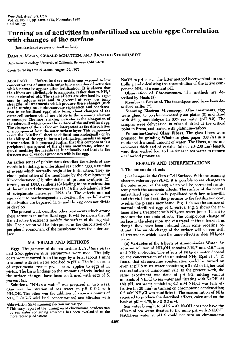
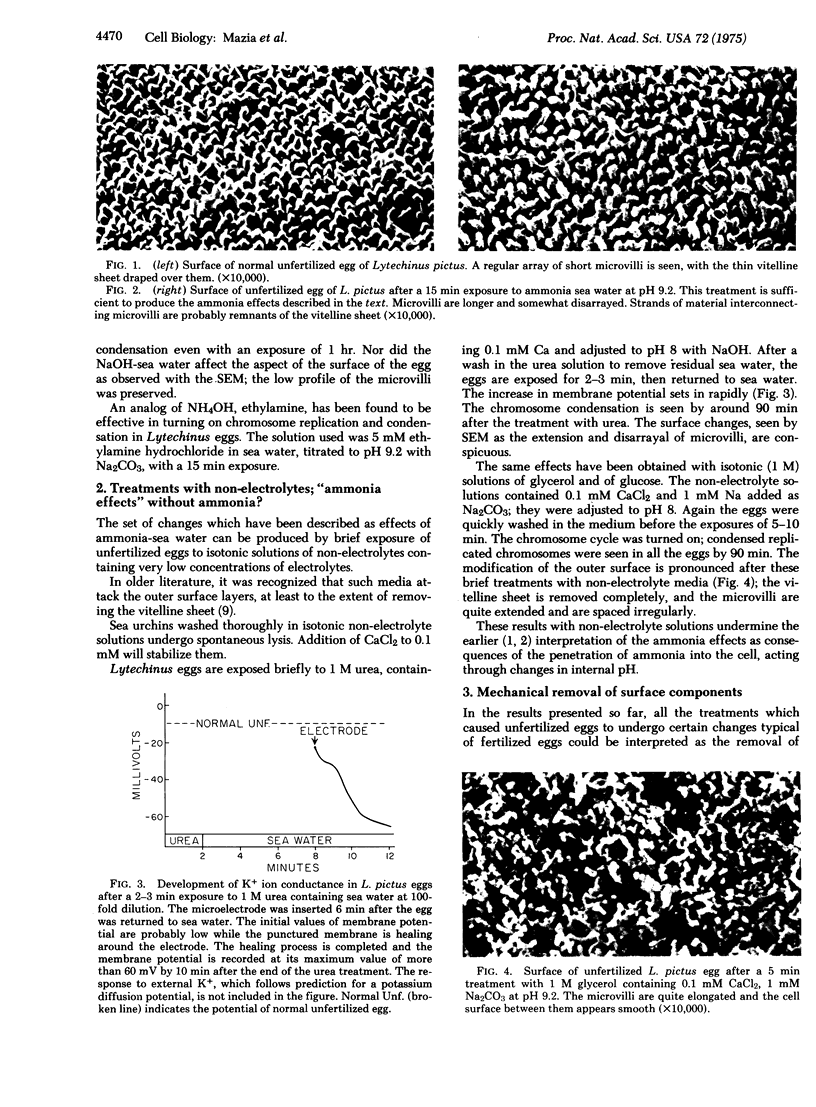
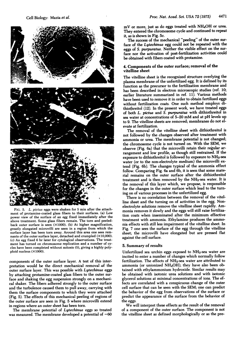
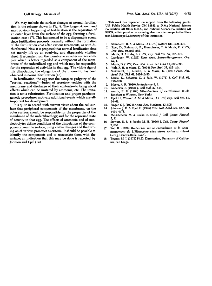
Unfertilized sea urchin eggs exposed to low concentrations of ammonia enter into a number of activities which normally appear after fertilization. It is shown that the effects are attributable to ammonia, rather than to NH4+ ions of elevated pH. The same effects are obtained by exposure to isotonic urea and to glycerol at very low ionic strengths. All treatments which produce these changes (such as the turning on of chromosome replication and condensation in unfertilized eggs) also bring about changes of the outer cell surface which are visible in the scanning electron microscope. The most striking indicator is the elongation of the microvilli which cover the surface of the unfertilized egg. The changes of the surface are interpreted as the dissociation of a component from the outer surface layer. This component is not the "vitelline" sheet as defined morphologically or by the ability of the egg to form a fertilization membrane upon insemination. It is proposed further that this component is a peripheral component of the plasma membrane, whose removal modifies the membrane functionally and leads to the derepression of various processes within the egg.
Full text
PDF

Images in this article
Selected References
These references are in PubMed. This may not be the complete list of references from this article.
- Anderson E. Oocyte differentiation in the sea urchin, Arbacia punctulata, with particular reference to the origin of cortical granules and their participation in the cortical reaction. J Cell Biol. 1968 May;37(2):514–539. doi: 10.1083/jcb.37.2.514. [DOI] [PMC free article] [PubMed] [Google Scholar]
- Epel D., Steinhardt R., Humphreys T., Mazia D. An analysis of the partial metabolic derepression of sea urchin eggs by ammonia: the existence of independent pathways. Dev Biol. 1974 Oct;40(2):245–255. doi: 10.1016/0012-1606(74)90127-4. [DOI] [PubMed] [Google Scholar]
- Epel D., Weaver A. M., Mazia D. Methods for revoval of the vitelline membrane of sea urchin eggs. I. Use of dithiothreitol (Cleland Reagent). Exp Cell Res. 1970 Jul;61(1):64–68. doi: 10.1016/0014-4827(70)90257-0. [DOI] [PubMed] [Google Scholar]
- Johnson J. D., Epel D. Relationship between release of surface proteins and metabolic activation of sea urchin eggs at fertilization. Proc Natl Acad Sci U S A. 1975 Nov;72(11):4474–4478. doi: 10.1073/pnas.72.11.4474. [DOI] [PMC free article] [PubMed] [Google Scholar]
- Mazia D. Chromosome cycles turned on in unfertilized sea urchin eggs exposed to NH4OH. Proc Natl Acad Sci U S A. 1974 Mar;71(3):690–693. doi: 10.1073/pnas.71.3.690. [DOI] [PMC free article] [PubMed] [Google Scholar]
- Mazia D., Ruby A. DNA synthesis turned on in unfertilized sea urchin eggs by treatment with NH4OH. Exp Cell Res. 1974 Mar 30;85(1):167–172. doi: 10.1016/0014-4827(74)90227-4. [DOI] [PubMed] [Google Scholar]
- Mazia D., Schatten G., Sale W. Adhesion of cells to surfaces coated with polylysine. Applications to electron microscopy. J Cell Biol. 1975 Jul;66(1):198–200. doi: 10.1083/jcb.66.1.198. [DOI] [PMC free article] [PubMed] [Google Scholar]
- Moore N. Harvey. Proc R Soc Med. 1916;9(SECT):9–20. doi: 10.1177/003591571600901602. [DOI] [PMC free article] [PubMed] [Google Scholar]
- Singer S. J. The molecular organization of membranes. Annu Rev Biochem. 1974;43(0):805–833. doi: 10.1146/annurev.bi.43.070174.004105. [DOI] [PubMed] [Google Scholar]
- Steinhardt R. A., Lundin L., Mazia D. Bioelectric responses of the echinoderm egg to fertilization. Proc Natl Acad Sci U S A. 1971 Oct;68(10):2426–2430. doi: 10.1073/pnas.68.10.2426. [DOI] [PMC free article] [PubMed] [Google Scholar]
- Steinhardt R. A., Mazia D. Development of K + -conductance and membrane potentials in unfertilized sea urchin eggs after exposure to NH 4 OH. Nature. 1973 Feb 9;241(5389):400–401. doi: 10.1038/241400a0. [DOI] [PubMed] [Google Scholar]
- Wilt F. H., Mazia D. The stimulation of cytoplasmic polyadenylylation in sea urchin eggs by ammonia. Dev Biol. 1974 Apr;37(2):422–424. doi: 10.1016/0012-1606(74)90158-4. [DOI] [PubMed] [Google Scholar]