Abstract
Resveratrol (RSV) is a natural polyphenol that is known as a powerful chemopreventive and chemotherapeutic anticancer molecule. This study focused on the effects of RSV on the activities and expression levels of antioxidant enzymes in the cancer cells. Prostate cancer PC-3 cells, hepatic cancer HepG2 cells, breast cancer MCF-7 cells and the non-cancerous HEK293T kidney epithelial cells were treated with a wide range of RSV concentrations (10–100 μM) for 24–72 h. Cell growth was estimated by trypan blue staining, activities of the antioxidant enzymes were measured spectrophotometrically, expression levels of the antioxidant enzymes were quantified by digitalizing the protein band intensities on Western blots, and the percentage of apoptotic cells was determined by flow cytometry. Treatment with a low concentration of RSV (25 μM) significantly increased superoxide dismutase (SOD) activity in PC-3, HepG2 and MCF-7 cells, but not in HEK293T cells. Catalase (CAT) activity was increased in HepG2 cells, but no effect was found on glutathione peroxidase (GPX) upon RSV treatment. RSV-induced SOD2 expression was observed in cancer cells, although the expression of SOD1, CAT and GPX1 was unaffected. Apoptosis increased upon RSV treatment of cancer cells, especially in PC-3 and HepG2 cells. Together, our data demonstrated that RSV inhibits cancer cell growth with minimal effects on non-cancerous cells. We postulate that the disproportional up-regulation of SOD, CAT and GPX expression and enzymatic activity in cancer cells results in the mitochondrial accumulation of H2O2, which in turn induces cancer cell apoptosis.
Keywords: Apoptosis, HepG2 cells, MCF-7 cells, PC-3 cells, resveratrol, superoxide dismutase
INTRODUCTION
Cancer is the second leading cause of death after myocardial infarction. Despite the great advances in modern medical science in the last century, most cancers are not yet curable. This is partly due to the complexity of the pathogenesis of cancer, and the difficulties in developing efficient treatments. Among the numerous factors, oxidative stress plays an important role in cancer initiation, promotion and progression by inducing the DNA damage and interfering with the intracellular signal transduction pathways (Klaunig and Kamendulis, 2004). Since antioxidant enzymes play crucial roles in protecting cells from oxidative stress, dysregulation or defects in the activity of antioxidant enzymes, such as superoxide dismutase (SOD), catalase (CAT) and glutathione-peroxidase (GPX), are associated with cancer (Khan et al., 2010). The decreased activities of antioxidant enzymes or the down-regulation of their expression were found to be associated with several types of cancers, including prostate cancer, bladder cancer, breast cancer, hepatic cancer, multiple myeloma (Arsova-Sarafinovska et al., 2009; De Craemer et al., 1993; Elchuri et al., 2005; Jeon et al., 2007; Kasapović et al., 2008; Sharma et al., 2009). Nevertheless, no change or even the higher expression and activities of antioxidant enzymes have also been reported in some cancers. For example, increased levels of SOD, with decreased CAT and unchanged GPX levels were reported in lung cancer tissues and in the A549 lung cancer cell line (Chung-man Ho et al., 2001). Therefore, maintaining the appropriate levels of antioxidant enzymatic activities may be essential for preventing cancer development. To this end, targeting antioxidant enzymatic activities could serve as an important strategy for developing therapeutic agents for cancer treatment. In addition, studying the effects of anticancer drugs on the activity and expression level of antioxidant enzymes will enhance the understanding of the mechanism in drug actions.
The preventive and therapeutic effects of copious natural products against cancer and other life threatening diseases have been documented. Resveratrol (RSV) [3, 5, 4′-trihydroxytrans-stilbene] has been shown to inhibit cancer initiation, promotion, and progression in numerous experimental models, including cancer cell lines, animal models, and even clinical trials (Harikumar and Aggarwal, 2008; Jang et al., 1997; Patel et al., 2011). Both in vitro and in vivo studies have demonstrated that RSV possesses anti-cancer potential against many types of cancers, including prostate, hepatic, breast, skin, colorectal, and pancreatic cancer (Benitez et al., 2007; Bishayee, 2009; Mo et al., 2012; Sengottuvelan et al., 2009). Mechanistically, different studies have revealed that RSV affects cancer cells by inducing apoptosis, altering the cell cycle, inhibiting angiogenesis, suppressing the signaling pathways of nuclear factor-kappa B (NF-κB) and cyclooxygenase, and activating the peroxisome proliferator-activated receptor (PPAR) (Benitez et al., 2007; Bishayee, 2009; Carbo et al., 1999; Chen et al., 2004; Mo et al., 2012; Nakata et al., 2012; Sengottuvelan et al., 2009; Zhou et al., 2005). Moreover, RSV inhibits the metabolic activation of carcinogens, and has antioxidant and anti-inflammatory properties. RSV also alters the expression of cancer related miRNAs in cancer cells (Bae et al., 2011). A recent study reported that RSV exerts its effects by increasing the activity of regulatory proteins, AMP-activated protein kinase and sirtuin through inhibition of cAMP-degrading phosphodiesterases (Park et al., 2012). However, the precise mechanisms underlying the effects of RSV action are far from fully understood.
A large number of studies have demonstrated that RSV can serve as either an antioxidant or pro-oxidant depending on the specific microenvironment. The specifics of what make RSV a protective agent for normal cells, and a radical generator with cytotoxicity against cancer cells is still debatable (Muqbil et al., 2012). Furthermore, the effects of RSV on the expression and activities of antioxidant enzymes in different cancers are contradictive. To dissect the mechanisms of RSV’s action on the anti-oxidative response, by using the non-cancerous cell HEK293T as a control, this study specifically focused on the RSV-mediated effects on the expression levels and activities of antioxidant enzymes in different cancer cell lines.
MATERIALS AND METHODS
Cell culture
Three cancer cell lines, PC-3 (prostate cancer), HepG2 (hepatic cancer) and MCF-7 (breast cancer), and as control, non-cancerous HEK293T (human embryonic kidney) cells were used in this study. PC-3 and HEK293T cells were cultured in RPMI1640 media [Invitrogen, USA], while HepG2 and MCF-7 cells were cultured in DMEM media [Thermo Scientific Hyclone, USA], supplemented with 10% fetal bovine serum [Hangzhou Sijiqing Biological Engineering Materials Co., Ltd., China] at 37°C in an atmosphere with 5% CO2. The cells were plated at a density of 1.0 × 10⁵ cells/ml in 24-well plate in 1 ml complete medium containing different concentrations (10, 25, 50 and 100 μM) of RSV [Sigma, USA]. After incubation for 24, 48, and 72 h, cells were harvested for subsequent experiments.
Cell growth analysis
Cell growth was assayed by trypan blue staining. Specifically, 0.8 mM of trypan blue [Solarbio, China] was prepared in phosphate buffered saline (PBS, pH 7.4). Cells were trypsinized, detached from the culture plates, and harvested. Then, an aliquot of cell culture was mixed with an equal volume of trypan blue solution. The viable cells, which excluded trypan blue, were then counted on a hemocytometer under the microscope.
Determination of the expression levels of antioxidant enzymes
The expression levels of enzymatic proteins were determined by electro-chemiluminescence (ECL) reactions. Specifically, cells were washed with PBS, and treated with radio immunoprecipitation assay buffer (RIPA) [Beyotime, China] and 1 mM phenylmethylsulfonyl fluoride (PMSF) for 30 min on ice. The cell lysate was collected as supernatant by centrifugation at 12, 000 × g for 15 min. Protein concentration was measured by the Bradford method (Bradford, 1976). Equal amount of proteins from each sample were then separated by SDS-PAGE. The separated proteins were transferred to nitrocellulose membranes by electro-blotting. The membrane was blocked for 2 h at room temperature in TBST (Tris-Buffered Saline and Tween 20: 50 mM Tris-HCl pH 7.5, 150 mM NaCl, 0.2% Tween-20) containing 5% nonfat milk. Blot was incubated with the primary antibody for 12 h followed by three washes with TBST. The membrane was then incubated with horseradish peroxidase (HRP)-conjugated-secondary antibody for 2 h, with gentle agitation, followed by another three washes with TBST. Protein bands were visualized after chemiluminescence and exposure to X-ray film. The primary antibodies used in this study were: anti-CAT, anti-SOD1, anti-SOD2, anti-GPX1, and anti-β-actin; anti-rabbit-IgG and anti-mouse-IgG were used as the secondary antibodies. Anti-SOD2 was purchased from Bios, Beijing Biosynthesis and Biotechnology Co. Ltd., China, and the other antibodies were purchased from Cell Signaling Technology, USA. The intensity of the bands was quantified using the ‘ImageJ’ software.
Determination of the activities of antioxidant enzymes
After centrifugation at 12, 000 × g for 15 min, the cell lysates were collected for the determination of enzyme activities. The activities of the antioxidant enzymes SOD, CAT, and GPX were measured as unit/mg protein by spectrophotometrical methods. SOD activity was assayed by the xanthine-oxidase method (Sun et al., 1988). CAT activity was assayed by the ammonium molybdate method (Góth, 1991), and GPX activity was assayed by the DTNB method (Rotruck et al., 1973). The commercial kits used in these assays were supplied by Nanjing Jiancheng Bioengineering Institute, China.
Determination of hydrogen peroxide content
Cells were collected by centrifugation (2, 000 × g for 5 min, at 4°C), washed with PBS and homogenized with PBS containing 1 mM EDTA (pH 7.0). The homogenized solution was centrifuged at 10, 000 × g for 15 min at 4°C and the supernatant was collected for the assay. Hydrogen peroxide (H2O2) content was determined by spectrophotometrical method (Graf and Penniston, 1980). The commercial kit used in this assay was supplied by Nanjing Jiancheng Bioengineering Institute, China.
Flow cytometric analysis of cell apoptosis
Cells were harvested after RSV treatment for 72 h, washed with PBS and fixed in 70% ethanol for overnight. Then, the percentage of apoptotic cells and cells in the different phases of the cell cycle were analyzed by flow cytometry.
Statistical analysis
Data were analyzed by one-way ANOVA followed by the LSD post-hoc test. Results were presented as mean ± SEM. P ≤ 0.05 was considered statistically significant. The SPSS16.0 statistical program was used for the analyses.
RESULTS
Resveratrol inhibits cancer cell growth in a dose-dependent manner
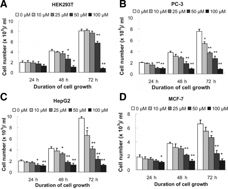
As shown in Fig. 1, after 24 h of RSV treatment at a concentration of less than 50 μM, the cell growth status did not signifycantly change. However, 48 h of treatment of 25 μM RSV led to significant growth inhibition in PC-3 and HepG2 cells, but not in MCF-7 cells. The growth of PC-3 and HepG2 cells was significantly inhibited by 10 μM or 25 μM RSV after 72 h of treatment. However, RSV treatment at a concentration less than 25 μM was ineffective in MCF-7 cells. In addition, 50 μM RSV showed significant inhibitory effects in all three cancer cell lines after 48 and 72 h of treatment, but at this concentration, RSV also exhibited significant growth inhibition of HEK293T cells. One hundred micromolar of RSV showed significant growth inhibitory effects on each of the tested cell lines. The dose of 25 μM RSV was identified as the most efficient dose in inhibiting cell growth of each tested cancer cell line, without affecting HEK293T cell.
Fig. 1.
Effects of RSV on the growth of cells. HEK293T (A), PC-3 (B), HepG2 (C) and MCF-7 (D) cells. Cells (1.0 × 05/ml) were treated with 0–100 μM of RSV for 24–72 h. After harvesting, trypan blue staining was performed for cell counting. Results in bars are presented as mean ± SEM (n = 5); **P ≤ 0.01 and *P ≤ 0.05, compared with control (0 μM RSV), analyzed by one-way ANOVA and LSD post-hoc test.
Resveratrol-mediated up-regulation of the activities and expression of antioxidant enzymes is disproportional in cancer cells
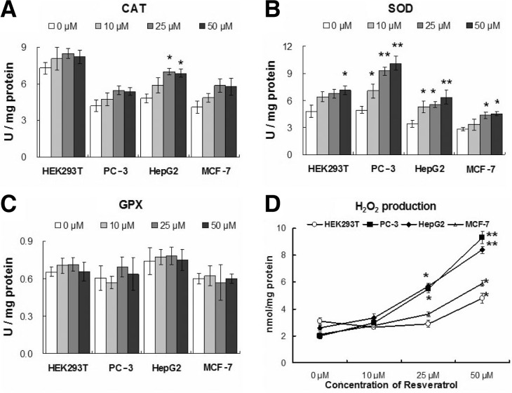
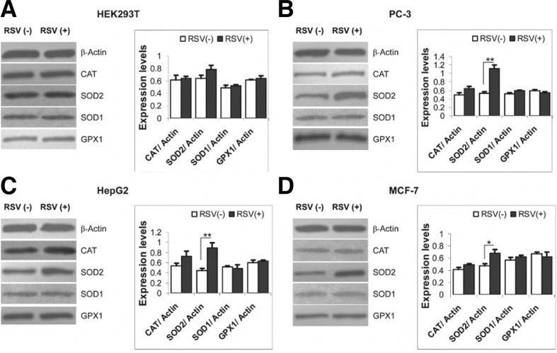
The SOD activity in PC-3 cells was found to be 4.94 unit/mg of protein, which significantly increased with RSV treatment for 72 h of 10 μM, 25 μM and 50 μM by 43%, 88% and 105%, respectively. In HepG2 cells, treatment of 10 μM, 25 μM and 50 μM RSV significantly increased SOD activity by 55%, 63% and 87% respectively compared to SOD activity in untreated HepG2 cells (3.41 unit/mg protein). Treatment with 25 μM and 50 μM RSV for 72 h significantly increased the SOD activity by 54% and 60%, respectively in MCF-7 cells, whereas the activity was 2.82 unit/mg protein in untreated cells. SOD activity in HEK293T cells was 4.8 unit/mg protein. RSV at lower concentrations (10 μM or 25 μM) showed no significant changes in SOD activity, but at high concentrations (≥ 50 μM), there was a 49% increase in SOD activity in HEK293T cells (Fig. 2A). Western blotting showed significantly increased levels of SOD2 in PC-3, HepG2 and MCF-7 cells (by 3-fold, 2.5-fold, and 1.5-fold, respectively) with treatment of 25 μM RSV for 72 h, but the effect on HEK293T cells was non-significant. The difference in the expression level of SOD1 in cells either treated or untreated with RSV was not significant (Fig. 3). CAT activity in PC-3, HepG2, MCF-7, and HEK293T cells was 4.20, 4.82, 4.08, and 7.28 unit/mg protein, respectively. The activity increased to some extent in each of the tested cell lines after RSV treatment for 72 h, and the effect in HepG2 cells was significant. In HepG2 cells, treatment with 25 μM or 50 μM RSV increased CAT activity by 45% and 42%, respectively. However the effect of RSV treatment on the CAT activity in other cells was non-significant (Fig. 2B). Western blot analyses did not show any significant changes in CAT protein expression in any cell lines treated with RSV, although a marginal increase was observed in the cancer cells treated with RSV (Fig. 3). The effect of RSV on GPX activity in the different cell lines is presented in Fig. 2C. In HEK293T, PC-3, HepG2, and MCF-7 cells, GPX activity was 0.65, 0.60, 0.74, and 0.60 unit/mg protein, respectively. Treatment with 10–50 μM RSV for 72 h showed no significant change in GPX activity. Consistent with this data, changes in GPX1 protein levels were minimal in RSV-treated and untreated cells (Fig. 3).
Fig. 2.
Effect of RSV treatment for 72 h on antioxidant enzyme activity. The effect of 72 h of RSV treatment on the activity of catalase (A), superoxide dismutase (B), and glutathione peroxidase (C) in PC-3, HepG2, MCF-7 and HEK293T cells. (D) presents the effect of RSV treatment on the production of H2O2 in PC-3, HepG2, MCF-7 and HEK293T cells. Results are presented as mean ± SEM (n = 5); **P ≤ 0.01 and *P ≤ 0.05, compared with control (0 μM RSV), analyzed by one-way ANOVA and LSD post-hoc test.
Fig. 3.
Effect of RSV treatment on the protein expression levels of antioxidant enzymes. The effect of RSV treatment (25 μM) for 72 h on the expression level of superoxide dismutase, catalase, and glutathione peroxidase in HEK293T (A), PC-3 (B), HepG2 (C), and MCF-7 (D) cells. Protein expression levels were determined by Western blot analyses. Expression levels were quantified by ImageJ software. Results are mean ± SEM (n = 3); RSV(−): non-treated; RSV(+): treated; **P ≤ 0.01 and *P ≤ 0.05, analyzed by one-way ANOVA and LSD post-hoc test.
Resveratrol induces hydrogen peroxide production in cancer cells
RSV showed significant effects on the production of H2O2 in cancer cells (Fig. 2D). Treatment of 10 μM RSV for 72 h slightly increased the H2O2 contents in cancer cells, meanwhile treatment with 25 μM RSV significantly increased H2O2 production in PC-3 and HepG2 cells (by 2.5-fold). In addition, a slight increase in H2O2 production was also noticed in MCF-7 cells. However, at these concentrations (10–25 μM), RSV had the opposite effect on HEK293T cells, in that it caused a decrease in H2O2 production. RSV concentrations at 50 μM resulted in a slight effect on H2O2 production in HEK293T and cancer cells. It is worth noting that the production of H2O2 in cancer cells was in accordance with the activity of SOD enzyme (Fig. 2A).
Resveratrol induces cancer cell apoptosis
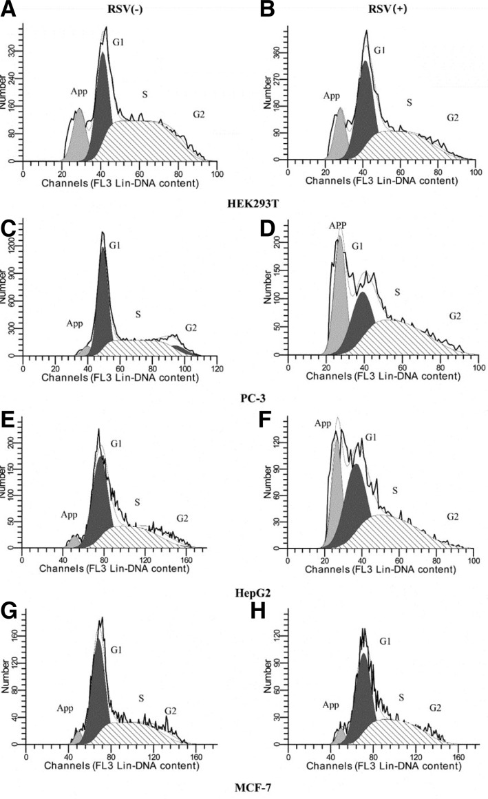
Flow cytometric analysis revealed that the percentage of apoptotic cells increased in cancer cell lines, especially in PC-3 and HepG2 cells (Fig. 4). The percentage of apoptotic cells was 4.40% in untreated PC-3 cells, and increased by nearly 7-fold (29.11%) in RSV-treated PC-3 cells. In untreated PC-3 cells, 50.72% cells were in G1 phase, 39.71% were in S phase and 9% were in G2 phase. On the other hand, in RSV-treated PC-3 cells, 41.33%, 58.66% and 0.01% of cells were in the G1, S and G2 phases, respectively. Similar to PC-3 cells, the percentage of apoptotic HepG2 cells was nearly 7-fold higher in RSV-treated HepG2 cells (22.89%) than that in untreated HepG2 cells (3.70%). In this case, in untreated HepG2 cells, 53.5% cells were in G1 phase, 39.89% were in S phase and 6.61% were in G2 phase, whereas, in RSV-treated HepG2 cells, 50.52% cells were in G1 phase and 49.48% in S phase, but no cells were in G2 phase. However RSV showed minimal effects on the percentage of apoptotic cells in MCF-7 cell line. The percentage of apoptotic cells was 3.60% in untreated MCF-7 cells that increased slightly (4.80%) in RSV-treated MCF-7 cells. In untreated MCF-7 cells, 53.02% were in G1, 42.04% were in S and 4.94% were in G2 phase, whereas, in RSV-treated MCF-7 cells, 53.73% cells were in G1, 46.27% in S phase, but no cells were in G2 phase. Unlike PC-3 and HepG2 cells, the percentage of apoptotic cells did not significantly increase after RSV treatment of MCF-7 cells. Treatment with 25 μM RSV for 72 h resulted in no change in the percentage of apoptotic cells, nor cell cycle distribution in HEK293T cells (the percentage of apoptotic cells was 14.48% in non-treated and 14.54% in the treated cells).
Fig. 4.
Flow cytometric analysis of apoptosis in RSV-treated cells. Cells were either left untreated or were treated with RSV (25 μM) for 72 h. The percentage of apoptotic cells was 14.48% in untreated HEK293T cells (A) and 14.54% in RSV-treated HEK293T cells (B); 4.40% in untreated PC-3 cells (C) and 29.11% in RSV-treated PC-3 cells (D); 3.70% in untreated HepG2 cells (E) and 22.89% in RSV-treated HepG2 cells (F); 3.60% in untreated MCF-7 cells (G) and 4.80% in RSV-treated MCF-7 cells (H). G1, G2 and S represent the stages of cell cycle.
DISCUSSION
It has been postulated that either decreased expression or lower activity of antioxidant enzymes is partially responsible for carcinogenesis (Arsova-Sarafinovska et al., 2009; De Craemer et al., 1993; Elchuri et al., 2005; Jeon et al., 2007; Kasapović et al., 2008; Khan et al., 2010; Sharma et al., 2009). Consequently, up-regulation of the expression of antioxidant enzymes or enhancement of their activities has been proposed as an effective strategy for both cancer prevention and therapy. For example, progestin induction of catalase activity has proven to be effective against breast cancer (Petit et al., 2009). Genistein, an anticancer isoflavonoid, was shown to up-regulate SOD2 and catalase expression in PC-3 and DU145 prostate cancer cells (Park et al., 2010). Taurine was found to increase the expression of SOD, GPX and CAT, and was thus effective in the B16F10 melanoma cell line (Yu and Kim, 2009).
RSV has been considered as an effective chemo-preventive agent against different cancers by hindering cancer initiation, promotion, and progression (Huang and Zhu, 2011; Jang et al., 1997). RSV is a natural polyphenol and possesses anti-oxidant potential (Frémont et al., 1999; Kovacic and Somanathan, 2010; Miller and Rice-Evans, 1995). It is therefore expected that RSV itself may serve as an antioxidant. However, the effect of RSV on the expression and activity of antioxidant enzymes in different cancers is controversial. This study was conducted specifically to dissect the mechanisms of RSV’s action on the anti-oxidative response in different cancers. We demonstrated that both the expression and enzymatic activity of SOD2 were significantly up-regulated by RSV in PC-3, HepG2 and MCF-7 cells. However, RSV only moderately up-regulated the expression levels and activities of CAT and SOD1, and showed no effect on the expression and activity of GPX1 in all tested cancer cells. More importantly, under low concentrations, the effects of RSV on the expression and activity of these enzymes in the non-cancerous HEK293T cells were marginal. This demonstrates that the antioxidative action of RSV is cancer-specific, and SOD2 is more responsive to RSV than other antioxidant enzymes.
Superoxide dismutase (SOD) is a class of enzymes that catalyzes the dismutation of superoxide into H2O2 and O2. Subsequently, CAT and GPX catalyze the decomposition of H2O2 to H2O and O2 (Khan et al., 2010). These consecutive reactions ensure that the cells effectively deal with oxidative stress, derived from reactive oxygen species. We demonstrated that although RSV simultaneously up-regulated SOD, CAT and GPX in different cancer cells, the up-regulation was disproportional to the dramatic increase of SOD expression and marginal elevation of CAT and GPX expression, as well as their enzymatic activities. The net result of this disproportional up-regulation was accumulation of H2O2. There are two SODs with different subcellular locations; namely SOD1 (also called Cu/Zn-SOD) and SOD2 (also known as Mn-SOD), which are located in the cytosol and mitochondria, respectively. Thus, it is expected that the dramatic up-regulation of SOD2 and the moderate elevation of CAT and GPX would result in the elevation of H2O2 concentrations in the mitochondria. H2O2 is not only a ‘reactive oxygen species’, but also an important signaling molecule (Giorgio et al., 2007). Multiple lines of evidence have indicated that mitochondrial H2O2 is a direct and potent apoptosis inducer (Giorgio et al., 2005; Kowaltowski et al., 2001). Consistent with the report that the up-regulation of SOD2 is important in the mechanism of RSV action in other human cells (Robb et al., 2008), we demonstrated that the treatment of cancer cells by RSV results in apoptosis, presumably due to the differentially regulated anti-oxidative enzymes. However, we only found this increase in apoptosis in the PC-3 and HepG2 cells when cells were treated with RSV, and this effect was not seen in MCF-7 cells. This indicates the pro-apoptotic effect of RSV is only specific to certain cancer cells. Therefore, we speculate that in MCF-7 cells, RSV induces growth inhibition and cell death through a non-apoptotic pathway. Indeed, previous studies have shown that RSV induces multiple pathways of MCF-7 cell death, including autophagy (Scarlatti et al., 2008).
In the present study, we used HEK293T as a control cell line to distinguish the effects of RSV on cancer cells from that on non-cancerous cells. We found that RSV, when used at low concentrations, inhibited the growth of multiple human cancer cell lines including prostate cancer (PC-3), hepatic cancer (HepG2) and breast cancer (MCF-7) cells, but without affecting the growth of the non-cancerous HEK293T cells. We also noticed that compared to CAT and GPX, SOD2 protein levels, as well as its enzymatic activity, were significantly up-regulated in all cancer cell lines, however in non-cancerous cells, these enzymes were all up-regulated in a relatively proportional manner. More importantly, no increase in apoptosis rate was observed in the non-cancerous cells. However, at a higher concentration (≥ 50 μM), RSV also exhibited an inhibitory effect on non-cancerous cell growth. Consistent with the findings from clinical trials (Patel et al., 2011; Scott et al., 2012), this suggests that higher doses of RSV may lead to adverse side effects. Although RSV was recently evaluated for safety clinical trials, and was found to be safe and reasonably well-tolerated at doses of up to 5 g/day for healthy adult males with a body weight of around 70 kg (Patel et al., 2011), we still caution that when RSV is used as either a preventive or therapeutic agent, the dosage should always be carefully considered. Further studies are recommended to investigate the mechanism underlying the up-regulation of SOD2 in cancer cells by RSV. In addition, more clinical investigations of RSV in regard to its safety as an anti-cancer drug, are necessary.
Acknowledgments
This work was supported by National Basic Research Program of China (No. 2011CB910700-704) and the Open-End Fund for the Valuable and Precision Instruments of Central South University (No. CSUZC2012008). We are thankful to Chinese Scholarship Council for providing Chinese Government Scholarship to Md. Asaduzzaman Khan.
REFERENCES
- Arsova-Sarafinovska Z, Eken A, Matevska N, Erdem O, Sayal A, Savaser A, Banev S, Petrovski D, Dzikova S, Georgiev V, et al. Increased oxidative/nitrosative stress and decreased antioxidant enzyme activities in prostate cancer. Clin Biochem. 2009;42:1228–1235. doi: 10.1016/j.clinbiochem.2009.05.009. [DOI] [PubMed] [Google Scholar]
- Bae S, Lee EM, Cha HJ, Kim K, Yoon Y, Lee H, Kim J, Kim YJ, Lee HG, Jeung HK, et al. Resveratrol alters microRNA expression profiles in A549 human non-small cell lung cancer cells. Mol. Cells. 2011;32:243–249. doi: 10.1007/s10059-011-1037-z. [DOI] [PMC free article] [PubMed] [Google Scholar]
- Benitez DA, Pozo-Guisado E, Alvarez-Barrientos A, Fernandez-Salguero PM, Castellón EA. Mechanisms involved in resveratrol-induced apoptosis and cell cycle arrest in prostate cancer-derived cell lines. J Androl. 2007;28:282–293. doi: 10.2164/jandrol.106.000968. [DOI] [PubMed] [Google Scholar]
- Bishayee A. Cancer prevention and treatment with resveratrol: from rodent studies to clinical trials. Cancer Prev. Res. (Phila) 2009;2:409–418. doi: 10.1158/1940-6207.CAPR-08-0160. [DOI] [PubMed] [Google Scholar]
- Bradford MM. A rapid and sensitive method for the quantitation of microgram quantities of protein utilizing the principle of protein-dye binding. Anal Biochem. 1976;72:248–254. doi: 10.1006/abio.1976.9999. [DOI] [PubMed] [Google Scholar]
- Carbó N, Costelli P, Baccino FM, López-Soriano FJ, Argilés JM. Resveratrol, a natural product present in wine, decreases tumour growth in a rat tumour model. Biochem Biophys Res Commun. 1999;254:739–743. doi: 10.1006/bbrc.1998.9916. [DOI] [PubMed] [Google Scholar]
- Chen Y, Tseng SH, Lai HS, Chen WJ. Resveratrol-induced cellular apoptosis and cell cycle arrest in neuroblastoma cells and antitumor effects on neuroblastoma in mice. Surgery. 2004;136:57–66. doi: 10.1016/j.surg.2004.01.017. [DOI] [PubMed] [Google Scholar]
- Chung-man Ho J, Zheng S, Comhair SA, Farver C, Erzurum SC. Differential expression of manganese superoxide dismutase and catalase in lung cancer. Cancer Res. 2001;61:8578–8585. [PubMed] [Google Scholar]
- De Craemer D, Pauwels M, Hautekeete M, Roels F. Alterations of hepatocellular peroxisomes in patients with cancer. Catalase cytochemistry and morphometry. Cancer. 1993;71:3851–3858. doi: 10.1002/1097-0142(19930615)71:12<3851::aid-cncr2820711210>3.0.co;2-l. [DOI] [PubMed] [Google Scholar]
- Elchuri S, Oberley TD, Qi W, Eisenstein RS, Jackson Roberts L, Van Remmen H, Epstein CJ, Huang TT. CuZnSOD deficiency leads to persistent and widespread oxidative damage and hepatocarcinogenesis later in life. Oncogene. 2005;24:367–380. doi: 10.1038/sj.onc.1208207. [DOI] [PubMed] [Google Scholar]
- Frémont L, Belguendouz L, Delpal S. Antioxidant activity of resveratrol and alcohol-free wine polyphenols related to LDL oxidation and polyunsaturated fatty acids. Life Sci. 1999;64:2511–2521. doi: 10.1016/s0024-3205(99)00209-x. [DOI] [PubMed] [Google Scholar]
- Giorgio M, Migliaccio E, Orsini F, Paolucci D, Moroni M, Contursi C, Pelliccia G, Luzi L, Minucci S, Marcaccio M, et al. Electron transfer between cytochrome c and p66Shc generates reactive oxygen species that trigger mitochondrial apoptosis. Cell. 2005;122:221–233. doi: 10.1016/j.cell.2005.05.011. [DOI] [PubMed] [Google Scholar]
- Giorgio M, Trinei M, Migliaccio E, Pelicci PG. Hydrogen peroxide: a metabolic by-product or a common mediator of ageing signals? Nat Rev Mol Cell Biol. 2007;8:722–728. doi: 10.1038/nrm2240. [DOI] [PubMed] [Google Scholar]
- Góth L. A simple method for determination of serum catalase activity and revision of reference range. Clin. Chim. Acta. 1991;196:143–151. doi: 10.1016/0009-8981(91)90067-m. [DOI] [PubMed] [Google Scholar]
- Graf E, Penniston JT. Method for determination of hydrogen peroxide, with its application illustrated by glucose assay. Clin Chem. 1980;26:658–660. [PubMed] [Google Scholar]
- Harikumar KB, Aggarwal BB. Resveratrol: a multi-targeted agent for age-associated chronic diseases. Cell Cycle. 2008;7:1020–1035. doi: 10.4161/cc.7.8.5740. [DOI] [PubMed] [Google Scholar]
- Huang X, Zhu HL. Resveratrol and its analogues: promising antitumor agents. Anticancer Agents Med Chem. 2011;11:479–490. doi: 10.2174/187152011795677427. [DOI] [PubMed] [Google Scholar]
- Jang M, Cai L, Udeani GO, Slowing KV, Thomas CF, Beecher CW, Fong HH, Farnsworth NR, Kinghorn AD, Mehta RG, et al. Cancer chemopreventive activity of resveratrol, a natural product derived from grapes. Science. 1997;275:218–220. doi: 10.1126/science.275.5297.218. [DOI] [PubMed] [Google Scholar]
- Jeon SH, Jae-Hoon Park JH, Chang SG. Expression of antioxidant enzymes (catalase, superoxide dismutase, and glutathione peroxidase) in human bladder cancer. Korean J Urol. 2007;48:921–926. [Google Scholar]
- Kasapović J, Pejić S, Todorović A, Stojiljković V, Pajović SB. Antioxidant status and lipid peroxidation in the blood of breast cancer patients of different ages. Cell Biochem Funct. 2008;26:723–730. doi: 10.1002/cbf.1499. [DOI] [PubMed] [Google Scholar]
- Khan MA, Tania M, Zhang DZ, Chen HC. Antioxidant enzymes and cancer. Chin J Cancer Res. 2010;22:87–92. [Google Scholar]
- Klaunig JE, Kamendulis LM. The role of oxidative stress in carcinogenesis. Annu Rev Pharmacol Toxicol. 2004;44:239–267. doi: 10.1146/annurev.pharmtox.44.101802.121851. [DOI] [PubMed] [Google Scholar]
- Kovacic P, Somanathan R. Multifaceted approach to resveratrol bioactivity: focus on antioxidant action, cell signaling and safety. Oxid Med Cell Longev. 2010;3:86–100. doi: 10.4161/oxim.3.2.3. [DOI] [PMC free article] [PubMed] [Google Scholar]
- Kowaltowski AJ, Castilho RF, Vercesi AE. Mitochondrial permeability transition and oxidative stress. FEBS Lett. 2001;495:12–15. doi: 10.1016/s0014-5793(01)02316-x. [DOI] [PubMed] [Google Scholar]
- Miller NJ, Rice-Evans CA. Antioxidant activity of resveratrol in red wine. Clin Chem. 1995;41:1789. [PubMed] [Google Scholar]
- Mo W, Xu X, Xu L, Wang F, Ke A, Wang X, Guo C. Resveratrol inhibits proliferation and induces apoptosis through the hedgehog signaling pathway in pancreatic cancer cell. Pancreatology. 2012;11:601–609. doi: 10.1159/000333542. [DOI] [PubMed] [Google Scholar]
- Muqbil I, Beck FW, Bao B, Sarkar FH, Mohammad RM, Hadi SM, Azmi AS. Old wine in a new bottle: the warburg effect and anticancer mechanisms of resveratrol. Curr Pharm Des. 2012;18:1645–1654. doi: 10.2174/138161212799958567. [DOI] [PubMed] [Google Scholar]
- Nakata R, Takahashi S, Inoue H. Recent advances in the study on resveratrol. Biol Pharm Bull. 2012;35:273–279. doi: 10.1248/bpb.35.273. [DOI] [PubMed] [Google Scholar]
- Park CE, Yun H, Lee EB, Min BI, Bae H, Choe W, Kang I, Kim SS, Ha J. The antioxidant effects of genistein are associated with AMP-activated protein kinase activation and PTEN induction in prostate cancer cells. J. Med. Food. 2010;13:815–820. doi: 10.1089/jmf.2009.1359. [DOI] [PubMed] [Google Scholar]
- Park SJ, Ahmad F, Philp A, Baar K, Williams T, Luo H, Ke H, Rehmann H, Taussig R, Brown AL, et al. Resveratrol ameliorates aging-related metabolic phenotypes by inhibiting cAMP phosphodiesterases. Cell. 2012;148:421–433. doi: 10.1016/j.cell.2012.01.017. [DOI] [PMC free article] [PubMed] [Google Scholar]
- Patel KR, Scott E, Brown VA, Gescher AJ, Steward WP, Brown K. Clinical trials of resveratrol. Ann N Y Acad Sci. 2011;1215:161–169. doi: 10.1111/j.1749-6632.2010.05853.x. [DOI] [PubMed] [Google Scholar]
- Petit E, Courtin A, Kloosterboer HJ, Rostène W, Forgez P, Gompel A. Progestins induce catalase activities in breast cancer cells through PRB isoform: correlation with cell growth inhibition. J Steroid Biochem Mol Biol. 2009;115:153–160. doi: 10.1016/j.jsbmb.2009.04.002. [DOI] [PubMed] [Google Scholar]
- Robb EL, Page MM, Wiens BE, Stuart JA. Molecular mechanisms of oxidative stress resistance induced by resveratrol: Specific and progressive induction of MnSOD. Biochem Biophys Res Commun. 2008;367:406–412. doi: 10.1016/j.bbrc.2007.12.138. [DOI] [PubMed] [Google Scholar]
- Rotruck JT, Pope AL, Ganther HE, Swanson AB, Hafeman DG, Hoekstra WG. Selenium: biochemical role as a component of glutathione peroxidase. Science. 1973;179:588–590. doi: 10.1126/science.179.4073.588. [DOI] [PubMed] [Google Scholar]
- Scarlatti F, Maffei R, Beau I, Codogno P, Ghidoni R. Role of non-canonical Beclin 1-independent autophagy in cell death induced by resveratrol in human breast cancer cells. Cell Death Differ. 2008;15:1318–1329. doi: 10.1038/cdd.2008.51. [DOI] [PubMed] [Google Scholar]
- Scott E, Steward WP, Gescher AJ, Brown K. Resveratrol in human cancer chemoprevention--choosing the ‘right’ dose. Mol Nutr Food Res. 2012;56:7–13. doi: 10.1002/mnfr.201100400. [DOI] [PubMed] [Google Scholar]
- Sengottuvelan M, Deeptha K, Nalini N. Resveratrol ameliorates DNA damage, prooxidant and antioxidant imbalance in 1, 2-dimethylhydrazine induced rat colon carcinogenesis. Chem Biol Interact. 2009;181:193–201. doi: 10.1016/j.cbi.2009.06.004. [DOI] [PubMed] [Google Scholar]
- Sharma A, Tripathi M, Satyam A, Kumar L. Study of antioxidant levels in patients with multiple myeloma. Leuk Lymphoma. 2009;50:809–815. doi: 10.1080/10428190902802323. [DOI] [PubMed] [Google Scholar]
- Sun YI, Oberley LW, Li Y. A simple method for clinical assay of superoxide dismutase. Clin Chem. 1988;34:497–500. [PubMed] [Google Scholar]
- Yu J, Kim AK. Effect of taurine on antioxidant enzyme system in B16F10 melanoma cells. Adv Exp Med Biol. 2009;643:491–499. doi: 10.1007/978-0-387-75681-3_51. [DOI] [PubMed] [Google Scholar]
- Zhou HB, Chen JJ, Wang WX, Cai JT, Du Q. Anticancer activity of resveratrol on implanted human primary gastric carcinoma cells in nude mice. World J Gastroenterol. 2005;11:280–284. doi: 10.3748/wjg.v11.i2.280. [DOI] [PMC free article] [PubMed] [Google Scholar]


