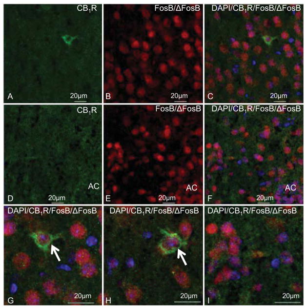
Figure 5.
Representative images showing CB1R-ir (green), FosB/ΔFosB-ir (red) and DAPI (blue) in the caudate-putamen and nucleus accumbens of mice that received repeated THC treatment. CB1R-ir fibers and puncta were seen in the caudate-putamen (A) and nucleus accumbens (B) and CB1R-ir cells were occasionally found in the caudate-putamen (A). FosB/ΔFosB-ir was localized to nuclei of cells in the caudate-putamen (B, C) and nucleus accumbens (E, F). FosB/ΔFosB-ir and DAPI were seen in a subset of cell nuclei that were surrounded by CB1R-ir puncta in the caudate-putamen (C, G, H) and nucleus accumbens (F, I). CB1R-ir was also seen in cells that contained FosB/ΔFosB-ir nuclei in the caudate-putamen (indicated by arrows in G, H). AC: anterior commissure

