Abstract
A case of posterior dislocation of the long head of biceps tendon, a rare occurrence following traumatic anterior glenohumeral dislocation, along with complete rotator cuff rupture and large haemarthrosis is presented with imaging and intra-operative findings. The interposed tendon prevented complete reduction. Appearances at MRI were diagnostic and directed the surgical approach.
Keywords: Posterior dislocation long head biceps tendon, MRI, traumatic anterior glenohumeral dislocation, failed glenohumeral reduction
CASE REPORT
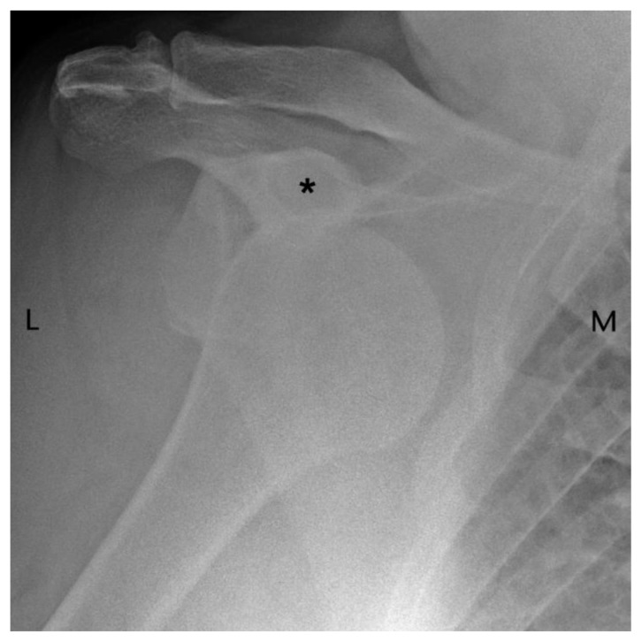
A 69 year old male on warfarin for atrial fibrillation, presented to the Emergency Department after falling and injuring his previously asymptomatic right shoulder. Clinically, there was marked soft tissue swelling and no neurovascular compromise. Initial plain radiographs demonstrated anterior glenohumeral joint dislocation with no apparent fracture (figure 1). His International Normalised Ratio (INR) was 4.8. Several attempts at closed reduction of the shoulder were made, including under general anaesthesia, however, further radiographs showed persistent anterior subluxation thought to be due to a large haemarthrosis.
Figure 1.
69 year old male presenting with traumatic anterior glenohumeral dislocation. Findings: the humeral head is seen to lie inferior to the coracoid process (asterisk) consistent with anterior shoulder dislocation. No definite fracture is seen. Technique: AP radiograph.
Imaging Findings
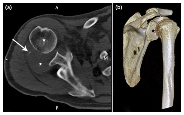
Unenhanced helical CT at day 1 post injury (Brilliance CT 64-channel, Philips, Surrey, UK) (figure 2) demonstrated anterior subluxation of the humeral head, a tiny avulsion fracture possibly from the greater tuberosity, extensive haemarthrosis with small fat component and suspected extensive rotator cuff tear.
Figure 2.
69 year old male with traumatic anterior glenohumeral dislocation. CT performed after failed attempts at reduction.
Findings:
a) axial image shows anterior subluxation at the glenohumeral joint, black asterisk marks humeral head. The course of infraspinatus is disrupted by a soft tissue “mass” posteriorly (posterior white asterisk) - a haemarthrosis within which lies a tiny avulsion fracture (arrow).
b) Posterolateral view of a 3D volume reconstruction showing the relationship of the humerus to the glenoid fossa (asterisk).
Technique:
Unenhanced MDCT, 125 mAs, 120kV, slice thickness 1mm, increment 0.5 mm (Brilliance CT 64-channel, Philips, Surrey, UK).
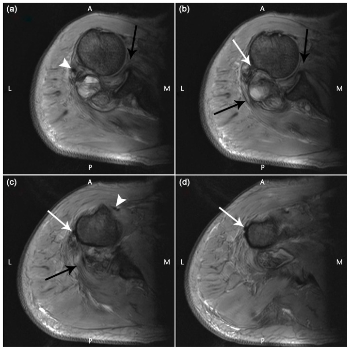
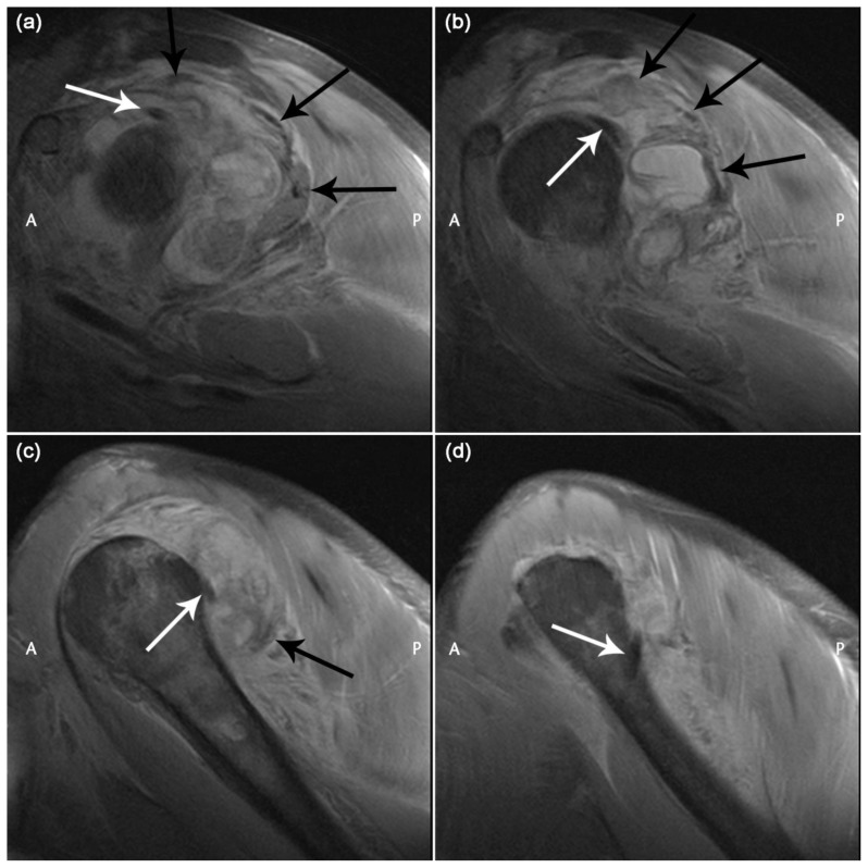
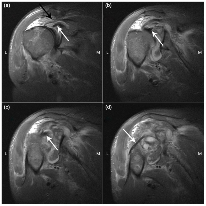
MRI at 1.5T (Magnetom Symphony, Siemens, Erlangen, Germany) was performed at day 3 post injury to look for possible anatomical causes of incomplete reduction and to further assess the integrity of the rotator cuff. This showed refractory anterior subluxation of the humeral head, marrow oedema in the humeral head consistent with microfractures, a large haemarthrosis with multiple fluid-fluid levels and complete full-thickness tears of supraspinatus, infraspinatus and teres minor tendons close to their insertions. There was also a complete full-thickness tear of the subscapularis tendon which took an abnormal course. The bicipital groove was empty and the long head of biceps tendon (LHBT) had dislocated posterolaterally. Axial gradient echo, coronal-oblique STIR and sagittal-oblique proton-density weighted sequences with fat saturation are presented (figure 3 – 5).
Figure 3.
69 year old male with traumatic anterior glenohumeral dislocation with failed reduction after multiple attempts, complete rotator cuff tear, posterior dislocation long head of biceps tendon and haemarthrosis.
Findings:
a) There is an abnormal course of subscapularis with the disrupted tendon insertion folded into the anterior joint space (black arrow). A haemarthrosis with fluid-fluid levels is seen posterior to the joint. The tiny avulsion fracture, (white arrowhead) seen as a small hypointense focus lies posterolaterally.
b) Disrupted subscapularis tendon and teres minor tendon are seen (anterior and posterior black arrows). The long head of biceps tendon has slipped over the greater tuberosity and lies posterior to the humeral head (white arrow).
c) Disrupted teres minor insertion is demonstrated (black arrow). The dislocated long head of biceps tendon lies against the lateral aspect of humerus (white arrow). Short head of biceps can be seen (white arrowhead).
d) The dislocated long head of biceps tendon is lying abnormally laterally at a more inferior level (white arrow).
Technique: Axial gradient echo MRI a–d cranial to caudal
1.5T; TR/TE/Flip angle: 1300ms/27ms/30 degrees; FOV 200; matrix 410 × 512; slice thickness 4 mm.
Figure 5.
69 year old male with traumatic anterior glenohumeral dislocation with failed reduction after multiple attempts, complete rotator cuff tear, posterior dislocation long head of biceps tendon and haemarthrosis.
Findings:
a) The long head of biceps tendon is seen just lateral to its origin (white arrow). The musculotendinous portions of the rotator cuff: supraspinatus, infraspinatus and teres minor are seen in a clockwise direction (black arrows).
b) The posteriorly dislocated long head of biceps tendon (white arrow) is seen. Torn, retracted supraspinatus demonstrated (superior black arrow) while infraspinatus and teres minor are stretched posteriorly due to the haemarthrosis (further black arrows).
c) The posteriorly dislocated long head of biceps tendon is again seen (white arrow). Supraspinatus and infraspinatus are no longer seen. Torn teres minor is demonstrated (black arrow).
d) The long head of biceps tendon is seen wrapping round the humeral shaft to come anteriorly (white arrow). No rotator cuff tendons are now visualised.
Technique: Sagittal oblique proton-density medial to lateral a–d
1.5T; TR/TE: 3570ms/15ms; FOV 160; matrix 512 × 512; slice thickness 4 mm
Management
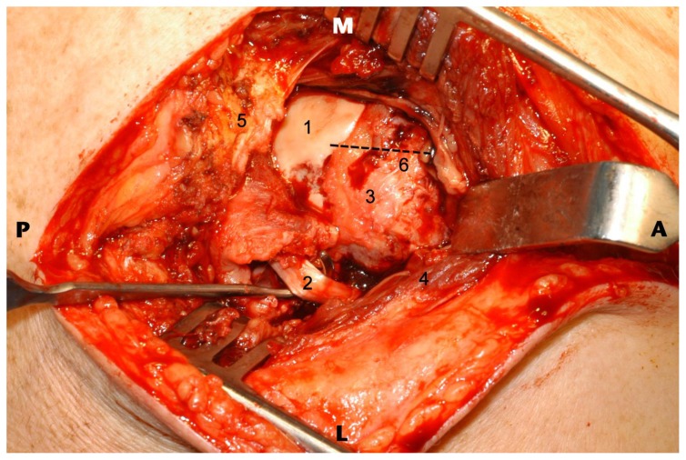
Information provided at MRI prompted a decision to perform an open operation rather than an attempt at an arthroscopic procedure. Surgical reduction was performed through a superior strap approach with deltoid split. Posterior dislocation of the long head of biceps was confirmed with an acute near circumferential avulsion of the rotator cuff. Following removal of haemarthrosis and biceps tendon tenotomy the humeral head reduced into anatomical position. Repair of the rotator cuff including subscapularis, supraspinatus, infraspinatus and teres minor muscles was performed using 4.5 mm TwinFix anchors (Smith and Nephew plc, UK). Posterior displacement of the LHBT and exposure of the superior articular surface of the humeral head due to rotator cuff tear can be seen in the intra-operative image (Figure 6).
Figure 6.
69 year old male following traumatic anterior glenohumeral dislocation with complete rotator cuff tear, posterior dislocation long head of biceps tendon and haemarthrosis at MRI. Intra-operative image seen as if from above, pre-reduction following removal of the haemarthrosis where tenotomy of long head of biceps tendon and rotator cuff repair were subsequently performed.
Findings:
1) The exposed articular surface of the humeral head secondary to rotator cuff tear is seen. The humeral head remains anteriorly translated relative to the glenoid (and long head of biceps origin).
2) Posteriorly displaced long head of biceps tendon is shown by the Langenbeck retractor.
3) Supraspinatus footprint region.
4) Deltoid muscle (reflected during exposure)
5) Acromion is here.
6) Line of the bicipital groove and hence anatomical position of long head of biceps tendon.
Technique: Superior strap approach with deltoid split.
Follow-up
The patient is making progress with intensive physiotherapy and gradually recovering rotator cuff function. There is minimal cosmetic deformity.
DISCUSSION
Anatomy
The LHBT originates from the supraglenoid tubercle of the scapula and superior glenoid labrum. It traverses the rotator interval and descends in the bicipital groove supported and stabilised by the structures of the biceps pulley: the coracohumeral ligament, superior glenohumeral ligament, superior fibres of the subscapularis tendon and the transverse humeral ligament (1, 2).
Etiology and Demographics
Traumatic anterior dislocation of the glenohumeral joint is relatively common. Associated injuries include greater tuberosity fractures, Hill-Sachs and/or bony Bankart defects, various labroligamentous lesions, rotator cuff tears and neurovascular damage. In a previous study where all subjects were assessed by ultrasound, 31.7% of patients with primary traumatic anterior dislocation were shown to have a full-thickness rotator cuff tear (3). A larger more recent study found rotator cuff tears in 10.1% when ultrasound was targeted at clinically suspicious cases (4).
Instability and medial dislocation of LHBT is a well-recognised entity secondary to subscapularis tendon tears and lesions of the biceps pulley. Posterolateral dislocation is a much rarer phenomenon - an exceptional complication following traumatic anterior glenohumeral dislocation. Literature on the subject is scarce and is limited to 8 case reports (5–12). It requires disruption of the structures of the biceps pulley and the posterolateral supporting structures of the shoulder joint with either rupture of supraspinatus and infraspinatus tendons close to their insertions on the greater tuberosity or a displaced fracture of the greater tuberosity.
Clinical and Imaging Findings
The MRI and MR arthrographic findings of medial LHBT subluxation and dislocation are well documented (13, 14).
With respect to posterior dislocation, imaging is extremely useful as clinical testing is not specific and even at open surgery it may not be recognized (10)].
Three previous case reports also highlight the MRI findings (5, 8, 10) while others made the pre-operative diagnosis with conventional (7, 9) or CT arthrography (5, 6) or unenhanced CT (11). Like us, Mullaney et al. performed non-contrast MRI in the immediate post injury period. A displaced greater tuberosity fracture was demonstrated along with disruption of infraspinatus, teres minor and the inferior glenohumeral ligament (8). Strobel et al. report the use of indirect MR arthrography 2 weeks following injury where rupture of supraspinatus and infraspinatus were seen along with partial subscapularis rupture and an undisplaced tuberosity fracture [10] while Bauer et al. used MRI to complement a diagnostic CT-arthrogram (5). In our case there was complete rotator cuff disruption, a tiny avulsion fracture and a large haemarthrosis secondary to warfarinisation. Other MRI findings common to all cases are an empty bicipital groove with LHBT seen displaced posterolateral to the humerus.
Treatment and Prognosis
Tenodesis, detaching LHBT origin from superior labrum and reattaching to humerus below the shoulder, or tenotomy, division of LHBT, is required to allow successful glenohumeral reduction. An element of rotator cuff repair will often be required assuming there is no pre-existing chronic rotator cuff tear with features to contraindicate this. Prognosis will depend on severity and presence of other associated injuries, the nature of any pre-existing pathology and compliance with post-operative physiotherapy.
Differential Diagnosis
There is no differential diagnosis for this condition.
TEACHING POINT
It is important to review the position of the long head of biceps tendon on imaging particularly following traumatic dislocation in the setting of incomplete reduction. Correct identification of this entity directs the surgical approach and procedure and if it is not identified, the interposed tendon will prevent adequate reduction and there will be long term dysfunction.
Figure 4.
69 year old male with traumatic anterior glenohumeral dislocation with failed reduction after multiple attempts, complete rotator cuff tear, posterior dislocation long head of biceps tendon and haemarthrosis.
Findings:
a) The long head of biceps tendon can be seen at its origin on the superior glenoid (white arrow).There is a full-thickness tear of supraspinatus with retraction (black arrow).
b) The intra-articular portion of the long head of biceps tendon is seen (white arrow).
c) The long head of biceps tendon is lying posteriorly with respect to the bicipital groove (white arrow).
d) The long head of biceps tendon can be seen dislocated posterior to the humerus (white arrow).
Technique: Coronal oblique STIR anterior to posterior a–d
1.5T; TR/TE/time to inversion: 4290ms/29ms/130ms; FOV 160; matrix 394 × 512; slice thickness 4 mm
Table 1.
Summary table of posterior dislocation long head of biceps tendon following traumatic anterior shoulder dislocation
| Etiology | Limited cases in literature. Posterior dislocation of long head of biceps tendon always associated with anterior glenohumeral dislocation. Requires disruption of the structures of the biceps pulley and the posterolateral supporting structures of the shoulder joint: either rupture of supraspinatus and infraspinatus tendons close to their insertions or a displaced fracture of the greater tuberosity. |
| Incidence | N/A |
| Gender ratio | Too few cases to comment |
| Age predilection | Too few cases to comment |
| Risk factors | Too few cases to comment |
| Treatment | Tenotomy or Tenodesis of dislocated tendon required to allow successful and complete glenohumeral reduction. A degree of rotator cuff repair usually required. |
| Prognosis | Depends on presence and severity of associated injuries, pre-existing pathology and compliance with post-operative physiotherapy. |
| Findings on imaging | Empty bicipital groove; long head of biceps tendon displaced posterolateral to the humerus; either tear of at least infraspinatus and supraspinatus tendons close to their insertion on the greater tuberosity or displaced greater tuberosity fracture. |
Table 2.
Types of Glenohumeral Dislocation
| Type | Frequency | Plain radiographic appearances |
|---|---|---|
| Anterior | 95% | Humeral head lies in a subcoracoid or subglenoid position on the frontal view or much more rarely subclavicular or intrathoracic; on the scapular Y view or axial view the humeral head is anterior to the glenoid. Associated findings are Hill-Sachs lesion and bony Bankart |
| Posterior | 2–4% | On frontal view: “lightbulb” sign due to humerus being fixed in internal rotation, loss of half-moon overlap between humeral head and glenoid due to lateral displacement, rim sign: >6 mm between medial edge of humeral head and anterior margin of glenoid and the trough-line sign – line paralleling medial humeral head due to impaction fracture anteromedial humeral head i.e. reverse Hill-Sachs. Associated finding is avulsion fracture of lesser tuberosity |
| Inferior (Luxatio erecta) | <1% | The patient’s arm is fixed in abduction; humeral head lies subcoracoid or subglenoid with humeral shaft parallel to spine of scapula on frontal view. Associated findings: fracture acromion, inferior glenoid and greater tuberosity |
| Superior | <1% | Humeral head driven upward through rotator cuff. On frontal view humeral head overlies clavicle and acromion. Associated findings: fracture of clavicle, acromion, coracoid or greater tuberosity |
ACKNOWLEDGEMENTS
Thanks to James Eyland, Medical Photographer for intra-operative photography and help with image preparation.
ABBREVIATIONS
- CT
Computed Tomography
- LHBT
Long head biceps tendon
- MRI
Magnetic Resonance Imaging
REFERENCES
- 1.Nakata W, Katou S, Fujita A, Nakata M, Lefor AT, Sugimoto H. Biceps pulley: normal anatomy and associated lesions at MR arthrography. Radiographics. 2011;31:791–810. doi: 10.1148/rg.313105507. [DOI] [PubMed] [Google Scholar]
- 2.Lee JC, Guy S, Connell D, Saifuddin A, Lambert S. MRI of the rotator interval of the shoulder. Clin Radiol. 2007;62:416–423. doi: 10.1016/j.crad.2006.11.017. [DOI] [PubMed] [Google Scholar]
- 3.Berbig R, Weishaupt D, Prim J, Shahin O. Primary anterior shoulder dislocation and rotator cuff tears. J Shoulder Elbow Surg. 1999;8:220–225. doi: 10.1016/s1058-2746(99)90132-5. [DOI] [PubMed] [Google Scholar]
- 4.Robinson CM, Shur N, Sharpe T, Ray A, Murray IR. Injuries associated with traumatic anterior glenohumeral dislocations. J Bone Joint Surg Am. 2012;94:18–26. doi: 10.2106/JBJS.J.01795. [DOI] [PubMed] [Google Scholar]
- 5.Bauer T, Vuillemin A, Hardy P, Rousselin B. Posterior dislocation of the long head of the biceps tendon: a case report. J Shoulder Elbow Surg. 2005;14:557–558. doi: 10.1016/j.jse.2004.10.013. [DOI] [PubMed] [Google Scholar]
- 6.Freeland AE, Higgins RW. Anterior shoulder dislocation with posterior displacement of the long head of the biceps tendon. Arthrographic findings. A case report. Orthopedics. 1985;8:468–469. doi: 10.3928/0147-7447-19850401-06. [DOI] [PubMed] [Google Scholar]
- 7.Inao S, Hirayama T, Takemitsu Y. Irreducible acute anterior dislocation of the shoulder: interposed bicipital tendon. J Bone Joint Surg Br. 1990;72:1079–1080. doi: 10.1302/0301-620X.72B6.2246296. [DOI] [PubMed] [Google Scholar]
- 8.Mullaney PJ, Bleakney R, Tuchscherer P, Boynton E, White L. Posterior dislocation of the long head of biceps tendon: case report and review of the literature. Skeletal Radiol. 2007;36:779–783. doi: 10.1007/s00256-007-0285-7. [DOI] [PubMed] [Google Scholar]
- 9.Rakofsky M, Arias C, Wagner JJ. Case report 633. Posterior dislocation of the long head of the biceps tendon without fracture of the tuberosity. Skeletal Radiol. 1990;19:532–534. doi: 10.1007/BF00202705. [DOI] [PubMed] [Google Scholar]
- 10.Strobel K, Treumann TC, Allgayer B. Posterior entrapment of the long biceps tendon after traumatic shoulder dislocation: findings on MR imaging. AJR Am J Roentgenol. 2002;178:238–239. doi: 10.2214/ajr.178.1.1780238. [DOI] [PubMed] [Google Scholar]
- 11.Day MS, Epstein DM, Young BH, Jazrawi LM. Irreducible anterior and posterior dislocation of the shoulder due to incarceration of the biceps tendon. Int J Shoulder Surg. 2010;4:83–85. doi: 10.4103/0973-6042.76970. [DOI] [PMC free article] [PubMed] [Google Scholar]
- 12.Janecki CJ, Barnett DC. Fracture-dislocation of the shoulder with biceps tendon interposition. J Bone Joint Surg Am. 1979;61:142–143. [PubMed] [Google Scholar]
- 13.Cervilla V, Schweitzer ME, Ho C, Motta A, Kerr R, Resnick D. Medial dislocation of the biceps brachii tendon: appearance at MR imaging. Radiology. 1991;180:523–526. doi: 10.1148/radiology.180.2.2068322. [DOI] [PubMed] [Google Scholar]
- 14.Morag Y, Jacobson JA, Shields G, et al. MR arthrography of rotator interval, long head of the biceps brachii, and biceps pulley of the shoulder. Radiology. 2005;235:21–30. doi: 10.1148/radiol.2351031455. [DOI] [PubMed] [Google Scholar]


