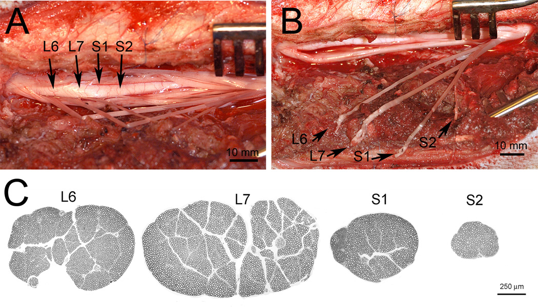
Figure 1.
Surgical display of left sided L6, L7, S1, and S2 ventral roots after a hemi-laminectomy and dural opening (A). The identification of the ventral roots were based on vertebral landmarks and characteristic differences in caliber for individual lumbosacral ventral roots. Sutures have been attached to each ventral root to assist with the identification and presentation. The L6-S2 ventral roots have been avulsed from the surface of the spinal cord (B). Note fine rootlets at the tip of the avulsed ventral root. Transverse sections of the L6, L7, S1, and S2 ventral roots harvested from the intact contralateral side at the end of experiment (C). Note the characteristic large size of the L6 and L7 ventral roots compared to the much smaller S1 and S2 ventral roots. Also note that ventral roots exhibit a very thin epineurium and that regions of densely packed myelinated fibers are separated by streaks of connective tissues.

