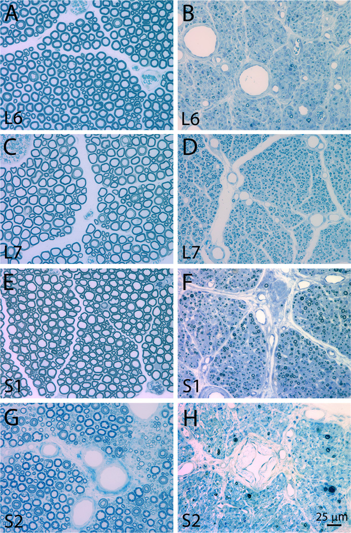
Figure 6.
Photomicrographs of representative plastic embedded and toluidine blue stained L6-S2 ventral roots at 6 months after a unilateral L6-S2 VRA injury. The intact L6, L7, S1, and S2 ventral roots are presented in A, C, E, and G, respectively. The avulsed L6, L7, S1, and S2 are presented in B, B, F, and H, respectively. Note the presence of a large number of small myelinated axons in the avulsed ventral roots.

