History
In December 1961, Letournel [10] published his original series on acetabular fracture classification and operative management. Before that time, acetabular fractures were classified as those associated with posterior hip dislocation and those associated with central hip dislocation. Many were treated without surgery, resulting in poor articular congruity of the hip. Citing an increasing incidence of these injuries with the increasing number of automobiles on the road and “disappointment with the closed treatment of these fractures, we [Judet et al.] decided to try open reduction” [8]. Their series included 173 patients, 129 of whom were treated surgically. Pelvic radiographs were correlated with surgical findings. The anterior and posterior columns of the acetabulum were defined based on the radiographic projections of normal and fractured acetabula on three views: AP pelvic film and internal and external 45° obliques (Judet views). Seven acetabular fracture patterns were described, divided into elementary and associated patterns.
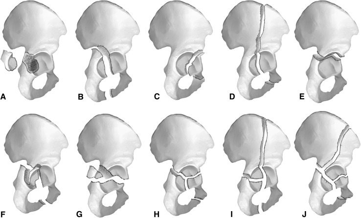
In 1980, using radiographic and surgical data from 647 acetabular fractures, of which 582 had undergone surgical fixation, Letournel [11] confirmed and updated his original description [8]. He divided acetabular fractures into 10 subtypes, five elementary patterns and five associated patterns (including more than one elementary pattern). Elementary patterns included posterior wall fractures, posterior column, anterior column, anterior wall, and transverse fracture patterns. Associated patterns include T-shaped, posterior column and posterior wall, transverse and posterior wall, anterior and hemitransverse, and fractures of both columns [11] (Fig. 1).
Fig. 1A–J.
Acetabular fracture patterns as described by Letournel. The simple patterns are posterior wall (A), posterior column (B), anterior wall (C), anterior column (D), and transverse (E). The associated patterns are posterior column with a posterior wall (F), transverse posterior wall (G), T-style (H), anterior column posterior hemitransverse (I), and both columns (J) [2]. Reprinted with permission, University of Washington Creative Services © 2013. All Rights Reserved.
Purpose
Letournel’s [11] classification system is an anatomic and radiographic description of fracture patterns correlated with operative findings. Specific fracture patterns emerged based on mechanism of injury, the vector of injury force application, anatomy of the innominate bone, and its mechanical properties. Despite dividing fractures into 10 subsets, Letournel [11] believed that acetabular fractures represent a spectrum of injury, because incomplete fracture lines and combined fracture patterns were common.
At the time of the original description, acetabular fractures were not commonly treated with open surgery, often leading to pain and posttraumatic arthritis. Letournel found that the quality of reduction of the articular surface improved clinical results and decreased the development of arthritis. He described arthritis rates of 5.4% in anatomically reduced patterns, jumping to 30.7% with imperfect reductions. Their followup ranged from 2 to 21 years with 75.7% very good results. Results correlated to the quality of reduction. Delays of surgical treatment of more than 3 weeks led to decreased ability to achieve satisfactory fracture reduction as determined by plain radiographs, increasing the risk for posttraumatic arthritis.
By treating a large number of acetabular fractures with open surgery, they were able to identify safe surgical approaches to address each fracture pattern. Unfortunately, there is no convenient universal approach that allows equally easy access to both columns of the acetabulum. The Kocher-Langenbeck approach is used for posterior wall, posterior column, and associated transverse patterns with central or posterior femoral head dislocation, whereas the ilioinguinal approach is used for anterior column, anterior wall, and anterior fractures with a posterior hemitransverse element. Surgeons treating transverse, transverse posterior wall, T-shaped, and both column patterns have a choice of either or both approaches. The surgeon must select the appropriate approach based on radiographic projection of the fracture before direct visualization of the injury in the operating room. This requires the fracture pattern to be fully understood before surgery, thus justifying the need for a detailed and accurate classification system.
In addition to the classification system, Letournel [11] developed and outlined principles for acetabular fracture reconstruction. He believed if both acetabular columns were restored anatomically, the acetabulum would also be restored, often without seeing the articular surface of the joint, assuming no incarcerated or impacted fragments exist. Dislocations were to be reduced after articular reduction was performed. He noted the importance of not accepting imperfect reductions when multiple fracture lines were present, because even slight displacements would be cumulative and result in an unacceptable final reduction. Lastly, after reduction and stabilization, the hip should be ranged through an arc of motion, assessing for hardware prominence and stability.
Validation
The integration of CT scans into common medical care of trauma patients has increased our ability to detect fractures of the quadrilateral space, sacrum, acetabular roof, and posterior acetabular lip; to identify loose bodies in the hip; and provides a more complete understanding of acetabular fracture characteristics [6, 20]. Letournel’s original classification system was based on plain radiographs, and there is debate whether addition of CT scans improves reliability of his classification system [4, 7, 15, 23].
Accurate and consistent classification of acetabular fractures is essential for surgical planning. Visutipol et al. [23] found that addition of a three-dimensional (3-D) CT scan did not improve the inter- or intraobserver reliability of Letournel’s classification with kappa values of 0.42 for plain films and 0.44 for 3-D CT evaluation. Other studies have shown improvements in reliability with implementation of two-dimensional (2-D) and 3-D CT scans. Hüfner et al. [7] showed improvement from 30% to 65% in correctly identifying fracture patterns by surgeons with limited experience when 3-D CT scans were used in addition to plain films and 2-D scans; however, experienced surgeons (> 10 years operative experience) were able to correctly diagnose fracture patterns with 2-D scans 76% of the time. Ohashi et al. [15] showed increased interobserver reliability with the addition of 2-D and 3-D scans. Garrett et al. [14] found the percent of accurate classification and interobserver reliability improved in both junior and senior residents when 3-D CT scans were used in addition to 2-D scans.
Experience plays a role in classification accuracy as well. Beaulé et al. [1] evaluated the inter- and intraobserver reliability of Letournel’s classification among three groups with varying levels of experience. They found kappa values, with and without CT scans, to be 0.70 and 0.74 for highly experienced, 0.71 and 0.69 for experienced, and 0.51 and 0.51 for inexperienced reviewers, respectively. The overall agreement between radiographic diagnosis and surgical findings was 74%. Hüfner et al. [7] found inexperienced orthopaedic surgeons accurately applied Letournel’s classification for acetabular fractures in only 11% of cases. In 2009, Prevezas et al. [18] proposed guidelines to increase the reliability and validity of Letournel’s classification. They suggested an algorithm for evaluating pelvic radiographs based first on examination of basic lines (ilioischial, iliopectineal), then further subdivision into Letournel’s 10 fracture patterns based on examination of four anatomic landmarks (teardrop, anterior and posterior acetabular rim, acetabular dome), the integrity of the obturator foramen, the level of fracture lines, and the presence or absence of a spur sign. They found implementation of their algorithm resulted in a kappa increase from 0.536 to 0.683 with a significant improvement for inexperienced orthopaedic surgeons [18].
Limitations
Although Letournel’s classification system provides a framework in which to understand acetabular fractures and plan surgical approaches, it does not address hip stability.
Within elementary and associated fracture patterns, there are many variations. For example, posterior wall acetabular fractures are present in three of 10 fracture patterns. The wall component of these fractures is not uniform with variable fracture sizes and soft tissue/capsular disruptions. Some hips are stable despite posterior wall fractures, whereas others are not. Radiographic and CT studies have evaluated posterior wall fractures and found instability may not be present unless more than 40% of the wall is fractured [3, 8]. These authors suggest nonoperative management to be reasonable if less than 20% to 45% of the joint surface is involved [9, 13, 20]. The integrity of the posterior capsule may be responsible for this retained stability [22]. Others have suggested hip stability can only be assessed with intraoperative fluoroscopy and patients generally do well, with or without surgery, as long as the hip is stable [5, 21]. However, biomechanical studies have found joint reaction forces increase as the size of the posterior wall fracture increases, leading to concern regarding long-term results [16].
In addition, Letournel [10] noted the importance of articular reduction after open reduction and internal fixation of acetabular fractures graded according to d’Aubigne and Postel. This observation has been substantiated by other reports, with many studies confirming that accurate reduction is associated with improved clinical outcomes [2, 14, 19, 24]. Letournel [10] also noted the importance of early operative interventions, within 3 weeks of injury, because the quality of reduction was much improved with early surgery.
Although anatomic fracture pattern is important when considering surgical intervention, many other factors (subchondral impaction, comminution, femoral head injury, intraarticular fracture fragments, etc) contribute to the decision to operate on patients with acetabular fractures. Murphy et al. [14] found associated patterns, local complications, and heterotopic ossification predict posttraumatic arthritis, need for THA, and worse clinical results. Rommens et al. [19] noted subchondral impaction, fracture comminution, and intraarticular fracture fragments negatively impacted Harris hip and Merle d’Aubdigné scores, regardless of articular reduction quality. Zha et al. [24] noted posterior wall comminution and femoral head injury predict worse Merle d’Aubigne-Postel score in elderly patients with acetabular fractures. Briffa et al. [2] found increasing age, delays in surgery, fracture pattern, and poor quality of reduction can predict worse d’Aubigne-Postel scores at 10 years and Lichte et al. [12] found articular reduction, initial displacement, and intraarticular fragments correlate with results at 30 years. Also, increasing body mass index has been found to predict complications after acetabular fracture surgery [17]. No additional acetabular fracture classification system has been developed that is used as frequently as Letournel’s.
Conclusions
Letournel’s acetabular fracture classification system divides injuries into 10 patterns, five elementary and five associated, based on surgical findings and plain radiograph characteristics. It also provides the fundamental description of the two columns of the acetabulum. The type of fracture is important for surgical planning because no single surgical approach allows convenient access to both columns without consequences.
Acetabular fractures are complicated injuries. The inter- and intraobserver reliability increases with surgeon experience. The addition of CT scans, both 2-D and 3-D, generally has been shown to improve the accuracy and reliability of this classification system, especially for the inexperienced surgeon.
Letournel recognized the importance of articular reduction, a principle that is important in terms of the prevention of posttraumatic arthritis, and surgical intervention within 3 weeks of injury. This classification system does not address hip stability and some other factors that predict patient outcomes. Nevertheless, the Letournel classification is the standard by which acetabular fractures are classified, discussed, and approached surgically.
Footnotes
Each author certifies that he or she, or a member of his or her immediate family, has no funding or commercial associations (eg, consultancies, stock ownership, equity interest, patent/licensing arrangements, etc) that might pose a conflict of interest in connection with the submitted article.
All ICMJE Conflict of Interest Forms for authors and Clinical Orthopaedics and Related Research editors and board members are on file with the publication and can be viewed on request.
References
- 1.Beaulé PE, Dorey FJ, Matta JM. Letournel classification for acetabular fractures. Assessment of interobserver and intraobserver reliability. J Bone Joint Surg Am. 2003;85:1704–1709. [PubMed] [Google Scholar]
- 2.Briffa N, Pearce R, Hill AM, Bircher M. Outcomes of acetabular fracture fixation with ten years’ follow-up. J Bone Joint Surg Br. 2011;93:229–236. doi: 10.1302/0301-620X.93B2.24056. [DOI] [PubMed] [Google Scholar]
- 3.Calkins MS, Zych G, Latta L, Borja FJ, Mnaymneh W. Computed tomography evaluation of stability in posterior fracture dislocation of the hip. Clin Orthop Relat Res. 1988;227:152–163. [PubMed] [Google Scholar]
- 4.Garrett J, Halvorson J, Carroll E, Webb LX. Value of 3-D CT in classifying acetabular fractures during orthopedic residency training. Orthopedics. 2012;35:e615–e620. doi: 10.3928/01477447-20120426-12. [DOI] [PubMed] [Google Scholar]
- 5.Grimshaw CS, Moed BR. Outcomes of posterior wall fractures of the acetabulum treated nonoperatively after diagnostic screening with dynamic stress examination under anesthesia. J Bone Joint Surg Am. 2010;92:2792–2800. doi: 10.2106/JBJS.J.00112. [DOI] [PubMed] [Google Scholar]
- 6.Harley JD, Mack LA, Winquist RA. CT of acetabular fractures: comparison with conventional radiography. AJR Am J Roentgenol. 1982;138:413–417. doi: 10.2214/ajr.138.3.413. [DOI] [PubMed] [Google Scholar]
- 7.Hüfner T, Pohlemann T, Gansslen A, Assassi P, Prokop M, Tscherne H. [The value of CT in classification and decision making in acetabulum fractures. A systematic analysis] [in German] Der Unfallchirurg. 1999;102:124–131. doi: 10.1007/s001130050383. [DOI] [PubMed] [Google Scholar]
- 8.Judet R, Judet J, Letournel E. Fractures of the acetabulum: classification and surgical approaches for open reduction. preliminary report. J Bone Joint Surg Am. 1964;46:1615–1646. [PubMed] [Google Scholar]
- 9.Keith JE, Jr, Brashear HR, Jr, Guilford WB. Stability of posterior fracture-dislocations of the hip. Quantitative assessment using computed tomography. J Bone Joint Surg Am. 1988;70:711–714. [PubMed] [Google Scholar]
- 10.Letournel E. [Fractures of the cotyloid cavity, study of a series of 75 cases] [in French] J Chronic Dis. 1961;82:47–87. [PubMed] [Google Scholar]
- 11.Letournel E. Acetabulum fractures: classification and management. Clin Orthop Relat Res. 1980;151:81–106. [PubMed] [Google Scholar]
- 12.Lichte P, Sellei RM, Kobbe P, Dombroski DG, Gansslen A, Pape HC. Predictors of poor outcome after both column acetabular fractures: a 30-year retrospective cohort study. Patient Saf Surg. 2013;7:9. doi: 10.1186/1754-9493-7-9. [DOI] [PMC free article] [PubMed] [Google Scholar]
- 13.Moed BR, Ajibade DA, Israel H. Computed tomography as a predictor of hip stability status in posterior wall fractures of the acetabulum. J Orthop Trauma. 2009;23:7–15. doi: 10.1097/BOT.0b013e31818f9a5c. [DOI] [PubMed] [Google Scholar]
- 14.Murphy D, Kaliszer M, Rice J, McElwain JP. Outcome after acetabular fracture. Prognostic factors and their inter-relationships. Injury. 2003;34:512–517. doi: 10.1016/S0020-1383(02)00349-2. [DOI] [PubMed] [Google Scholar]
- 15.Ohashi K, El-Khoury GY, Abu-Zahra KW, Berbaum KS. Interobserver agreement for Letournel acetabular fracture classification with multidetector CT: are standard Judet radiographs necessary? Radiology. 2006;241:386–391. doi: 10.1148/radiol.2412050960. [DOI] [PubMed] [Google Scholar]
- 16.Olson SA, Bay BK, Pollak AN, Sharkey NA, Lee T. The effect of variable size posterior wall acetabular fractures on contact characteristics of the hip joint. J Orthop Trauma. 1996;10:395–402. doi: 10.1097/00005131-199608000-00006. [DOI] [PubMed] [Google Scholar]
- 17.Porter SE, Russell GV, Dews RC, Qin Z, Woodall J, Jr, Graves ML. Complications of acetabular fracture surgery in morbidly obese patients. J Orthop Trauma. 2008;22:589–594. doi: 10.1097/BOT.0b013e318188d6c3. [DOI] [PubMed] [Google Scholar]
- 18.Prevezas N, Antypas G, Louverdis D, Konstas A, Papasotiriou A, Sbonias G. Proposed guidelines for increasing the reliability and validity of Letournel classification system. Injury. 2009;40:1098–1103. doi: 10.1016/j.injury.2009.06.005. [DOI] [PubMed] [Google Scholar]
- 19.Rommens PM, Ingelfinger P, Nowak TE, Kuhn S, Hessmann MH. Traumatic damage to the cartilage influences outcome of anatomically reduced acetabular fractures: a medium-term retrospective analysis. Injury. 2011;42:1043–1048. doi: 10.1016/j.injury.2011.03.058. [DOI] [PubMed] [Google Scholar]
- 20.Shirkhoda A, Brashear HR, Staab EV. Computed tomography of acetabular fractures. Radiology. 1980;134:683–688. doi: 10.1148/radiology.134.3.7355218. [DOI] [PubMed] [Google Scholar]
- 21.Tornetta P., 3rd Non-operative management of acetabular fractures. The use of dynamic stress views. J Bone Joint Surg Br. 1999;81:67–70. doi: 10.1302/0301-620X.81B1.8805. [DOI] [PubMed] [Google Scholar]
- 22.Vailas JC, Hurwitz S, Wiesel SW. Posterior acetabular fracture-dislocations: fragment size, joint capsule, and stability. J Trauma. 1989;29:1494–1496. doi: 10.1097/00005373-198911000-00007. [DOI] [PubMed] [Google Scholar]
- 23.Visutipol B, Chobtangsin P, Ketmalasiri B, Pattarabanjird N, Varodompun N. Evaluation of Letournel and Judet classification of acetabular fracture with plain radiographs and three-dimensional computerized tomographic scan. J Orthop Surg (Hong Kong). 2000;8:33–37. doi: 10.1177/230949900000800107. [DOI] [PubMed] [Google Scholar]
- 24.Zha GC, Sun JY, Dong SJ. Predictors of clinical outcomes after surgical treatment of displaced acetabular fractures in the elderly. J Orthop Res. 2013;31:588–595. doi: 10.1002/jor.22279. [DOI] [PubMed] [Google Scholar]


