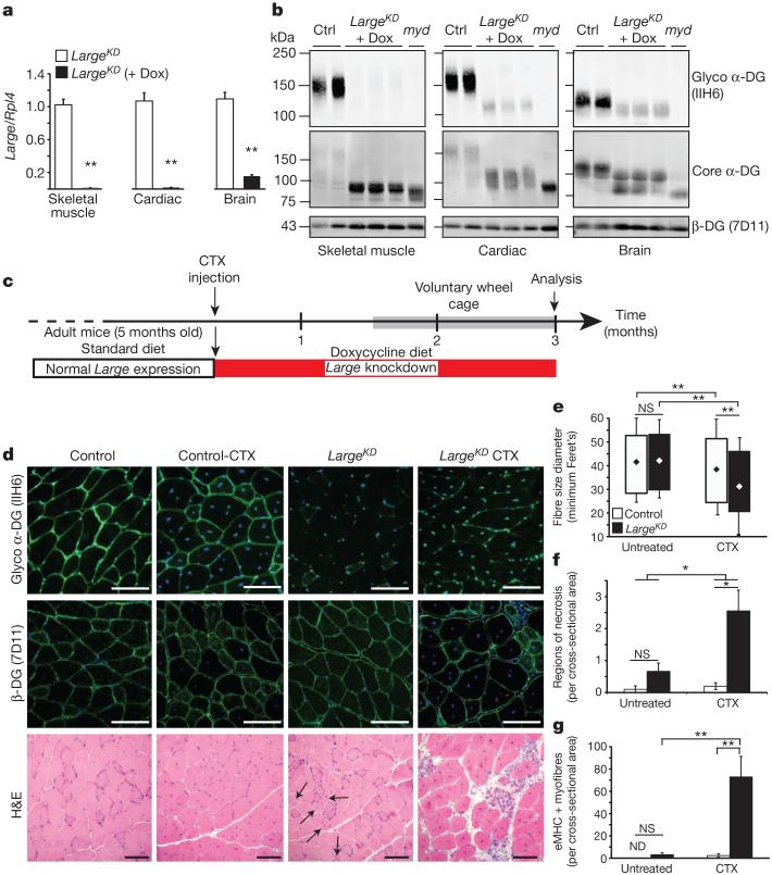
Figure 1. Muscle generated during Large knockdown is predisposed to aggressive dystrophy.
a, b, Quantification of Large mRNA expression in LargeKD mice on doxycycline (dox) induction (n=3 animals per group, 2 experimental replicates) (a), and western blot-based determination of LARGE glycosylation 3 months after shRNA induction, and in littermate controls (ctrl) and LARGE-null negative control (myd) (b). Each lane is a sample from an individual animal. c, Experimental outline. Six weeks after CTX injury and simultaneous induction of Large knockdown, the mice were housed in cages with exercise wheels to circumvent the typical sedentary behaviour of laboratory mice (grey bar). Two experimental replicates for all analysis. d, Representative images of IIH6 and β-DG reactivity in immunofluorescence- and haematoxylin-and-eosin (H&E)-stained sections (black arrows, centrally nucleated myofibres). e–g, Tibialis anterior muscle sections (4 sections/muscle) of control and LargeKD mice (white and black bars, respectively, LargeKD mouse, n=3; control, n=4 biological replicates) were assessed for: e, fibre diameter (interaction P<0.001, ~2,000 fibres per group; diamonds represent mean fibre diameter); f, average regions of necrosis (interaction P=0.057); and g, average number of recently regenerated, embryonic myosin-positive fibres (interaction P=0.007). Error bars represent s.d. in e, and s.e.m. elsewhere. Post-hoc comparisons *P<0.05, ** P<0.0001; NS, not significant; ND, not detected. Scale bars, 100 μm.

