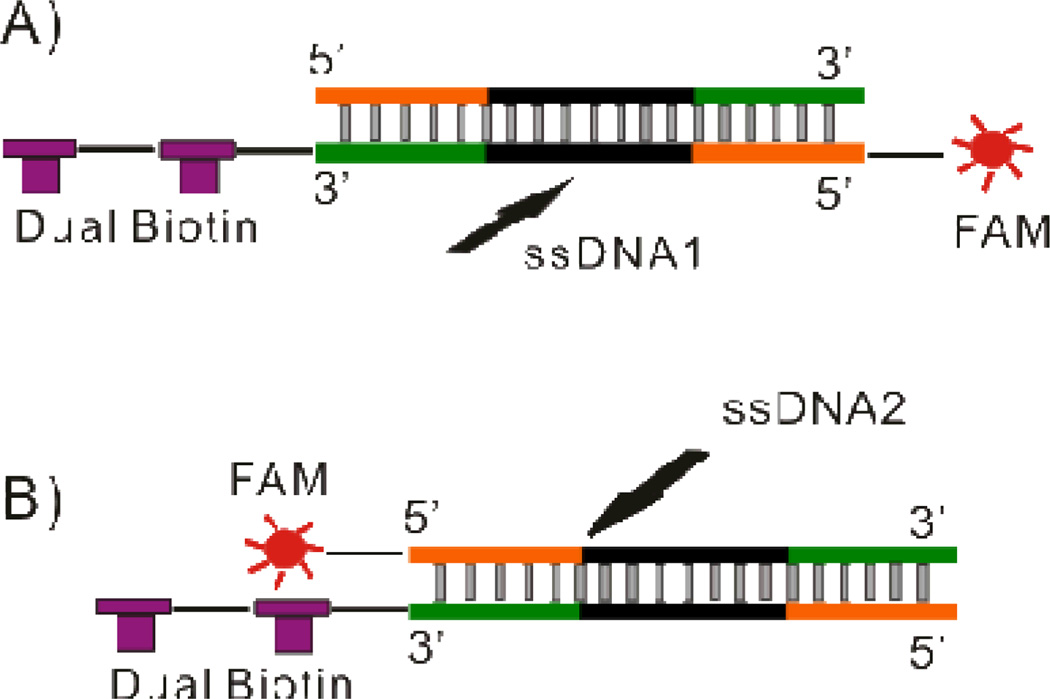
Figure 1.
Schematic demonstrating the labeling configuration of (A) dsDNA1 and (B) dsDNA2. In dsDNA1 dual biotin and FAM were labeled on the same strand allowing the complementary strand to be followed using fluorescence. In dsDNA2 dual biotin and FAM were labeled on opposite strands allowing the forward sequence to be followed.

