

Abstract
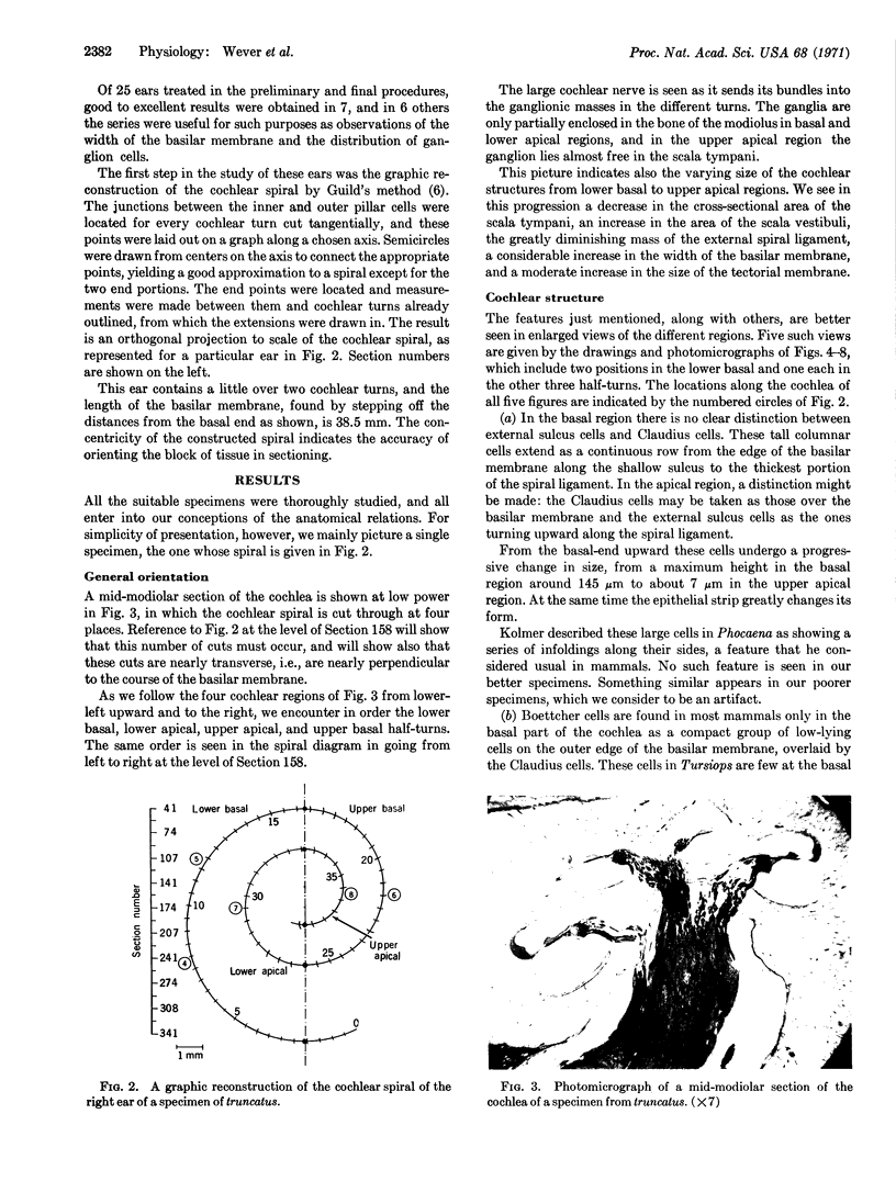
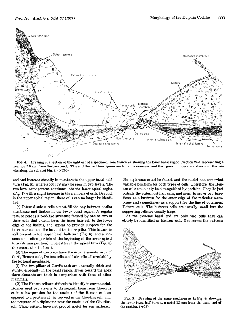
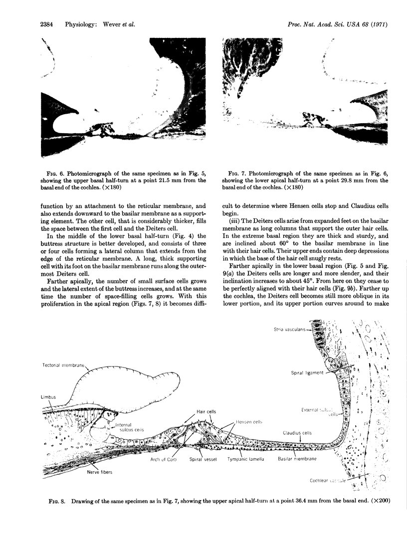
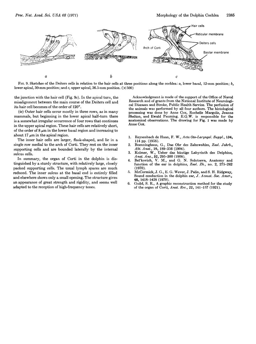
The anatomy of the cochlea of the dolphin Tursiops truncatus was studied in a number of specimens after fixation by vital perfusion, cellodin embedding, and serial sectioning. The results reveal the general structural relations and cellular detail up to the limits of light microscopy. A description is given of the variations of structure along the course of the cochlea, in which there are many departures from the typical mammalian form, especially in the compact quality of the tissues and the sturdiness of its elements. Apparently these features represent an adaptation of the cetacean ear to the reception of high-frequency sounds.
Keywords: organ of Corti, ear, light microscopy
Full text
PDF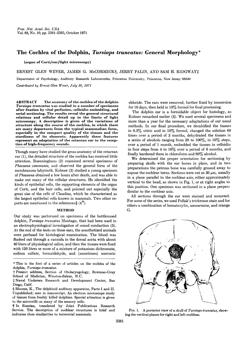
Images in this article
Selected References
These references are in PubMed. This may not be the complete list of references from this article.
- McCormick J. G., Wever E. G., Palin J. Sound conduction in the dolphin ear. J Acoust Soc Am. 1970 Dec;48(6 Suppl):1418+–1418+. doi: 10.1121/1.1912302. [DOI] [PubMed] [Google Scholar]