Abstract
Background: Alendronate is a bisphosphonate that is approved to reduce bone loss in glucocorticoid treated patients. In this paper, we present a case of femoral fracture following the use of Alendronate.
Case presentation: A- 46 year old woman who was a known case of hemolytic anemia has been treated by prednisolone (with different doses from 7.5 to 75 mg/day), calcium-D 500 mg/day and alendronate 70 mg/week for 3 years. Despite improvement of bone density, she experienced a low truama femoral shaft fracture.
Conclusion: This case shows a rare complication of treatment by alendronate. It may be needed to evaluate patients with long term usage of bisphosphonates for cortical thickness.
Key Words: Alendronate, Bisphosphonate, Fracture, Prednisolon, Hemolytic anemia
Alendronate is a bisphosphonate that is approved by the FDA to reduce bone loss in glucocorticoid treated patients. According to the recommendation for the prevention and treatment of glucocorticoid-induced osteoporosis, it is one of the first choices for these patients with T score <-1 (1). The evidence derived from the literature suggests that there is long term efficacy and safety with bisphosphonates such as alendronate and risedronate in the treatment of osteoporosis in postmenopausal women (2). After using bisphosphonates in millions of patients in the world, some unexpected possible adverse effects have been reported, including osteonecrosis of jaw, atypical femur fractures, atrial fibrillation, and esophageal cancer (3). There were some reports of atypical fractures in patients who were treated by bisphosphonates (4-9). In this article, we present a 46-year-old woman with a history of right atypical femoral fracture after 3 years of alendronate therapy.
Case history
A forty six year old woman presented with severe thigh pain due to fracture of femoral shaft after falling down in September 2011. She was a known case of hemolytic anemia (coombs positive) from three years ago and her treatment was started by high dose glucocorticoids (prednisolone 60 milligrams per day) and calcium-D (500mg-200IU/day) Bone marrow aspiration and biopsy showed leucocytoblastic reaction and hyper- cellular marrow. CT scan of lung, abdomen and pelvic did not show any abnormal finding. She was referred to a rheumatologist for the management of osteoporosis. The patient denied any musculoskeletal complaint and history for any bone fracture, spontaneous abortion, uveitis, dyspnea or Reynaud phenomenon.
Laboratory data included normal complete blood cell count (CBC) and thyroid function test, erythrocyte sedimentation rate (ESR): 44mm/h, serum calcium: 9.4 mg/dl, serum phosphor: 2.6 mg/dl, blood urine nitrogen (BUN): 24 mg/dl and serum creatinine: 0.9 mg/dl. Urine calcium in 24 hours was 291 mg. Bone mineral densitometry (BMD) by Hologic Explorer S/N 84109 showed osteopenia in spine and femoral neck (T score= -1.04 and -1.41, respectively).
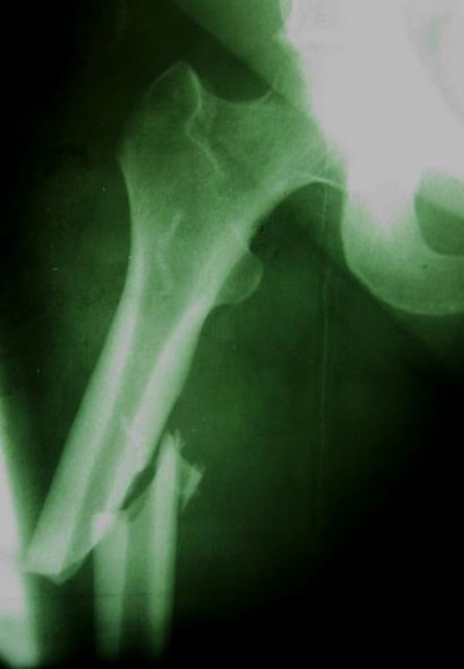
Because of osteopenia and glucocorticoid usage, treatment with alendronate (70 mg weekly) was added to calcium-D and prednisolone dosage was adjusted. She was visited every 3 months and laboratory evaluation including serum calcium, phosphate, alkalin phosphatase, 25(OH) D, 24 hours urine calcium were done in every visit. Two years later, BMD was repeated and showed improvement as a T score equal -1.1 in spine and -0.7 in femur. Serum and urine biochemistry followed and a dosage of steroid was adjusted according to CBC. During her treatment period, doses of prednisolone were between 7.5 to 75 mg per day according to hemolysis state. She had several attacks of hemolytic anemia. Three years after starting alendronate, she experienced a femoral shaft fracture when she was walking in the street and after a simple falling down. Radiograph showed an atypical femoral shaft fracture and cortical thickening (Figure 1). BMD demonstrated a T score equal-1 in spine and -0.9 in femur in this time. Alendronate was changed to raloxifen.
Figure 1.
Radiography demonstrating a right femoral shaft fracture with displacement and cortical thickness
Discussion
This article presents a 46-year old woman with a low trauma femoral fracture during treatment by alendronate. She undertook glucocorticoid therapy and despite the improvement of bone density in femoral area, experienced a low trauma fracture. Atypical femoral fractures are defined as fractures located in the subtrochanteric region and femoral shaft transverse or short oblique orientation, occurring, spontaneously or after minimal trauma, possessing a medial spike, absence of comminuting. Glucocorticoids, proton-pump inhibitor therapy and bisphosphonates may be risk factors (3).
Atypical fractures represented 30.3% of all diaphyseal and subtrochanteric femoral fractures. Ninety percent of all atypical fractures were associated with bisphosphonate usage. Atypical fractures were happened in younger, more active patients and were independent prior to fracture and experienced significant complications and self-reported level of function and health declines after fracture (4, 10). Cortical thickening may be an early sign of fracture risk and a simple, transverse fracture with a unicortical beak in an area of cortical hypertrophy is specific to alendronate users with atypical fractures (5, 6, 12 and 14). It may be related to long duration of drug use and prolonged suppression of bone turnover, which can lead to the accumulation of microdamage and development of hypermineralized bone (7). Bisphosphonates decrease bone turn over and increase bone mineral density by inhibiting osteoclast-mediated bone resorption and in long term, may result to an adynamic brittle bone (15). It varied from 3 to 12 years (11-12). There were some reports with more than one low trauma fracture (4, 12)
Despite these reports of association of bisphosphonates and atypical fractures, there is no rationale for their discontinuation in patients with osteoporosis, but continued use of them beyond a treatment period of 3 to 5 years should be reevaluated annually (9). It is recommended a drug holiday after 5-10 years of bisphosphonate treatment. The patients at mild risk may stop treatment after 5 years and remain off as long as bone mineral density is stable and no fractures occur. High-risk patients should be treated for 10 years, have a holiday of no more than a year or two, and perhaps be on a non-bisphosphonate treatment during that time (13). In summary, this case shows a rare complication of treatment by alendronate that experienced femoral fracture following low trauma.
Acknowledgments
The author would like to thank Dr Ghasem Janbabai for hematologic management.
Conflict of interest: There was no conflict of Interest.
References
- 1.Lane NE. Metabolic bone disease. In: Firestein GS, Budd RC, Harris ED, McInnes IB, Ruddy Sh, Sergent JS, editors. Kelley's text book of rheumatology. 18th ed. Philadelphia: Saunders Elsevier; 2009. p. 1590. [Google Scholar]
- 2.Iwamoto J, Takeda T, Sato Y. Efficacy and safety of alendronate and risedronate for postmenopausal osteoporosis. Curr Med Res Opin. 2006;22:919–28. doi: 10.1185/030079906X100276. [DOI] [PubMed] [Google Scholar]
- 3.McClung M, Harris ST, Miller PD, et al. Bisphosphonate Therapy for Osteoporosis: Benefits, Risks, and Drug Holiday. Am J Med. 2013;126:13–20. doi: 10.1016/j.amjmed.2012.06.023. [DOI] [PubMed] [Google Scholar]
- 4.Goh SK, Yang KY, Koh JS, et al. Subtrochanteric insufficiency fractures in patients on alendronate therapy: A caution. J Bone Joint Surg Br. 2007;89:349–353. doi: 10.1302/0301-620X.89B3.18146. [DOI] [PubMed] [Google Scholar]
- 5.Kwek EB, Goh SK, Koh JS, Png MA, Howe TS. An emerging pattern of subtrochanteric stress fractures: A long-term complication of alendronate therapy? Injury. 2008;39:224–31. doi: 10.1016/j.injury.2007.08.036. [DOI] [PubMed] [Google Scholar]
- 6.Neviaser AS, Lane JM, Lenart BA, Edobor-Osula F, Lorich DG. Low-energy femoral shaft fractures associated with alendronate use. J Orthop Trauma. 2008;22:346–50. doi: 10.1097/BOT.0b013e318172841c. [DOI] [PubMed] [Google Scholar]
- 7.Odvina CV, Levy S, Rao S, Zerwekh JE, Rao DS. Unusual mid-shaft fractures during long-term bisphosphonate therapy. Clin Endocrinol (Oxf) 2010;72:161–8. doi: 10.1111/j.1365-2265.2009.03581.x. [DOI] [PubMed] [Google Scholar]
- 8.Visekruna M, Wilson D, McKiernan FE. Severely suppressed bone turnover and atypical skeletal fragility. J Clin Endocrinol Metab. 2008;93:2948–52. doi: 10.1210/jc.2007-2803. [DOI] [PubMed] [Google Scholar]
- 9.Gunawardena I, Baxter M, Rasekh Y. Bisphosphonate-related subtrochanteric femoral fractures. Am J Geriatr Pharmacother. 2011;9:194–8. doi: 10.1016/j.amjopharm.2011.02.009. [DOI] [PubMed] [Google Scholar]
- 10.Shkolnikova J, Flynn J, Choong P. Burden of bisphosphonate-associated femoral fractures. ANZ J Surg. 2013;83:175–81. doi: 10.1111/ans.12018. [DOI] [PubMed] [Google Scholar]
- 11.Kao CM, Huang PJ, Chen CH, Chen SJ, Cheng YM. Atypical femoral fracture after long-term alendronate treatment: report of a case evidenced with magnetic resonance imaging. Kaohsiung J Med Sci. 2012;28:555–8. doi: 10.1016/j.kjms.2012.04.019. [DOI] [PMC free article] [PubMed] [Google Scholar]
- 12.Goddard MS, Reid KR, Johnston JC, Khanuja HS. Atraumatic bilateral femur fracture in long-term bisphosphonate use. Orthopedics. 2009:32. doi: 10.3928/01477447-20090624-27. [DOI] [PubMed] [Google Scholar]
- 13.Watts NB, Diab DL. Long-term use of bisphosphonates in osteoporosis. J Clin Endocrinol Metab. 2010;95:1555–65. doi: 10.1210/jc.2009-1947. [DOI] [PubMed] [Google Scholar]
- 14.Nieves JW, Cosman F. Atypical subtrochanteric and femoral shaft fractures and possible association with bisphosphonates. Curr Osteoporos Rep. 2010;8:34–9. doi: 10.1007/s11914-010-0007-2. [DOI] [PubMed] [Google Scholar]
- 15.Kim SY, Schneeweiss S, Katz JN, Levin R, Solomon DH. Oral Bisphosphonates and risk of subtrochanteric or diaphyseal femur fractures in a population- based cohort. J Bone Miner Res. 2011;26:993–1001. doi: 10.1002/jbmr.288. [DOI] [PMC free article] [PubMed] [Google Scholar]


