Abstract
The striatum is a brain area implicated in the pharmacological action of drugs of abuse. Adenosine A2A receptors (A2ARs) are highly expressed in the striatum and mediate, at least in part, cocaine-induced psychomotor effects in vivo. Here we studied the synaptic mechanisms implicated in the pharmacological action of cocaine in the striatum and investigated the influence of A2ARs. We found that synaptic transmission was depressed in corticostriatal slices after perfusion with cocaine (10 μM). This effect was reduced by the A2AR antagonist ZM241385 and almost abolished in striatal A2AR-knockout mice (mice lacking A2ARs in striatal neurons, stA2ARKO). The effect of cocaine on synaptic transmission was also prevented by the protein tyrosine phosphatases (PTPs) inhibitor sodium orthovanadate (Na3VO4). In synaptosomes prepared from striatal slices, we found that the activity of striatal-enriched protein tyrosine phosphatase (STEP) was upregulated by cocaine, prevented by ZM241385, and absent in synaptosomes from stA2ARKO. The role played by STEP in cocaine modulation of synaptic transmission was investigated in whole-cell voltage clamp recordings from medium spiny neurons of the striatum. We found that TAT-STEP, a peptide that renders STEP enzymatically inactive, prevented cocaine-induced reduction in AMPA- and NMDA-mediated excitatory post-synaptic currents, whereas the control peptide, TAT-myc, had no effect. These results demonstrate that striatal A2ARs modulate cocaine-induced synaptic depression in the striatum and highlight the potential role of PTPs and specifically STEP in the effects of cocaine.
Keywords: cocaine, striatum, A2A receptor, synaptic transmission, STEP
INTRODUCTION
The study of the molecular mechanisms underlying the effects of drugs of abuse is an important research field in neuroscience. The involvement of the nucleus accumbens and ventral tegmental area in mediating the behavioral effect of drugs of abuse is well-known (Lüscher and Malenka, 2011). In the last years, however, the role of the dorsal striatum has emerged (Gerdeman et al, 2003) and has been implicated in mediating some of the psychomotor effects of psychostimulant drugs (Rebec, 2006; Borgkvist and Fisone, 2007).
Cocaine, a widely used substance of abuse, acts by blocking the dopamine transporter and thus elevating extracellular dopamine levels (Kuhar et al, 1991). In addition, cocaine interacts directly with sodium channels, and thereby has an effect on firing activity independent of transmitter release (Kiyatkin and Rebec, 2000). The pharmacological actions of cocaine also involve additional neurotransmitter systems. For example, acute cocaine administration inhibits the serotonin and noradrenaline membrane transporters, thus increasing their extracellular levels (Uhl et al, 2002).
Cocaine increases cerebral extracellular levels of adenosine (Herrera-Marschitz et al, 1994; Fiorillo and Williams, 2000), an endogenous purine nucleoside that modulates dopaminergic neurotransmission (Ferrè et al, 1997). In particular, in the fetal brain, a direct correlation was shown between cocaine, adenosine increase, and adenosine A2A receptor (A2AR) stimulation (Kubrusly and Bhide, 2010). Among the different adenosine receptors, the A2AR subtype is highly expressed in the striatum, where it is involved in the locomotor, sensitizing, and rewarding properties of cocaine (Shen et al, 2008; Filip et al, 2006; Soria et al, 2006; Justinova et al, 2003; Chen et al, 2000). However, the cellular and synaptic mechanisms through which A2ARs modulate cocaine effects are unknown. In the present study, we address this important issue by biochemical and electrophysiological experiments in mouse brain slices. As a role of protein tyrosine kinases (PTKs) in mediating cocaine-induced effects in basal ganglia has recently emerged (Schumann et al, 2009; Pascoli et al, 2011), the possible involvement of protein tyrosine phosphatases (PTPs) was also investigated. In particular, we examine the role played by the striatal-enriched protein tyrosine phosphatase (STEP), a brain-specific phosphatase that is highly expressed in the striatum and that has been implicated in the pathophysiology of several neuropsychiatric diseases when present at inappropriate high levels (Goebel-Goody et al, 2012).
MATERIALS AND METHODS
Chemicals
Cocaine (Istituto Superiore di Sanità drug of abuse depository), raclopride (Tocris, Bristol, UK), SCH23390 (Sigma-Aldrich, Milan, Italy), and sodium orthovanadate (Na3VO4, Sigma Chemical, St Louis, MO, USA) were dissolved in H2O, whereas ZM241385 and FK506 (Tocris) in 0.1% DMSO. PP2 (Alexis, Enzo Life Science, Farmingdale, NY, USA) was dissolved in 2.5% DMSO. We used the following antibodies: polyclonal anti-STEP (Cell Signaling Technology (Danvers, MA, USA), monoclonal anti-β actin (Calbiochem, EDM Chemical, Merck, Darmstadt, Germany), goat anti-GAPDH (Everest Biotech, Oxfordshire, UK), polyclonal anti-GluN2B phosphoTyr1472, and anti-GluN2B (Millipore Bioscience Research Reagent, Billerica, MA, USA) and peroxidase-conjugated goat anti-mouse and goat anti-rabbit (Bio-Rad, Hercules, CA, USA). Protein A/G PLUS-agarose was from Santa Cruz Biotechnology (Santa Cruz, CA, USA) and Trysacryl-immobilized Protein A from Thermo Scientific (Waltham, MA, USA). Nitrocellulose was from Schleicher and Schuell Bioscience Inc. (Dassel, Germany) and p-nitrophenyl phosphate (p-NPP) from Sigma Chemical (St Louis, MO, USA). Complete protease inhibitor cocktail, EDTA-free, was from Roche Diagnostics (Basel, Switzerland).
The TAT-STEP46 was affinity purified as described by Paul et al (2007); it has a point mutation in its catalytic domain (C300S) that renders it enzymatically inactive, and thus acts as a substrate-trapping mutant. TAT-myc was obtained through the addition of a myelocytomatosis virus (myc) to the C-terminus of TAT and was used as control peptide (Paul et al, 2007).
Animals
Male C57Bl/6 (3–4 months old, Harlan, Italy) and stA2ARKO (and aged-matched wild-type) mice were used. To generate stA2ARKO, the Cre-loxP strategy was used as previously described (Bastia et al, 2005; Shen et al, 2008). The animals were kept under standardized temperature, humidity, and lighting conditions with free access to water and food. All procedures met the European guidelines for the care and use of laboratory animals (86/609/ECC) and those of the Italian Ministry of Health (DL 116/92).
Electrophysiology
Corticostriatal slices were prepared as previously described (Chiodi et al, 2012). Briefly, mice were decapitated under ether anesthesia, coronal slices (300 μm) were cut with a vibratome, and incubated for 1 h in artificial cerebrospinal fluid (ACSF). Single slices were transferred to a submerged recording chamber and superfused with ACSF at 32–33 °C. Drugs were applied by bath perfusion with ACSF.
Extracellular field potentials (FPs) were recorded in the dorsomedial striatum with a glass microelectrode and evoked at the frequency of 0.05 Hz by stimulating the white matter with a bipolar platinum/iridium concentric electrode (FHC, Bowdoin, ME, USA). Signals were acquired with the DAM-80 AC differential amplifier (WPI Instruments, Sarasota, FL, USA) and analyzed with WinLTP software (Anderson and Collingridge, 2007).
Whole-cell voltage-clamp recordings were made with borosilicate glass pipettes (4–6 MΩ), (WPI, Berlin, Germany) filled with (in mM) 125 Cs-methanesulfonate, 3 KCl, 4 NaCl, 1 MgCl2, 5 MgATP, 9 EGTA, 8 HEPES, 10 phosphocreatine (pH 7.2 with CsOH). AMPA- and NMDA-mediated excitatory post-synaptic currents (AMPA-EPSC and NMDA-EPSC, respectively) were evoked by extracellular stimulation at the frequency of 0.05 Hz with a bipolar platinum/iridium concentric electrode (FHC) placed in the white matter. Signals were amplified with a PC-501A patch clamp amplifier (Warner Instrument Corp). Data were filtered at 1 kHz, digitized at 10 kHz, stored on a computer, and analyzed off-line using the WinWCP (Strathclyde Electrophysiology Software). Medium-sized spiny neurons (MSNs) in dorsomedial striatum were visualized using a × 60 water-immersion objective and infrared differential interference optics video microscopy, and identified by small somata and basic membrane properties. Passive membrane properties of MSN were determined in voltage-clamp mode by applying a depolarized step voltage command (10 mV) and using the cell test function integrated in the WinWCP program. Series resistance, not compensated, was <20 MΩ and was checked periodically (changes >30% led to cell discard). For AMPA-EPSC, cells were clamped at −70 mV in ACSF containing 10 μM bicuculline and 50 μM APV (to block GABAA and NMDA receptors, respectively), whereas for NMDA-EPSC cells were held at+40 mV in ACSF containing 10 μM bicuculline and 5 μM NBQX (to block AMPA receptors). At the end of the experiments, slices were perfused with NBQX or APV to verify the specificity of the recorded currents. For both extracellular and patch-clamp recordings, three consecutive responses were averaged and at least 10 min of stable baseline recording preceded drug application. Mean basal amplitude was calculated from the 5 min immediately preceding drug application. Drug effects (average of the last 3 min of drug application) were expressed as percentage of basal.
Preparation of Striatal Synaptosomes
We made striatal slices by removing the cortex from corticostriatal slices and prepared a crude synaptosomal fraction as described previously (Huttner et al, 1983). Briefly, the striata were homogenized in ice-cold buffer (in mM): 320 sucrose, 5 Hepes-NaOH, pH 7.4, 0.5 EDTA, 0.1 phenylmethylsulphonyl fluoride (PMSF), 1 Na3VO4, 10 NaF, and protease inhibitor mixture (Complete, Roche) using a Teflon-glass grinder. The homogenates were centrifuged at 800 g at 4 °C for 10 min, and the supernatants were collected and centrifuged at 9200 g for 15 min. Pellets were washed in homogenization buffer and centrifuged at 10200 g for 15 min to obtain a crude synaptosomal fraction. We prepared samples for determination of STEP activity in the same buffer without phosphatase inhibitors. Protein content was determined using the bicinchoninic acid assay (BCA kit, Thermo Scientific).
STEP Activity and Western Blot Analysis
Striatal synaptosomes were solubilized by incubation for 30 min at 0 °C with 4X RIPA buffer (in mM): 100 Tris-HCl, pH 7.5, 600 NaCl, 4% (w/v) Triton X-100, 4% (v/v) sodium deoxycholate, 0.4% SDS (v/v), 0.4 PMSF, and protease inhibitors (Complete). After centrifugation at 16 000 g for 30 min at 4 °C, the supernatant was incubated with 25 μl 50% (w/v) protein A/G PLUS agarose beads for 2 h at 4 °C, clarified by centrifugation, and incubated overnight at 4 °C in a rotating wheel with a polyclonal anti-STEP antibody. The immunocomplex was precipitated by the addition of 50% (w/v) Trysacryl-immobilized Protein A beads. To measure the activity of STEP, the immunoprecipitates were suspended in 200 μl of assay buffer (in mM: 25 Hepes, pH 7.0, 20 MgCl2, 0.1 PMSF) containing 15 mM para-nitrophenyl phosphate (p-NPP) and incubated 60 min at 30 °C under gentle stir. The activity was determined in the clarified supernatants by measuring the absorbance at 405 nm of p-nitrophenol.
For western blot, samples were resolved on 10% SDS–PAGE, and proteins were transferred to nitrocellulose (Mallozzi et al, 2013). The immunoreactive bands were detected by chemiluminescence coupled to peroxidase activity (ECL kit, Thermo Scientific) and quantified using a Bio-Rad ChemiDoc XRS system.
PTP Activity
Total PTP activity was detected in striatal synaptosomes using p-NPP as substrate, according to the procedure described previously (Mallozzi et al, 1997). Briefly, synaptosomes were suspended in assay buffer containing 15 mM p-NPP and incubated at 37 °C for 30 min. The reaction was stopped by the addition of 0.1 mM NaOH. Samples were centrifuged, and the release of p-nitrophenol from p-NPP was measured in the supernatant at 405 nm.
Statistical Analysis
All data were presented as means±SEM Student's t-test was used for single comparisons. Differences among multiple groups were analyzed by one-way ANOVA (followed by the Bonferroni post hoc test) when the assumptions of normality and homogeneity of variance were met (by using the Kolmogorov–Smirnov test and Bartlett test, respectively) or, in the other cases, by the Kruskall–Wallis nonparametric analysis of variance (followed by Dunn's test for multiple comparisons). A p-value <0.05 indicated statistically significant differences.
RESULTS
Cocaine-Induced Inhibition of Synaptic Transmission is Mediated by A2ARs Expressed on Striatal Neurons
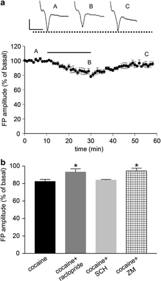
Corticostriatal slice perfusion with cocaine (10 μM, 20 min) transiently depressed synaptic transmission as shown by a reduction in the FP amplitude (83.13±1.24% of basal, Figures 1a and b), an effect attenuated by the D2 receptor antagonist raclopride (10 μM, 93.23±3.7% of basal p<0.05 vs cocaine alone) but not by the D1 receptor antagonist SCH23390 (10 μM, 83.94±0.71% of basal p>0.05 vs cocaine alone) (Figure 1b).
Figure 1.
Electrophysiological experiments showing the effect of cocaine on synaptic transmission in corticostriatal slices. (a) A dosage of 10 μM cocaine induced a transient reduction in field potential (FP) amplitude. Each point represents the mean of three responses. Insets show FPs recorded in basal condition (A), 20 min after cocaine application (B) and at the wash-out (C). The horizontal bar indicates the period of drug application. Calibration bars: 0.5 mV, 5 ms. (b) The effect of cocaine (N=10) was significantly reduced by raclopride (N=3) and ZM241385 (N=4), whereas it was unaffected by SCH23390 (N=4). *p<0.05, significantly different from cocaine alone, (the Kruskal–Wallis test). ZM=ZM241385; SCH=SCH23390.
To verify whether the effect of cocaine was mediated by presynaptic mechanisms, we used a protocol of paired-pulse stimulation by delivering two stimuli (50 ms apart) that evoked two consecutive FPs, and the ratio between the amplitude of the second and the first FP was calculated. Cocaine induced a significant increase in paired-pulse ratio values (1.08±0.03 and 1.23±0.03 in basal condition and after cocaine, respectively, N=4, p<0.05 paired Student's t-test, data not shown), suggesting that cocaine-induced reduction of synaptic transmission involves a presynaptic mechanism of action.
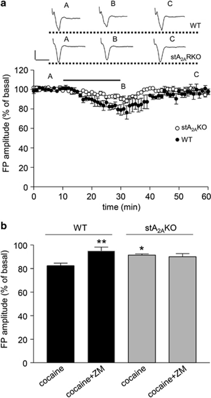
To investigate whether A2ARs contributed to the cocaine-induced synaptic effects, we applied the selective A2AR antagonist ZM241385 (100 nM) 10 min before and during cocaine administration. As shown in Figure 1b, ZM241385 significantly reversed the effects of cocaine (94.36±3.2% of basal, p<0.05 vs cocaine alone). Notably, ZM241385 also reversed cocaine-induced increase in paired-pulse ratio (1.137±0.023 and 1.161±0.055, in basal condition and after cocaine, respectively, N=4, p>0.05, paired Student's t-test). To confirm the involvement of A2ARs, we studied the synaptic effects of cocaine in corticostriatal slices from stA2ARKO, where the A2AR is selectively deleted in striatal neurons (Lazarus et al, 2011). Cocaine-induced synaptic depression was significantly reduced in stA2ARKO as compared with WT mice (91.39±1.2% and 82.49±2.1% of basal, respectively, p<0.05, Figure 2a). In addition, although ZM241385 reduced cocaine-induced synaptic depression in WT (94.98±3.3% of basal, p<0.01 vs cocaine alone), it had no effects on stA2ARKO (90.03±2.7% of basal p>0.05 vs cocaine alone), confirming the involvement of striatal A2ARs in the synaptic effects of cocaine. (Figure 2b).
Figure 2.
Effects of cocaine on synaptic transmission in WT and stA2AKO mice. (a) cocaine-induced depression in FP amplitude is reduced in stA2AKO compared with WT mice. Each point represents the mean of three responses. Insets show FPs recorded in basal condition (A), 20 min after cocaine application (B) and at the wash-out (C), in WT and stA2AKO mice. The horizontal bar indicates the period of drug application. Calibration bars: 0.5 mV, 5 ms. (b) cocaine-induced reduction in FP amplitude in stA2AKO mice (N=6) is significantly reduced with respect to WT mice (N=6). ZM241385 prevents the effect of cocaine in WT mice (N=5) but not in stA2AKO mice (N=4). *p<0.05 and **p<0.01, significantly different from WT cocaine (the Kruskal–Wallis test). ZM=ZM241385.
Cocaine-Induced Synaptic Depression is Prevented by PTPs, but not by src-PTKs Inhibitors
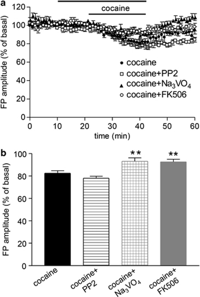
As tyrosine phosphorylation signaling is involved in some of the synaptic effects of cocaine (Schumann et al, 2009), we evaluated the effect of the src-PTKs inhibitor PP2 and of the PTP inhibitor sodium orthovanadate (Na3VO4). We perfused slices with 10 μM PP2 or 1 mM Na3VO4 10 min before and during cocaine application (Figure 3a). Cocaine-induced synaptic depression was unaffected by PP2 (79.14±1.2% of basal, p>0.05 vs cocaine alone, Figure 3b), whereas it was reduced by Na3VO4 (93.15±3.06% of basal, p<0.01 vs cocaine alone, Figure 3b), suggesting that cocaine reduces synaptic transmission through activation of tyrosine phosphatases. Slice perfusion with 1 μM FK506, a calcineurin inhibitor, also significantly reduced cocaine-induced synaptic depression (92.64±2.3% of basal, p<0.01 vs cocaine, Figure 3b), suggesting that the effect of cocaine on synaptic transmission was also controlled by calcineurin, a serine/threonine calcium-dependent phosphatase. These results confirm that the effects of cocaine involve the activation of protein phosphatases.
Figure 3.
The synaptic effect of cocaine is modulated by protein phosphatase inhibitors. (a) Time course of cocaine-induced reduction in FP amplitude alone and in the presence of the protein tyrosine phosphatase inhibitor Na3VO4, the calcineurin inhibitor FK506, and the protein tyrosine kinase inhibitor PP2. The horizontal bars indicate the period of drug application. (b) The effect of cocaine (N=10) was significantly reduced by Na3VO4 (N=5) and FK506 (N=5) but not by PP2 (N=5). **p<0.01, significantly different from cocaine alone (one-way ANOVA).
Cocaine Stimulates Tyrosine Phosphatase Activity in Striatal Slices, an Effect Controlled by A2ARs
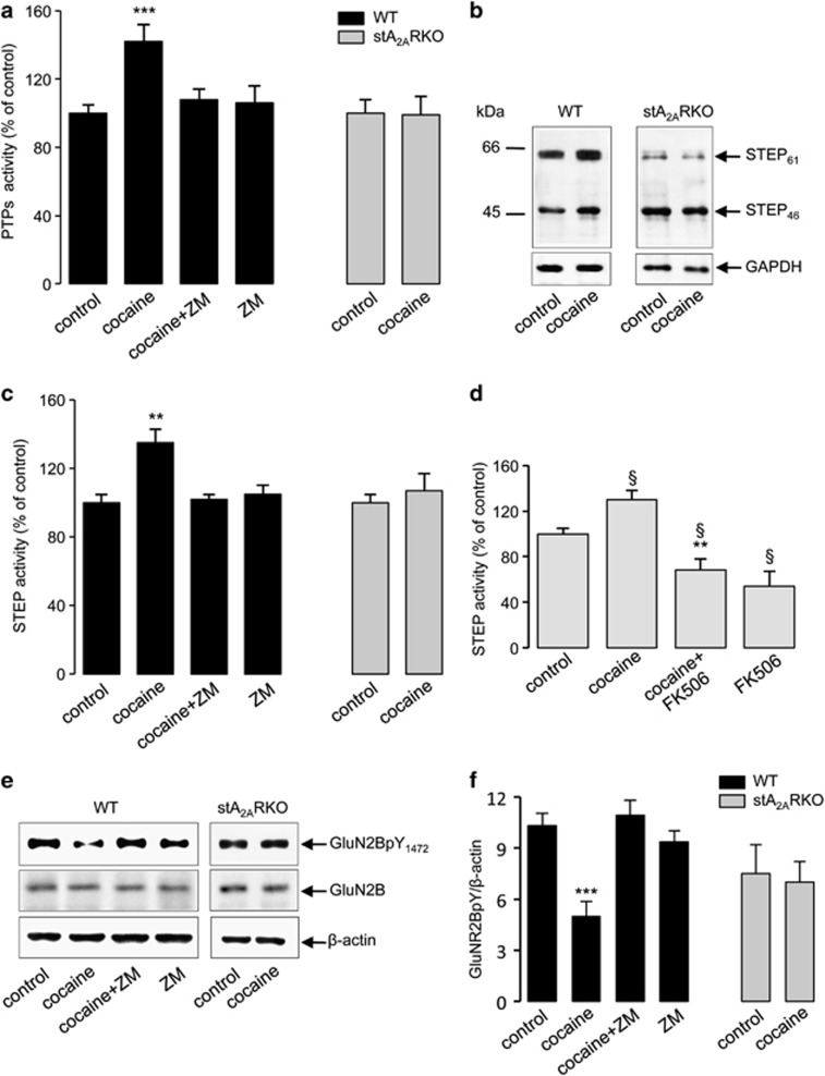
Cocaine (10 μM for 10 min) upregulated total PTP activity in striatal synaptosomes (142±10% of control, p<0.001), and such an effect depended on A2AR stimulation, as ZM241385 (100 nM) prevented it (Figure 4a). Moreover, cocaine failed to stimulate PTP activity in slices from stA2ARKO (Figure 4a), confirming the involvement of striatal A2ARs in the effects of cocaine.
Figure 4.
The effect of cocaine on tyrosine phosphatase activity is mediated by A2ARs. (a) PTP activity was measured in synaptosomes obtained from WT (black columns, N=10) and stA2ARKO striatal slices (gray columns, N=4) treated with 10 μM cocaine for 10 min, with or without ZM241385. The activity is expressed as percentage of the control. The bar graph represents the means±SEM. ***significantly different from control (p<0.001, Kruskal–Wallis test). (b) Western blot analysis with an anti-STEP monoclonal antibody of synaptosomes from WT and stA2ARKO striatal slices treated with cocaine. The molecular mass markers in kDa are indicated on the left. The nitrocellulose was also probed with an anti-GAPDH antibody to evaluate the amount of loaded proteins (lower panels). The immunoreactive bands were detected by chemiluminescence coupled to peroxidase activity (ECL). The results shown are representative of three independent experiments. (c) Effect of cocaine on STEP activity. STEP was immunoprecipitated by a specific polyclonal antibody from solubilized synaptosomes prepared from WT (N=5) and stA2ARKO striatal slices (N=4) (black and gray columns, respectively) treated as described in (a). The phosphatase activity of STEP-immunocomplex is expressed as percentage of the value measured in control (100%). The bar graph represents the means±SEM. **significantly different from control (p<0.01, the Kruskal–Wallis test). (d) Effect of the calcineurin inhibitor FK506 on STEP phosphatase activity. Striatal slices (N=5) were incubated for 2-3 h with 1 μM FK506 and then treated with cocaine in the presence of FK506. STEP activity was measured in the immunocomplex obtained from solubilized synaptosomes. The bar graph represents the means±SEM (§p<0.05 vs control; **p<0.01 vs cocaine, the Kruskal–Wallis test). (e) Western blot analysis with anti-GluN2BpY1472 and anti-GluN2B antibodies of synaptosomes from WT and stA2ARKO striatal slices treated as described in (a). The nitrocellulose was also probed with an anti-β-actin antibody to evaluate the amount of loaded proteins (lower panels). The immunoreactive bands were detected by chemiluminescence coupled to peroxidase activity (ECL). (f) Quantification by densitometric analysis of GluN2BpY1472 band intensity relative to β-actin. The bar graph represents the means±SEM of 5 and 3 independent experiments for WT and stA2ARKO, respectively (***p<0.001 vs control, the Kruskal–Wallis test). ZM=ZM241385.
Cocaine Activates STEP Through the Involvement of A2AR
STEP is highly enriched in the striatum and regulates AMPA receptor internalization (Zhang et al, 2008). The two major isoforms, STEP46 and STEP61, are revealed by western blot analysis in striatal slices, and cocaine (10 μM, 10 min) induced an increase in protein levels of both isoforms (Figure 4b). STEP61 and STEP46 were identified also in stA2ARKO, but, in these mice, cocaine treatment did not change STEP protein levels (Figure 4b). These results indicate that A2ARs control cocaine-induced upregulation of STEP in the striatum.
Next, we determined whether the phosphatase activity of STEP was changed by cocaine. As shown in Figure 4c, cocaine increased STEP activity (135±8%, p<0.01 vs control), an effect significantly decreased by ZM241385. As expected, Na3VO4 blocked PTP activity (results not shown). Cocaine did not increase STEP activity in stA2ARKO (Figure 4c), strongly implying A2ARs in cocaine-induced modulation of STEP activity.
The Activity of STEP is Controlled by Calcineurin
In electrophysiology experiments, inhibiting calcineurin activity prevented cocaine-induced synaptic depression. We therefore studied the effect of FK506 on cocaine-induced STEP activation. Striatal slices were incubated for 2–3 h with 1 μM FK506 and then treated for 10 min with cocaine plus FK506. In this condition, not only did cocaine fail to increase STEP activity, but FK506 decreased STEP activity to below control values (p<0.01 vs cocaine alone and p<0.05 vs control, Figure 4d). In addition, FK506 alone reduced STEP activity by about 50% (p<0.05 vs control, Figure 4d), suggesting that calcineurin not only mediates cocaine effects but also tonically controls STEP activity.
The STEP Substrate GluN2B is Dephosphorylated by Cocaine Treatment
Active STEP dephosphorylates tyrosine residues on its substrates causing their inactivation and, in the case of glutamate receptors, promoting their internalization from synaptosomal surface membranes (Zhang et al, 2008; Zhang et al, 2010, 2011). Substrates of STEP include the GluN2B subunit of the NMDA receptor. In striatal slices treated with cocaine, the tyrosine phosphorylation of GluN2B subunit (Tyr1472) was significantly downregulated (p<0.001 vs control), an effect prevented by ZM241385 (Figure 4e and f). Noteworthy, cocaine did not inhibit GluN2B tyrosine phosphorylation in stA2ARKO (Figure 4e and f), in line with its inability to induce STEP activation in the absence of A2ARs (Figure 4c). Immunoblotting of GluN2B did not reveal protein level changes in the different experimental conditions (Figure 4e).
STEP Modulates Cocaine-Induced Synaptic Depression
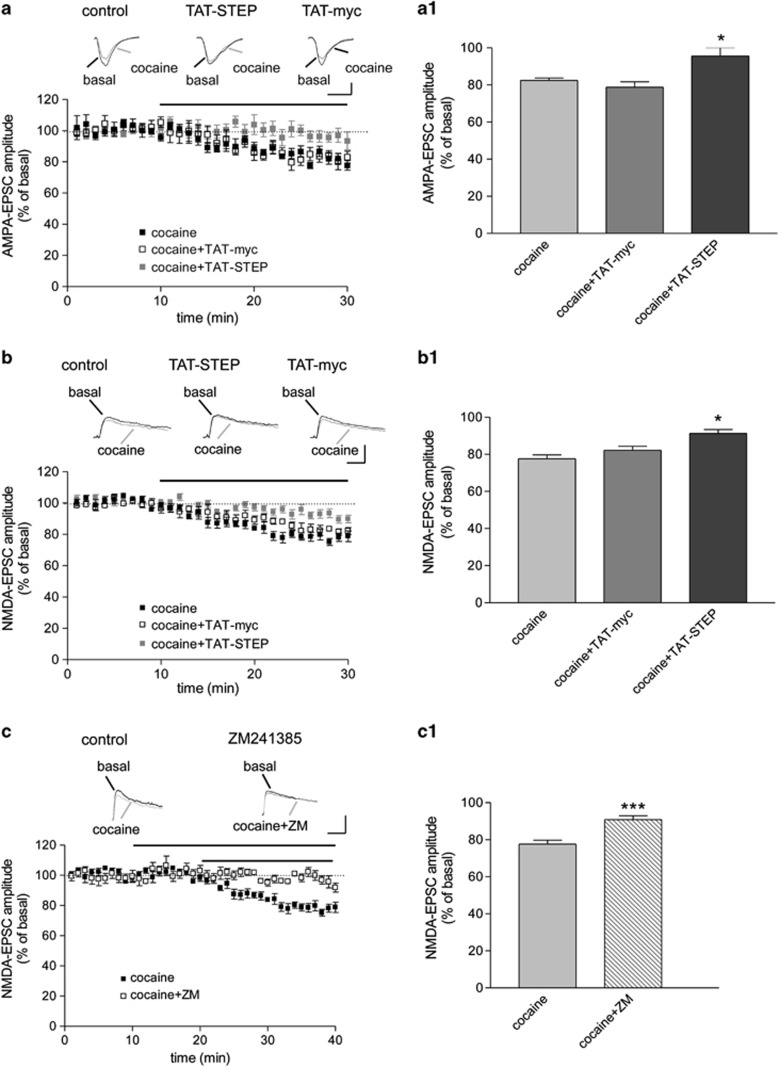
Having found that the synaptic effects of cocaine are dependent on PTP activation and that cocaine increases STEP activity, we investigated whether STEP was involved in cocaine-mediate synaptic depression. The substrate-trapping mutant TAT-STEP (C300S) failed (up to 300 nM) to influence the reduction in FP induced by cocaine (data not shown). As this lack of effect could be due to a poor penetration of the peptide into the cells, we performed whole-cell voltage clamp experiments in MSNs to record excitatory synaptic transmission, and TAT-STEP (100 nM) was added to the intracellular solution of the patch pipette. In the striatum, the majority of cells (95%) are MSNs that exhibited negative resting membrane potentials and passive membrane properties similar to those previously reported (Cepeda et al, 2008). Twenty-minute slice perfusion with cocaine (10 μM) caused an inhibition of AMPA-EPSC (83.67±1.3% of basal Figure 5a and a1), in accordance with the results obtained with extracellular recordings. In the presence of TAT-STEP (100 nM) in the recording pipette, the effect of cocaine on AMPA-EPSC was significantly attenuated (95.66±4.45% of basal, p<0.05, Figure 5a and a1). On the contrary, the control peptide TAT-myc did not change cocaine-induced reduction in AMPA-EPSC (77.8±3.68% of basal, Figure 5a and a1, p>0.05 vs cocaine, p<0.05 vs cocaine+TAT-STEP). Cocaine reduced NMDA-EPSC amplitude (77.66±2.02% of basal, Figure 5b and b1), an effect significantly reduced by TAT-STEP (91.33±2.02%, p<0.05, Figure 5b and b1), but not by the control peptide TAT-myc (82.25±2.11 of basal values, p>0.05 vs cocaine, p<0.05 vs cocaine+TAT-STEP, Figure 5b and b1). Notably, although the overall effect was significant on average for both AMPA and NMDA currents, the addition of TAT-STEP completely abolished cocaine effect in about 50% of the neurons, whereas in the remaining cells cocaine effects were largely unaffected.
Figure 5.
The effect of cocaine on excitatory synaptic transmission in MSNs is modulated by STEP and A2ARs. (a) A dosage of 10 μM cocaine reduced AMPA-EPSC (N=7); the effect of cocaine was reduced in the presence of the substrate trapping peptide TAT-STEP 100 nM in the recording pipette (N=10), while the addition of the control peptide TAT-myc (N=8) was ineffective. Each point represents the mean of three responses; insets show AMPA-EPSCs before and after cocaine application in control condition and in the presence of TAT-myc and TAT-STEP in the patch pipette. The horizontal bar indicates the period of cocaine application. Calibration bars: 50 ms, 50 pA. (a1) Bar graph shows the effect of cocaine on the amplitude of AMPA-EPSC 20 min after drug application (expressed as percentage of basal values). *p<0.05 vs cocaine and cocaine+TAT-myc, one-way ANOVA. (b) 10 μM cocaine reduced NMDA-EPSC (N=8); the effect of cocaine was significantly reduced by the addition of TAT-STEP in the recording pipette (N=15) but not by the control peptide TAT-myc (N=8). Each point represents the mean of three responses; insets show NMDA-EPSCs before and after cocaine application in control condition and in the presence of TAT-myc and TAT-STEP in the patch pipette. The horizontal bar indicates the period of cocaine application. Calibration bars: 100 ms, 100 pA. (b1) Bar graph shows the effect of cocaine on the amplitude of NMDA-EPSC 20 min after drug application (expressed as percentage of basal values). *p<0.05 vs cocaine and cocaine+TAT-myc, one-way ANOVA. (c) The effect of cocaine on NMDA-EPSC (N=8) is reduced by ZM241385 (100 nM) applied 10 min before and along with cocaine (N=8). Each point represents the mean of three responses; insets show NMDA-EPSCs before and after cocaine application in control condition and in the presence of ZM241385. The horizontal bars indicate the period of drugs application. Calibration bars: 100 ms, 100 pA. (c1) Bar graph shows the effect of cocaine, alone and in combination with ZM241385, on the amplitude of NMDA-EPSC 20 min after cocaine application (expressed as percentage of basal values). ***p<0.001, the Student t-test. ZM=ZM241385.
A2ARs Control Cocaine-Induced Reduction in NMDA-EPSC
Having found that cocaine reduces the tyrosine phosphorylation state of the NMDA receptor subunit GluN2B (that can be related to NMDA receptor endocytosis) and that this effect is controlled by A2ARs, we explored the hypothesis that A2AR activation is required for cocaine effects on NMDA currents. Ten-minute slice pretreatment with ZM241385 (100 nM) significantly reduced cocaine-induced decrease in NMDA-EPSC (cocaine alone: 77.66±2.02 of basal; cocaine+ ZM241385: 90.86±2.09 of basal; p<0.05, Figure 5c and c1). ZM241385 blocked the effect of cocaine in four out of the eight patched cells, whereas in the others it did not have effects.
DISCUSSION
The present results demonstrate the following: (i) cocaine-induced depression in excitatory synaptic transmission in the dorsal striatum is dependent on D2 receptors and requires the recruitment of striatal A2ARs; (ii) STEP is an important mediator of cocaine effects; and (iii) cocaine increased STEP activity through the involvement of striatal A2ARs.
The main effect of cocaine is through the blockade of transporter-mediated dopamine reuptake, leading to an increase in dopamine levels and stimulation of dopamine receptors. In agreement with other studies demonstrating that acute cocaine application in corticostriatal slices affects synaptic transmission through D2 receptors (Centonze et al, 2002; Wu et al, 2007; Tozzi et al, 2012), we found that the effect of cocaine on synaptic transmission is prevented by D2 receptor blockade.
In line with previous studies concerning psychomotor and reinforcing effects of cocaine (Chen et al, 2000; Soria et al, 2006; Shen et al, 2008), we found that cocaine-induced depression in synaptic transmission is significantly reduced when A2ARs are pharmacologically blocked or genetically knocked out. According with the results obtained in slices from stA2ARKO, striatal A2ARs seem to have a major role in modulating cocaine effects.
A role for src family PTKs has been identified as mediating some of the effects of cocaine (Schumann et al, 2009; Pascoli et al, 2011). We found that cocaine-induced reduction in FP was unaffected by the src family PTKs inhibitor PP2, whereas the non-specific PTP inhibitor Na3VO4 prevented it. These data suggested the involvement of PTPs in cocaine-induced effects and, indeed, cocaine stimulates PTP activity in striatal synaptosomes. Interestingly, ZM241385 abolished cocaine-induced increase in PTP activity, whereas cocaine failed to increase PTP activity in synaptosomes from stA2ARKO, strongly suggesting the involvement of A2ARs.
We identified STEP as an important mediator of cocaine effects in the striatum. STEP is a brain-specific tyrosine phosphatase that is upregulated and over active in several neuropsychiatric disorders (Kurup et al, 2010; Zhang et al, 2011; Carty et al, 2012; reviewed in Goebel-Goody et al 2012). Here we found that the effect of cocaine on AMPA- and NMDA-EPSC was prevented by the blockade of STEP substrates by TAT-STEP. However, TAT-STEP completely abolished cocaine effects only in a subset of neurons. This finding was not unexpected considering the involvement of A2ARs in cocaine-induced increase of STEP activity and the fact that A2ARs are expressed in only a subpopulation of MSNs (those projecting to the external part of the globus pallidus) (Ferrè et al, 1997; Schiffmann et al, 2007). Accordingly, the A2AR antagonist prevented cocaine-induced reduction in NMDA-EPSC only in some neurons. Although in the present study we did not distinguish between the two neuronal populations, it is likely that the effect of TAT-STEP and of the A2AR antagonist was observable only in neurons expressing A2ARs. Thus, our hypothesis is that, in those cells expressing A2ARs, cocaine causes the activation of A2ARs and STEP that contribute to its synaptic effect.
The finding that cocaine reduces excitatory synaptic transmission has been already reported in different brain areas (Nicola et al, 1996; Wu et al, 2007; Kombian et al, 2009). In contrast with our findings, it has recently been reported that in the striatum cocaine induced excitatory synaptic depression only with the blockade of A2ARs, an effect mediated by A2AR located on cholinergic interneurons (Tozzi et al, 2012). Although the reasons of this discrepancy are not obvious, differences in the preparation and/or the recording conditions could have contributed to it. For instance, probably because of a different degree of basal synaptic activity, cocaine (10 μM) significantly depressed synaptic transmission in our study, whereas it did not in the other. Different effects of cocaine can induce different states of A2AR activation and, probably, an altered pre- vs post-synaptic balance (Blum et al, 2003; Popoli et al, 2004; Tebano et al, 2004), which is recognized as an important discriminating factor in the induction of opposite effects of A2AR blockade toward psychostimulat-induced effects (Shen et al, 2008). Under our experimental conditions, the occurrence of a permissive role of A2ARs on cocaine-induced synaptic effects is strongly supported by the fact that cocaine effects are reduced in the absence of striatal A2ARs. As in stA2AKO mice, the deletion of the receptor occurs not only in MSNs but also in the cholinergic interneurons (see Lazarus et al, 2011), we cannot rule out an involvement of these cells in A2AR modulation of cocaine effects.
STEP mediates AMPA and NMDA receptor subunit endocytosis through tyrosine dephosphorylation of their subunits (Zhang et al, 2008; Zhang et al, 2010, 2011; Kurup et al, 2010). In agreement with a recent study (Sun et al, 2013), we found that cocaine reduced the phosphorylation level of the NMDAR subunit GluN2B, an effect prevented by ZM241385 and absent in stA2ARKO. This finding suggests that cocaine influences synaptic transmission through STEP activation and the recruitment of A2ARs. As calcineurin is an activator of STEP (Paul et al, 2003), the significant reduction in cocaine effects by the calcineurin inhibitor FK506 supports the involvement of STEP in the synaptic effects of cocaine. The model is further strengthened by the inhibitory effect of TAT-STEP on cocaine-induced depression of AMPA- and NMDA-EPSC. Importantly, we found that ZM241385 prevented cocaine-induced changes in paired-pulse ratio, suggesting that cocaine-induced presynaptic reduction in glutamate release involves A2ARs. Thus, we hypothesize that there is the involvement of presynaptic A2ARs in mediating the effects of cocaine, which modulate glutamate release, in concert with post-synaptic A2AR-mediated activation of STEP. An important conclusion of this study is that a tonic activation of A2ARs is required to generate the increase in STEP activity as well as the synaptic depression induced by cocaine. It will be important to understand the mechanisms through which cocaine, A2ARs, and STEP interact, and, more importantly, how and to what extent these in vitro findings are relevant to cocaine use/abuse/addiction. Interestingly, a previous study demonstrated that striatal infusion of TAT-STEP prevents amphetamine-induced behavioral stereotypes (Tashev et al, 2009), suggesting that the modulation of STEP activity could be relevant for the in vivo effects of other psychostimulants, including cocaine. Indeed, activation of STEP occurs during early withdrawal from long access cocaine self-administration, identifying STEP as a critical phosphatase that regulates tyrosine phosphorylation of substrates involved in drug addiction (Sun et al, 2013). However, further studies using STEP knockout mice or STEP inhibitors will clarify the role of STEP in cocaine use and, more in general, in drug addiction.
FUNDING AND DISCLOSURE
This work was supported in part by the National Institute of Health, grant MH052711 (Lombroso, PJ). The authors decalre no conflict of interest.
Acknowledgments
We thank Dr Teodora Macchia for providing cocaine and Adriano Urcioli and Alessio Gugliotta for assistance with animal work.
References
- Anderson WW, Collingridge GL. Capabilities of the WinLTP data acquisition program extending beyond basic LTP experimental functions. J Neurosci Methods. 2007;162:346–356. doi: 10.1016/j.jneumeth.2006.12.018. [DOI] [PubMed] [Google Scholar]
- Bastia E, Xu YH, Scibelli AC, Day YJ, Linden J, Chen JF, et al. A crucial role for forebrain adenosine A(2A) receptors in amphetamine sensitization. Neuropsychopharmacology. 2005;30:891–900. doi: 10.1038/sj.npp.1300630. [DOI] [PubMed] [Google Scholar]
- Blum D, Galas MC, Pintor A, Brouillet E, Ledent C, Muller CE, et al. A dual role of adenosine A2A receptors in 3-nitropropionic acid-induced striatal lesions: implications for the neuroprotective potential of A2A antagonists. J Neurosci. 2003;23:5361–5369. doi: 10.1523/JNEUROSCI.23-12-05361.2003. [DOI] [PMC free article] [PubMed] [Google Scholar]
- Borgkvist A, Fisone G. Psychoactive drugs and regulation of the cAMP/PKA/DARPP-32 cascade in striatal medium spiny neurons. Neurosci Biobehav Rev. 2007;31:79–88. doi: 10.1016/j.neubiorev.2006.03.003. [DOI] [PubMed] [Google Scholar]
- Carty NC, Xu J, Kurup P, Brouillette J, Goebel-Goody SM, Austin DR, et al. The tyrosine phosphatase STEP: implications in schizophrenia and the molecular mechanism underlying antipsychotic medications. Transl Psychiatry. 2012;2:e137. doi: 10.1038/tp.2012.63. [DOI] [PMC free article] [PubMed] [Google Scholar]
- Centonze D, Picconi B, Baunez C, Borrelli E, Pisani A, Bernardi G, et al. Cocaine and amphetamine depress striatal GABAergic synaptic transmission through D2 dopamine receptors. Neuropsychopharmacology. 2002;26:164–175. doi: 10.1016/S0893-133X(01)00299-8. [DOI] [PubMed] [Google Scholar]
- Cepeda C, André VM, Yamazaki I, Wu N, Kleiman-Weiner M, Levine MS. Differential electrophysiological properties of dopamine D1 and D2 receptor-containing striatal medium-sized spiny neurons. Eur J Neurosci. 2008;27:671–682. doi: 10.1111/j.1460-9568.2008.06038.x. [DOI] [PubMed] [Google Scholar]
- Chen JF, Beilstein M, Xu YH, Turner TJ, Moratalla R, Standaert DG, et al. Selective attenuation of psychostimulant-induced behavioral responses in mice lacking A(2A) adenosine receptors. Neuroscience. 2000;97:195–204. doi: 10.1016/s0306-4522(99)00604-1. [DOI] [PubMed] [Google Scholar]
- Chiodi V, Uchigashima M, Beggiato S, Ferrante A, Armida M, Martire A, et al. Unbalance of CB1 receptors expressed in GABAergic and glutamatergic neurons in a transgenic mouse model of Huntington's disease. Neurobiol Dis. 2012;45:983–991. doi: 10.1016/j.nbd.2011.12.017. [DOI] [PubMed] [Google Scholar]
- Ferrè S, Fredholm BB, Morelli M, Popoli P, Fuxe K. Adenosine-dopamine receptor-receptor interaction as an integrative mechanism in the basal ganglia. Trends Neurosci. 1997;20:482–487. doi: 10.1016/s0166-2236(97)01096-5. [DOI] [PubMed] [Google Scholar]
- Filip M, Frankowska M, Zaniewska M, Przegaliński E, Muller CE, Agnati L, et al. Involvement of adenosine A2A and dopamine receptors in the locomotor and sensitizing effects of cocaine. Brain Res. 2006;1077:67–80. doi: 10.1016/j.brainres.2006.01.038. [DOI] [PubMed] [Google Scholar]
- Fiorillo CD, Williams JT. Selective inhibition by adenosine of mGluR IPSPs in dopamine neurons after cocaine treatment. J Neurophysiol. 2000;83:1307–1314. doi: 10.1152/jn.2000.83.3.1307. [DOI] [PubMed] [Google Scholar]
- Gerdeman G, Partridge JG, Lupica CR, Lovinger DM. It could be habit forming: drugs of abuse and striatal synaptic plasticity. Trends Neurosci. 2003;26:184–192. doi: 10.1016/S0166-2236(03)00065-1. [DOI] [PubMed] [Google Scholar]
- Goebel-Goody SM, Baum M, Paspalas CD, Fernandez SM, Carty NC, Kurup P, et al. Therapeutic implications for striatal-enriched protein tyrosine phosphatase (STEP) in neuropsychiatric disorders. Pharmacol Rev. 2012;64:65–87. doi: 10.1124/pr.110.003053. [DOI] [PMC free article] [PubMed] [Google Scholar]
- Herrera-Marschitz M, Loidl CF, You ZB, Andersson K, Silveira R, O'Connor WT, et al. Neurocircuitry of the basal ganglia studied by monitoring neurotransmitter release. Effects of intracerebral and perinatal asphyctic lesions. Mol Neurobiol. 1994;9:171–182. doi: 10.1007/BF02816117. [DOI] [PubMed] [Google Scholar]
- Huttner WB, Schiebler W, Greengard P, De Camilli P. Synapsin I (protein I), a nerve terminal-specific phosphoprotein. III. Its association with synaptic vesicles studied in a highly purified synaptic vesicle preparation. J Cell Biol. 1983;96:1374–1388. doi: 10.1083/jcb.96.5.1374. [DOI] [PMC free article] [PubMed] [Google Scholar]
- Justinova Z, Ferre S, Segal PN, Antoniou K, Solinas M, Pappas LA, et al. Involvement of adenosine A1 and A2A receptors in the adenosinergic modulation of the discriminative-stimulus effects of cocaine and methamphetamine in rats. J Pharmacol Exp Ther. 2003;307:977–986. doi: 10.1124/jpet.103.056762. [DOI] [PubMed] [Google Scholar]
- Kiyatkin EA, Rebec GV. Dopamine-independent action of cocaine on striatal and accumbal neurons. Eur J Neurosci. 2000;12:1789–1800. doi: 10.1046/j.1460-9568.2000.00066.x. [DOI] [PubMed] [Google Scholar]
- Kombian SB, Ananthalakshmi KV, Zidichouski JA, Saleh TM. Substance P and cocaine employ convergent mechanisms to depress excitatory synaptic transmission in the rat nucleus accumbens in vitro. Eur J Neurosci. 2009;8:1579–1587. doi: 10.1111/j.1460-9568.2009.06704.x. [DOI] [PubMed] [Google Scholar]
- Kubrusly RC, Bhide PG. Cocaine exposure modulates dopamine and adenosine signaling in the fetal brain. Neuropharmacology. 2010;58:436–443. doi: 10.1016/j.neuropharm.2009.09.007. [DOI] [PMC free article] [PubMed] [Google Scholar]
- Kuhar MJ, Ritz MC, Boja JW. The dopamine hypothesis of the reinforcing properties of cocaine. Trends Neurosci. 1991;14:299–302. doi: 10.1016/0166-2236(91)90141-g. [DOI] [PubMed] [Google Scholar]
- Kurup P, Zhang Y, Xu J, Venkitaramani DV, Haroutunian V, Greengard P, et al. Abeta-mediated NMDA receptor endocytosis in Alzheimer's disease involves ubiquitination of the tyrosine phosphatase STEP61. J Neurosci. 2010;30:5948–5957. doi: 10.1523/JNEUROSCI.0157-10.2010. [DOI] [PMC free article] [PubMed] [Google Scholar]
- Lazarus M, Shen HY, Cherasse Y, Qu WM, Huang ZL, Bass CE, et al. Arousal effect of caffeine depends on adenosine A2A receptors in the shell of the nucleus accumbens. J Neurosci. 2011;31:10067–10075. doi: 10.1523/JNEUROSCI.6730-10.2011. [DOI] [PMC free article] [PubMed] [Google Scholar]
- Lüscher C, Malenka RC. Drug-evoked synaptic plasticity in addiction: from molecular changes to circuit remodeling. Neuron. 2011;69:650–663. doi: 10.1016/j.neuron.2011.01.017. [DOI] [PMC free article] [PubMed] [Google Scholar]
- Mallozzi C, Di Stasi AM, Minetti M. Peroxynitrite modulates tyrosine-dependent signal transduction pathway of human erythrocyte band 3. FASEB J. 1997;11:1281–1290. doi: 10.1096/fasebj.11.14.9409547. [DOI] [PubMed] [Google Scholar]
- Mallozzi C, D'Amore C, Camerini S, Macchia G, Crescenzi M, Petrucci TC, et al. Phosphorylation and nitration of tyrosine residues affect functional properties of Synaptophysin and Dynamin I, two proteins involved in exo-endocytosis of synaptic vesicles. Biochim Biophys Acta. 2013;1833:110–121. doi: 10.1016/j.bbamcr.2012.10.022. [DOI] [PubMed] [Google Scholar]
- Nicola SM, Kombian SB, Malenka RC. Psychostimulants depress excitatory synaptic transmission in the nucleus accumbens via presynaptic D1-like dopamine receptors. J Neurosci. 1996;16:1591–1604. doi: 10.1523/JNEUROSCI.16-05-01591.1996. [DOI] [PMC free article] [PubMed] [Google Scholar]
- Pascoli V, Besnard A, Hervé D, Pagès C, Heck N, Girault JA, et al. Cyclic adenosine monophosphate–independent tyrosine phosphorylation of NR2B mediates cocaine-induced extracellular signal-regulated kinase activation. Biol Psychiatry. 2011;69:218–227. doi: 10.1016/j.biopsych.2010.08.031. [DOI] [PubMed] [Google Scholar]
- Paul S, Nairn AC, Wang P, Lombroso PJ. NMDA-mediated activation of the tyrosine phosphatase STEP regulates the duration of ERK signaling. Nat Neurosci. 2003;6:34–42. doi: 10.1038/nn989. [DOI] [PubMed] [Google Scholar]
- Paul S, Olausson P, Venkitaramani DV, Ruchkina I, Moran TD, Tronson N, et al. The protein tyrosine phosphatase STEP gates long-term potentiation and fear memory in the lateral amygdala. Biol Psychiatry. 2007;61:1049–1061. doi: 10.1016/j.biopsych.2006.08.005. [DOI] [PMC free article] [PubMed] [Google Scholar]
- Popoli P, Minghetti L, Tebano MT, Pintor A, Domenici MR, Massotti M. Adenosine A2A receptor antagonism and neuroprotection: mechanisms, lights, and shadows. Crit Rev Neurobiol. 2004;16:99–106. doi: 10.1615/critrevneurobiol.v16.i12.110. [DOI] [PubMed] [Google Scholar]
- Rebec GV. Behavioral electrophysiology of psychostimulant. Neuropsychopharmacology. 2006;31:2341–2348. doi: 10.1038/sj.npp.1301160. [DOI] [PubMed] [Google Scholar]
- Schumann J, Michaeli A, Yaka R. Src-protein tyrosine kinases are required for cocaine-induced increase in the expression and function of the NMDA receptor in the ventral tegmental area. J Neurochem. 2009;108:697–706. doi: 10.1111/j.1471-4159.2008.05794.x. [DOI] [PubMed] [Google Scholar]
- Schiffmann SN, Fisone G, Moresco R, Cunha RA, Ferre S. Adenosine A2A receptors and basal ganglia physiology. Prog Neurobiol. 2007;83:277–292. doi: 10.1016/j.pneurobio.2007.05.001. [DOI] [PMC free article] [PubMed] [Google Scholar]
- Shen HY, Coelho JE, Ohtsuka N, Canas PM, Day YJ, Huang QY, et al. A critical role of the adenosine A2A receptor in extrastriatal neurons in modulating psychomotor activity as revealed by opposite phenotypes of striatum and forebrain A2A receptor knock-outs. J Neurosci. 2008;28:2970–2975. doi: 10.1523/JNEUROSCI.5255-07.2008. [DOI] [PMC free article] [PubMed] [Google Scholar]
- Soria G, Castañé A, Ledent C, Parmentier M, Maldonado R, Valverde O. The lack of A2A adenosine receptors diminishes the reinforcing efficacy of cocaine. Neuropsychopharmacology. 2006;31:978–987. doi: 10.1038/sj.npp.1300876. [DOI] [PubMed] [Google Scholar]
- Sun WL, Zelek-Molik A, McGinty JF.2013Short and long access to cocaine self-administration activates tyrosine phosphatase STEP and attenuates GluN expression but differentially regulates GluA expression in the prefrontal cortex Psychopharmacology (Berl)doi: 10.1007/s00213-013-3118-5 [DOI] [PMC free article] [PubMed]
- Tashev R, Moura PJ, Venkitaramani DV, Prosperetti C, Centonze D, Paul S, Lombroso PJ. A substrate trapping mutant form of striatal-enriched protein tyrosine phosphatase prevents amphetamine-induced stereotypies and long-term potentiation in the striatum. Biol Psychiatry. 2009;65:637–645. doi: 10.1016/j.biopsych.2008.10.008. [DOI] [PMC free article] [PubMed] [Google Scholar]
- Tebano MT, Pintor A, Frank C, Domenici MR, Martire A, Pepponi R, et al. Adenosine A2A receptor blockade differentially influences excitotoxic mechanisms at pre- and postsynaptic sites in the rat striatum. J Neurosci Res. 2004;77:100–107. doi: 10.1002/jnr.20138. [DOI] [PubMed] [Google Scholar]
- Tozzi A, de Iure A, Marsili V, Romano R, Tantucci M, Di Filippo M, et al. A2A adenosine receptor antagonism enhances synaptic and motor effects of cocaine via CB1 cannabinoid receptor activation. PLoS One. 2012;7:e38312. doi: 10.1371/journal.pone.0038312. [DOI] [PMC free article] [PubMed] [Google Scholar]
- Uhl GR, Hall FS, Sora I. Cocaine, reward, movement and monoamine transporters. Mol Psychiatry. 2002;7:21–26. doi: 10.1038/sj.mp.4000964. [DOI] [PubMed] [Google Scholar]
- Wu N, Cepeda C, Zhuang X, Levine MS. Altered corticostriatal neurotransmission and modulation in dopamine transporter knock-down mice. J Neurophysiol. 2007;98:423–432. doi: 10.1152/jn.00971.2006. [DOI] [PubMed] [Google Scholar]
- Zhang Y, Venkitaramani DV, Gladding CM, Zhang Y, Kurup P, Molnar E, et al. The tyrosine phosphatase STEP mediates AMPA receptor endocytosis after metabotropic glutamate receptor stimulation. J Neurosci. 2008;28:10561–10566. doi: 10.1523/JNEUROSCI.2666-08.2008. [DOI] [PMC free article] [PubMed] [Google Scholar]
- Zhang YF, Kurup P, Xu J, Carty N, Fernandez S, Nygaard HB, et al. Genetic reduction of the tyrosine phosphatase STEP reverses cognitive and cellular deficits in a mouse model of Alzheimer's disease. Proc Natl Acad Sci USA. 2010;107:19014–19019. doi: 10.1073/pnas.1013543107. [DOI] [PMC free article] [PubMed] [Google Scholar]
- Zhang Y, Kurup P, Xu J, Anderson GM, Greengard P, Nairn AC, et al. Reduced levels of the tyrosine phosphatase STEP block beta amyloid-mediated GluA1/GluA2 receptor internalization. J Neurochem. 2011;119:664–672. doi: 10.1111/j.1471-4159.2011.07450.x. [DOI] [PMC free article] [PubMed] [Google Scholar]


