Abstract
There is an accelerated resorption in the first six months after the extraction of the dental element, both horizontally and vertically. These clinical changes normally undertake the aesthetic result of prosthetic rehabilitation, and implant installation after the extraction can be a resource to decrease resorption. The clinical case described in this paper demonstrates a sequence of clinical atraumatic extraction, and then the Immediate installation provisionalization. It is concluded that when carefully indicated and planned, this technique can provide an immediate result promising with maintaining the tooth gingival contour. How to cite this article: Tavarez RR, Machado dos Reis WL, Rocha AT, Firoozmand LM, Bandéca MC, Tonetto MR, Malheiros AS. Atraumatic extraction and immediate implant installation: The importance of maintaining the contour gingival tissues. J Int Oral Health 2013; 5(6):113-8 .
Key words: : Atraumatic extraction, immediate implant, prostheses and implants
Introduction
Tooth loss by caries, periodontal diseases or fractures are common in daily practice. Given the dental loss is critical that the professional acts with the intention of providing information to patients about different treatment options for replacement of tooth loss. 1 In anterior teeth, the esthetic involvement is increased, where a careful planning is required to maintain the contour of the gingival tissue, especially when the implants are used. 2 , 3
The tooth removal brings as a consequence, a rapid resorption of the alveolar ridge in the first months after the extraction, both in vertical and horizontal. 4 - 6 In anterior teeth, decreased tissue promotes aesthetic changes that hinder the prosthetic rehabilitation. The decrease in the thickness of the edge, change gingival contour and loss of dental papilla with the appearance of black spaces are found in these cases. 7 The atraumatic extractions, 8 implant installation in the alveoli of the extracted tooth 9 and immediate provisionalization have been proposed as alternatives to maintain the volume and contour tissue, decrease costs and time treatment. 10
Preservation of bone margins during the extraction, the establishment of the primary stability of the implant in the apical portion of the socket, the careful control of the flap tissue, adaptation and polishing of the provisional in the implant and peri-implant tissues are factors of great importance for the longevity of the treatment and clinical results. 11 , 12 The careful control of biofilm by the patient during the healing period is also considered a major factor for the positive outcomes of implants placed in the alveoli immediately after atraumatic extraction. 13
Thus, this paper aims to present a clinical case where the extraction was performed using atraumatic extractor with implant placement and immediate provisionalization in a maxillary lateral incisor.
Clinical Case Report
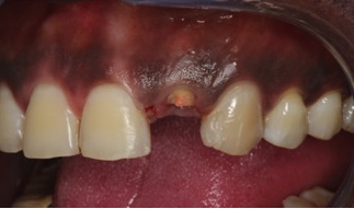
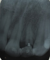
A male patient, 40, complained of the left maxillary lateral incisor with horizontal fracture at the level of the marginal gingiva. When clinical and radiographic examination, it was observed that the root canal had to narrow with little remaining tooth and unfavorable prognosis for prosthetic rehabilitation ( Figure 1 ).
Figure 1: Initial clinical case where one observes amount of remaining reduced tooth.
After thorough analysis of the case study, it was evaluated the different treatment alternatives, opting for root extraction and installation of dental implant and immediate provisionalization. It was verified the systemic condition of the patient and planned atraumatic extraction of the root with the aid of dental extractor Neodent (Neodent, Curitiba, Paraná, Brazil) ( Figure 2 ).
Figure 2: Initial radiograph.
The atraumatic dental extraction technique was initiated by sindesmotomia ( Figure 3 ) and subsequently the root canal has been prepared for fixation of the pin tractor, selected according to the diameter of the root canal ( Figure 4 ). A digital key was
Figure 3: Sindesmotomia with minimal trauma.
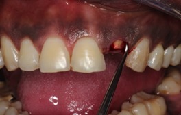
Figure 4: Preparation of conduct for fixing pin tractor.
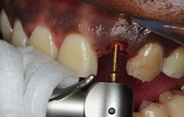
used to position the pin tractor inside the root ( Figure 5 ).
Figure 5: Pin tractor fixed in the root canal.
Subsequently, the conical tips of the steel cable was seated in the tractor pin. The cable was stretched until they fit into one of the hooks of the drive axle puller
Figure 9: alveolus after extraction with minimal trauma.
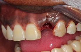
tooth ( Figure 6 ). The traction was done according to the direction of the long axis of the tooth. With this, there was obtained the periodontal ligament rupture and extracting roots ( Figures 7 and 8 ) with maximum preservation of the alveolar bone and surrounding soft tissues ( Figure 9 ).
Figure 6: Extractor dental positioned and fixed to the pin tractor.
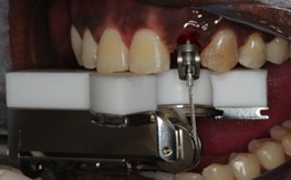
Figure 7: Extrusion of tooth with root retractor.
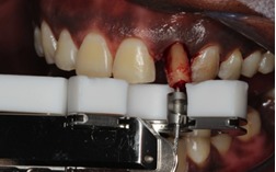
Figure 8: Extruded tooth root retractor.
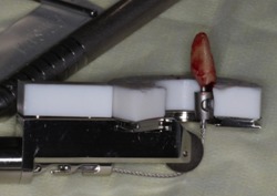
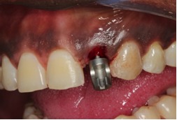
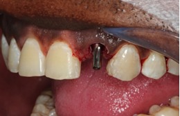
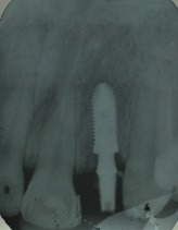
Hobbing was performed and immediate implant installation Alvin Morse Taper (Neodent, Curitiba, Paraná, Brazil) 3.75 x 11.5mm with torque above 50 Ncm ( Figures 10 and 11 ). Implant was placed in a trunnion universal ( Figure 12 ) titanium (Neodent, Neodent, Curitiba, Paraná, Brazil) and immediately made a temporary crown (Figures 13, 14 and 15 ).
Figure 10: Milling performed post extraction.
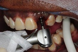
Figure 11: Implant installation - Neodent morse taper 3.75 x 11.5.
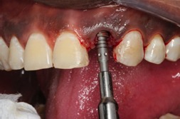
Figure 13: Placement of immediate provisional restoration.
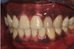
Figure 14: Post placement of provisional restoration.
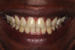
Figure 12: trunnion universal Neodent.
Discussion
The achievement extraction atraumatic is a surgical technique that can present major clinical advantages in the final outcome of prosthetic rehabilitation, it provides greater tissue preservation alveolar bone and adjacent soft tissue. 6 , 9 This has resulted in a lower possibility of changing the volume and contour of the tissues and, consequently, satisfactory aesthetic results. The method used for extraction and the manner in which the alveolus after the extraction is treated can influence the degree of preservation of the alveolar
Figure 15: Radiographs after implant installation.
bone. 14 Various techniques have been proposed for this purpose, 8 , 15 - 17 with the use of dental extractor is a method which allows in a simple way and with a minimum of trauma to extract the tooth while maintaining the integrity alveolar. The literature shows that the atraumatic extraction may be indicated especially when there is a thin thickness of bone tissue.
In the same way, implant placement immediately after tooth extraction has been proposed in order to avoid reabsorption and breakdown of tissues after extraction, 12 , 18 and decrease time to treatment. 19 The determination of the prognosis of the tooth to be implanted, the causes of tooth loss, length and width alveolar beyond the area to be implanted, should be evaluated for the indication of the technique.
In the case of immediate implants in aesthetic areas, ideally there should be a minimum distance of 5mm from the bone crest to contact point for obtaining papillae that fill the interproximal space. 20 The platform of the implant should be placed a minimum of three millimeters apical line cemento-enamel of the adjacent teeth and the apical crystal interproximal bone. These maneuvers will ensure an adequate emergence profile and facilitate the acquisition of aesthetics.
Another aspect of great importance, after immediate implant placement, consists in proper preparation and installation of the temporary restoration. The immediate provisionalization, has also been reported as an important procedure for the stability of peri-implant tissues and the aesthetic result of isolated implants in the maxilla. 7 , 10 , 18 , 21 , 22
Thus, in order to obtain successful treatment of atraumatic extraction, installation and provisionalization immediate, it must be made an appropriate choice of the case, surgical and prosthetic planning, not neglecting the postoperative care. 2
Conclusion
From the clinical case presented and the literature reviewed is posssivel conclude that with an adequate surgical-prosthetic planning associated with an accurate selection of the case, it is observed that the atraumatic extraction associated with immediate implant installation that presents clinical results that allow maintaining harmony and aesthetics of the gum line.
Contributor Information
Rudys Rodolfo De Jesus Tavarez, Post Graduation Program in Dentistry, Department of Restorative Dentistry, School of Dentistry, CEUMA University, São Luis, MA, Brazil.
Washigton Luís Machado dos Reis, Department of Restorative Dentistry, School of Dentistry, CEUMA University, São Luis, MA, Brazil.
Adrycila Teixeira Rocha, Department of Restorative Dentistry, School of Dentistry, CEUMA University, São Luis, MA, Brazil.
Leily Macedo Firoozmand, Post Graduation Program in Dentistry, Department of Restorative Dentistry, School of Dentistry, CEUMA University, São Luis, MA, Brazil.
Matheus Coêlho Bandéca, Post Graduation Program in Dentistry, Department of Restorative Dentistry, School of Dentistry, CEUMA University, São Luis, MA, Brazil.
Mateus Rodrigues Tonetto, Post Graduation Program in Dentistry, Department of Integrated Dental Science, University of Cuiaba, Cuiaba, MT, Brazil.
Adriana Santos Malheiros, Post Graduation Program in Dentistry, Department of Restorative Dentistry, School of Dentistry, CEUMA University, São Luis, MA, Brazil.
References
- 1.B Suprakash, AR Ahammed, A Thareja, R Kandaswamy, K Nilesh, S Bhondwe Mahajan. Knowledge and attitude of patients toward dental implants as an option forreplacement of missing teeth. J Contemp Dent Pract. 2013;14(1):115–118. doi: 10.5005/jp-journals-10024-1282. [DOI] [PubMed] [Google Scholar]
- 2.W Becker, M Goldstein. Immediate implant placement: treatment planning and surgical steps for successful outcome. Periodontol 2000. 2008;47:79–89. doi: 10.1111/j.1600-0757.2007.00242.x. [DOI] [PubMed] [Google Scholar]
- 3.B Mummidi, ChH Rao, AL Prasanna, M Vijay, KB Reddy, M Raju. Esthetic dentistry in patients with bilaterally missing maxillary lateral incisors: a multidisciplinary case report. J Contemp Dent Pract. 2013;14(2):348–354. doi: 10.5005/jp-journals-10024-1326. [DOI] [PubMed] [Google Scholar]
- 4.B Leblebicioglu, M Salas, Y Ort, A Johnson, VO Yildiz, DG Kim, S Agarwal, DN Tatakis. Determinants of alveolar ridge preservation differ by anatomic location. J Clin Periodontol. 2013;40(4):387–395. doi: 10.1111/jcpe.12065. [DOI] [PMC free article] [PubMed] [Google Scholar]
- 5.R Horowitz, D Holtzclaw, PS Rosen. A review on alveolar ridge preservation following tooth extraction. J Evid Based Dent Pract. 2012;12(3 Suppl):149–160. doi: 10.1016/S1532-3382(12)70029-5. [DOI] [PubMed] [Google Scholar]
- 6.M Kubilius, R Kubilius, A Gleiznys. The preservation of alveolar bone ridge during tooth extraction. Stomatologija. 2012;14(1):3–11. [PubMed] [Google Scholar]
- 7.R Noelken, BA Neffe, M Kunkel, W Wagner. Maintenance of marginal bone support and soft tissue esthetics at immediately provisionalized OsseoSpeed‚ NC implants placed into extraction sites: 2-year results. Clin Oral Implants Res. 2013 doi: 10.1111/clr.12069. [DOI] [PubMed] [Google Scholar]
- 8.E Muska, C Walter, A Knight, P Taneja, Y Bulsara, M Hahn, M Desai, T Dietrich. Atraumatic vertical tooth extraction: a proof of principle clinical study of a novel system. Oral Surg Oral Med Oral Pathol Oral Radiol. 2013;116(5):303–310. doi: 10.1016/j.oooo.2011.11.037. [DOI] [PubMed] [Google Scholar]
- 9.JB Park. Immediate placement of dental implants into fresh extraction socket in the maxillary anterior region: a case report. J Oral Implantol. 2010;36(2):153–157. doi: 10.1563/AAID-JOI-D-09-00045. [DOI] [PubMed] [Google Scholar]
- 10.I Turkyilmaz, JC Suarez, AM Company. Immediate implant placement and provisional crown fabrication after a minimally invasive extraction of a peg-shaped maxillary lateral incisor: a clinical report. J Contemp Dent Pract. 2009;10(5):73–80. [PubMed] [Google Scholar]
- 11.ML Kolinski, JE Cherry, BS McAllister, KD Parrish, DW Pumphrey, RL Schroering. Evaluation of a Variable-Thread Tapered Implant in Extraction Sites With Immediate Temporization: A 3-Year Multi-Center Clinical Study. J Periodontol. 2013 doi: 10.1902/jop.2013.120638. [DOI] [PubMed] [Google Scholar]
- 12.T De Rouck, K Collys, I Wyn, J Cosyn. Instant provisionalization of immediate single-tooth implants is essential to optimize esthetic treatment outcome. Clin Oral Implants Res. 2009;20(6):566–570. doi: 10.1111/j.1600-0501.2008.01674.x. [DOI] [PubMed] [Google Scholar]
- 13.D Botticelli, T Berglundh, J Lindhe. Hard-tissue alterations following immediate implant placement in extraction sites. J Clin Periodontol. 2004;31(10):820–828. doi: 10.1111/j.1600-051X.2004.00565.x. [DOI] [PubMed] [Google Scholar]
- 14.AA Oghli, H Steveling. Ridge preservation following tooth extraction: a comparison between atraumatic extraction and socket seal surgery. Quintessence Int. 2010;41(7):605–609. [PubMed] [Google Scholar]
- 15.DR Meneses. Exodontia Atraumatica e Previsibilidade em Reabilitacao Oral com Implantes Osseointegraveis - Relato de Casos clinicos Aplicando o Sistema Brasileiro de Exodontia Atraumática Xt Lifting®. Rev Port Estomatol Cir Maxilofac. 2009;50:11–17. [Google Scholar]
- 16.L Mahesh, TV Narayan, P Bali, S Shukla. Socket preservation with alloplast: discussion and a descriptive case. J Contemp Dent Pract. 2012;13(6):934–937. doi: 10.5005/jp-journals-10024-1257. [DOI] [PubMed] [Google Scholar]
- 17.DE Papadimitriou, A Geminiani, T Zahavi, C Ercoli. Sonosurgery for atraumatic tooth extraction: a clinical report. J Prosthet Dent. 2012;108(6):339–343. doi: 10.1016/S0022-3913(12)00169-2. [DOI] [PubMed] [Google Scholar]
- 18.JY Kan, K Rungcharassaeng, J Lozada. Immediate placement and provisionalization of maxillary anterior single implants: 1-year prospective study. Int J Oral Maxillofac Implants. 2003;18(1):31–39. [PubMed] [Google Scholar]
- 19.L Younis, A Taher, MI Abu-Hassan, O Tin. Evaluation of bone healing following immediate and delayed dental implant placement. J Contemp Dent Pract. 2009;10(4):35–42. [PubMed] [Google Scholar]
- 20.DP Tarnow, AW Magner, P Fletcher. The effect of the distance from the contact point to the crest of bone on the presence or absence of the interproximal dental papilla. J Periodontol. 1992;63(12):995–996. doi: 10.1902/jop.1992.63.12.995. [DOI] [PubMed] [Google Scholar]
- 21.P Malo, B Rangert, L Dvarsater. Immediate function of Branemark implants in the esthetic zone: a retrospective clinical study with 6 months to 4 years of follow-up. Clin Implant Dent Relat Res. 2000;2(3):138–146. doi: 10.1111/j.1708-8208.2000.tb00004.x. [DOI] [PubMed] [Google Scholar]
- 22.I Turkyilmaz, JC Suarez. An alternative method for flapless implant placement and an immediate provisional crown: a case report. J Contemp Dent Pract. 2009;10(3):89–95. [PubMed] [Google Scholar]


