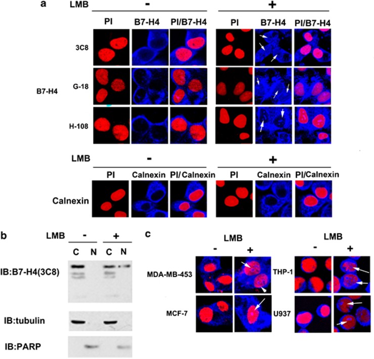
Figure 3.
Subcellular localization of B7-H4 in different cancer cell lines. (a) Confocal immunofluorescent microscopy demonstrated a nuclear translocation (indicated by white arrow) of B7-H4 in the presence of LMB. Anti-B7-H4 mAb 3C8, polyclonal antibodies G-18 and H-108 were used. Calnexin was used as a cytoplasmic marker (PI (red, DNA) and cy5 (blue, B7-H4)). (b) 20 μg total protein from each fraction (C or N) was blotted with anti-B7-H4 3C8, anti-α-tubulin or anti-PARP. (Anti-α-tubulin and anti-PARP were used for testing the house keeping protein or nuclear protein, respectively, for confirming equivalent loading). (c) B7-H4 nuclear translocation (white arrows indicate nuclear B7-H4) was detected in MDA-MB-453, MCF-7, THP-1 and U937 cells by confocal immunofluorescence microscopy. Cells were treated with solvent alone (−) or 10 ng/ml LMB (+) for 24 h and immunostained using anti-B7-H4 3C8. (PI (red,DNA) and cy5 (blue, B7-H4)).

