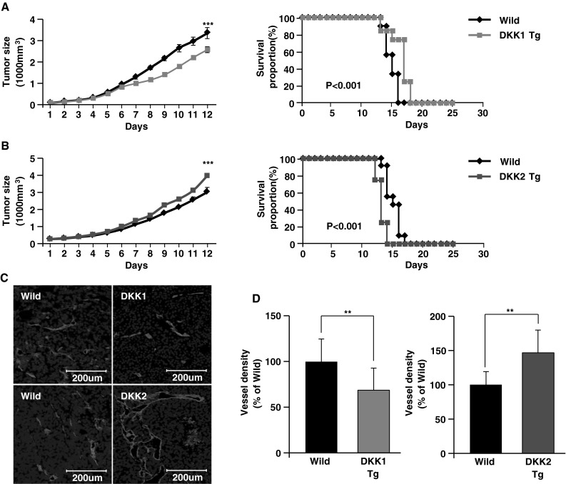
Fig. 2.

Endothelial-specific DKK1 or DKK2 expression in mouse tumors showed consistent results with the virus model. a–b B16F10 murine melanoma cells were injected subcutaneously into the abdominal space of transgenic mice that endothelial-specifically expressed DKK1 (DKK1 Tg, n = 10) or DKK2 (DKK2 Tg, n = 11), or their wild-type littermates (n = 10 and n = 11, respectively). Tumor growth was monitored over time, and data are presented as mean ± SE (left panels). The percentage of surviving mice was determined by monitoring the tumor growth-related events (tumor size >3,000 mm3) over a period of 25 days (right panels). Melanoma tumors (1,000 mm3) were prepared, excised, and stained with anti-CD31 antibody, an EC marker. Confocal imaging revealed (c) and quantified (d) CD31+ tumor vessels in virus-infected tumor sections. Nuclei were stained with DAPI. Data are mean ± SD (**p < 0.01; ***p < 0.001)
