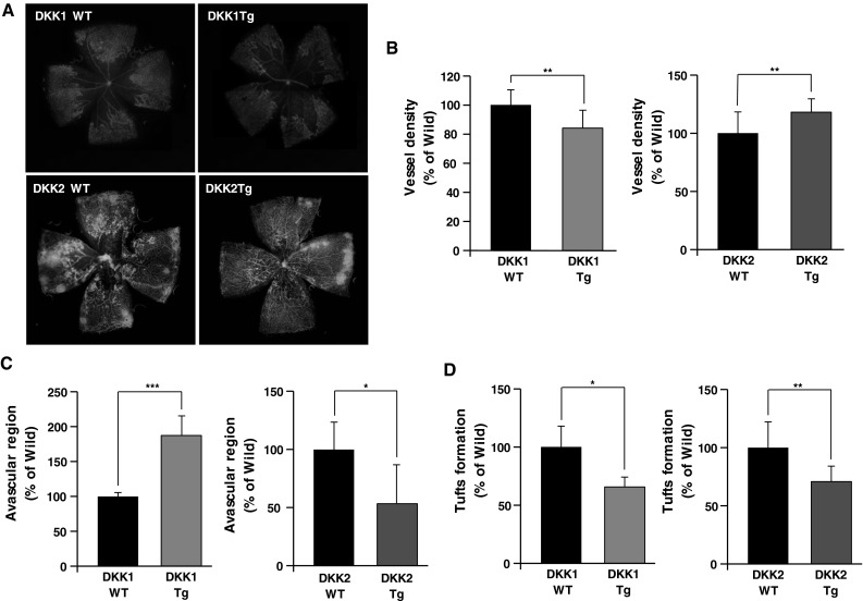
Fig. 7.

Vessel abnormality caused by oxygen-induced retinopathy (OIR) is decreased in both DKK1 Tg mice and DKK2 Tg mice, compared to wild-type littermates. a DKK1 Tg (n = 4), DKK2 Tg (n = 4), and their wild-type littermates (n = 4 in each comparison) were exposed to a 75 % O2 environment for 5 days, followed by return to normoxia (i.e., relative hypoxia) for another 5 days. Retinas were assessed after animal sacrifice and enucleation. Fluorescence-conjugated isolectin B4 staining of OIR retina vessels is presented. Vessel density (b), avascular region (c), and tuft formation (d) were quantified. Data are mean ± SD (*p < 0.05, **p < 0.01)
