Abstract
Background:
Sirtuin1 is an enzyme that deacetylates histones and several non-histone proteins including P53 during the stress. P300 is a member of the histone acetyl transferase family and enzyme that acetylates histones. Hereby, this study describes the potency combination of Salermide as a Sirtuin1 inhibitor and cholera toxin B (CTB) as a P300 activator to induce apoptosis Michigan Cancer Foundation-7 (MCF-7) and MRC-5.
Methods:
Cells were cultured and treated with a combination of Salermide and CTB respectively at concentrations of 80.56 and 85.43 μmol/L based on inhibitory concentration 50 indexes at different times. The percentage of apoptotic cells were measured by flow cytometry. Real-time polymerase chain reaction was performed to estimate the messenger ribonucleic acid expression of Sirtuin1 and P300 in cells. Enzyme linked immunosorbent assay and Bradford protein techniques were used to detect the endogenous levels of total and acetylated P53 protein generated in both cell lines.
Results:
Our findings indicated that the combination of two drugs could effectively induced apoptosis in MCF-7 significantly higher than MRC-5. We showed that expression of Sirtuin1 and P300 was dramatically down-regulated with increasing time by the combination of Salermide and CTB treatment in MCF-7, but not MRC-5. The acetylated and total P53 protein levels were increased more in MCF-7 than MRC-5 with incubated combination of drugs at different times. Combination of CTB and Salermide in 72 h through decreasing expression of Sirtuin1 and P300 genes induced acetylation of P53 protein and consequently showed the most apoptosis in MCF-7 cells, but it could be well-tolerated in MRC-5.
Conclusion:
Therefore, combination of drugs could be used as an anticancer agent.
Keywords: Apoptosis, cholera toxin B, Michigan Cancer Foundation-7, MRC-5, Salermide
INTRODUCTION
The histone acetyltransferases (HATs) induce transfer of acetyl group to lysine amino acid residues, which present the histone protein tails from acetyl coenzyme A to form ε-N-acetyl lysine.[1] Therefore, they facilitate the accessibility of transcription factors to deoxyribonucleic acid (DNA).[2] Histone deacetylases (HDACs) comprise a super family of enzymes involved in regulating the lifespan, which include regulation of transcription.[3] On the other hand, the HDACs, despite HATs, cause increase in lifespan.[4] Class III HDACs were discovered more recently and this group of deacetylases was named “Sirtuins” (silent information regulators).[5] The Sirtuins have a nicotine adenine dinucleotide as a unique cofactor to this family that is necessary for the removal of the acetyl group from the lysine residues (deacetylases function).[6] P300 is a member of the mammalian HAT protein family, which suggests that this molecule is competent of acetylating all core histone proteins and it is an important transcriptional co-activator, which may play a distinct role in regulation of a wide range of biological processes such as survival and apoptosis through histone acetylation.[7] Importantly, alteration of gene expression in cancer based on interaction of these epigenetic modifications (post-translation), play a significant role in tumorogenesis.[8] In diseases like cancer, often there occurs an imbalance between the expression of transcriptional co-activator proteins that contain HATs and HDACs families.[9] In human cancers, it has been shown that HAT activities were disrupted.[10] The HATs would often down-regulate and Sirtuin1 often up-regulate in several types of tumors.[11,12] Histones are not the only proteins that can be alteration, P300 and Sirtuin1 presumably can also catalyze acetylation and deacetylation of several non-histone proteins such as P53 (the most important tumor suppressor gene. Activation of P53 can lead to cell cycle arrest, DNA repair and apoptosis.[13] Inactivation of P300 mediates deacetylation of P53 and negatively regulates the activity of this protein.[14] Sirtuin1 mediates deacetylation of P53 and negatively regulates the activity of this protein.[15] In normal cells, P53 is a short-lived protein due to activity of mouse double minute 2 homolog (Mdm2) as a ubiquitin ligase, to inhibit and destabilize P53, so P53 levels are undetectable and inactive to induce apoptosis.[16] In response to various types and stress levels, which cause DNA damage, HATs family mediate acetylation of P53 in C terminus and blocks some of the major P53 ubiquitination sites by Mdm2.[17] This function leads to P53 protein stabilization and activation of P53 protein in human cells.[18] Hyperacetylation of P53 can also cause the hyperactivity of this protein.[19] It seems that P300 is able to acetylated and activate P53 and induce apoptosis in response to DNA damage in some cancer cells.[20] On the other hand, Sirtuin1 is able to deacetylase and inhibit P53 activity and suppress the induction of apoptosis in a number of cancer cells.[21] The balance of P53 acetylation and deacetylation, respectively mediated by the HATs (particularly P300) and HDACs (particularly Sirtuin1), is usually well-regulated, but the balance often gets upset in diseases like cancer.[22] Studies suggest that pharmacologic inhibition of Sirtuin1 may promote apoptosis by direct acetylation of P53 in some cells and can be used as an anticancer strategy.[23] Inactivation of P300 and activation of Sirtuin1 are encountered in several types of tumors such as in certain types of human cancers breast carcinomas.[24,25] The human breast carcinoma cell line Michigan Cancer Foundation-7 (MCF-7) has a wild-type P53, but this tumor suppressor gene is responsible for epigenetic event is not functional and cannot induce apoptosis.[26] These effects seem to be reversed in cancer cells by activation of P300 and inactivation of Sirtuin1.[27,28] The studies suggest that pharmacologic activation of P300 and inhibition of Sirtuin1 may promote apoptosis by direct hyperacetylation of P53 in cancer cells and could be used as an anti-cancer strategy.[29,30] Salermide is a Sirtuin1 inhibitor and cholera toxin B (CTB) is a small molecule activator of P300.[31,32] We know there are no such reports on effects combination of Salermide and CTB as an anti-tumor to induce P53 acetylation in cancer cell lines. We assumed that the apoptotic effect of these drugs is different in normal and cancer cells.[32,33] In this study, we investigated of apoptotic effects of combination Salermide as a Sirtuin1 inhibitor and CTB as a P300 activator to induce P53 protein acetylation and consequent apoptosis in MCF-7 and MRC-5 (lung fibroblasts as non-tumorigenic) cell lines.
METHODS
Cell lines, drug, treatment and culture condition
Human breast cancer MCF-7 and human lung fibroblasts MRC-5 were purchased from the National Cell Bank of Iran-Pasteur Institute. CTB (N-[4-chloro-3-trifluoromethyl-phenyl]-2-ethoxy-benzamide), an activator of P300 and Salermide (N-{3-[(2-hydroxynaphthalen-1-ylmethylene) -amino]-phenyl}-2-phenylpropionamidea) as the inhibitor of Sirtuin1 were purchased from sigma (USA). All cell lines used in the present study were cultured in Roswell Park Memorial Institute (RPMI)-1640 medium (sigma) supplemented with 10% fetal bovine serum (sigma) and 1% penicillin-streptomycin (sigma) and incubated at 37°C and in a humidified atmosphere containing 5% CO2. Drugs were dissolved in stock solutions and for treatments the compounds were diluted in dimethyl sulfoxide (DMSO) to the appropriate concentrations according to reported procedures.[16] After the cells were >80% confluent and growing exponentially in 10 cm diameter culture dishes, 105 cells (MCF-7 or MRC-5) were counted and plated in 3 cm diameter culture dishes and kept in RPMI-1640 culture medium for 24 h, which were then incubated with certain concentrations of CTB and Salermide combination, based on inhibitory concentration (IC50) index, at different times (24, 48 and 72 h).[35]
IC50 assay
The IC50 values for the CTB and Salermide in MCF-7 groups were acquired after 24 h of treatment. Briefly, 104 cells (MCF-7) were counted and placed into each well of a 24-well micro plate and were treated with various drugs concentrations (0, 6.25, 12.5, 25, 50, 100, 150, 200 μM doses) for 24 h and the Methyl Tiazolyl Tetrazolium (MTT)survival assay was then carried out for evaluating the cell viability with different drugs concentrations of MCF-7 groups. A graph of viability versus drugs concentration were used to calculate IC50 values for MCF-7 cell line.[2,27]
Flow-cytometric analysis
The percentage of apoptotic cells was measured by flow cytometry following AnnexinV flow cytometry1-histogram (FL1-H) and propidium iodide (PI) (FL2-H) labeling. A minimum of 5 × 10⁵ cells/ml were analyzed for each sample. Cells were treated with a combination of CTB (85.43 μmol/L) and Salermide (80.56 μmol/L) for 24, 48 and 72 h and then washed in phosphate-buffered saline (PBS) and re-suspended in binding buffer (10×; 5 μl). AnnexinV-FITC was added to 195 μl cell suspensions and then analysis was carried out according to the manufacturer's protocol (BMS500F1/100CE AnnexinV-FITC, eBioscience, USA). Finally, the apoptotic cells were counted by FACScan flow cytometry (Becton Dickinson, Heidelberg, Germany). These experiments were carried out in triplicate and were, independently, repeated at least 3 times.[18,36]
Reverse transcription and real-time polymer chain reaction analysis
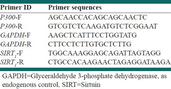
RT quantitative real-time polymerase chain reaction (Real time - PCR) was performed to quantitatively estimate the messenger ribonucleic acid (mRNA) expression of P300 and Sirtuin1 in MCF-7 and MRC-5 cells before and after treatment with a combination of CTB and Salermide at different times. Total RNA was isolated by RNeasy mini kit (Qiagen), treated by RNase-free DNase set (Qiagen) to eliminate the genomic DNA. The RNA concentration was determined using a biophotometer (Eppendorf). Total RNA (100 ng) was reverse-transcribed to complementary DNA (cDNA) by using the RevertAid™ first strand cDNA synthesis kit (Fermentas) according to the manufacturer's instructions. The maxima SYBR Green Rox qPCR master mix kit (Fermentas) was used for RT-PCR. Primer sequences are shown in Table 1. RT-PCR reactions were performed using step one plus (Applied Biosystem). The PCR amplification conditions consisted of 10 min at 95°C followed by 40 cycles of denaturation step at 95°C for 15 s and annealing and extension for 1 min at 60°C. Data were analyzed using the comparative Ct (ΔΔct) method. The relative expression level of P300 and Sirtuin1 were calculated by determining a ratio between the amount of P300 and Sirtuin1 and that of endogenous control. Melting curve analysis (60°C → 95°C increment of 0.3°C) was used to determine the melting temperature of specific amplification products and primer dimmers. These experiments were carried out in triplicate and were independently repeated at least 3 times.[37]
Table 1.
Primers used in real-time polymerase chain reaction

Bradford protein assay
Total (intracellular) protein concentration was determined by Bradford method. Bradford protein quantification assay is an accurate procedure for determining the concentration of protein in solution based on binding of Coomassie Blue dye to proteins. This method was carried out before enzyme linked immunosorbent assay (ELISA) assay. Total proteins extracted from MCF-7 and MRC-5 cells before and after combination of CTB and Salermide treatment will be described later in ELISA assay. Bovine serum albumin was used at 9 different concentrations (0.25, 0.5, 1, 1.5, 2, 3, 4, 5 and 6 mg/ml) to prepare a protein standard. After diluting the protein standards, the stock dye reagent was prepared (500 mg Coomassie Blue was dissolved in 500 ml methanol and was added to 100 ml phosphoric acid and 50 ml double-distilled water H2O [ddH2O]) that was diluted in 8 ml ddH2O. A total of 2 ml of dye reagent was added to each tube of protein standard and was incubated at room temperature for at least 5 min. Absorbance of the protein standards and experimental samples were carried out by the spectrophotometry (Bausch and Lomb, Germany) at 595 nm and finally, a standard curve was plotted.[38]
Acetylated and total P53 sandwich ELISA assay
ELISA was used to specifically detect endogenous levels of total and acetylated P53 protein generation in MCF-7 and MRC-5 cells in the presence or absence combination of CTB and Salermide at different times (24, 48 and 72 h). Acetylated and total P53 ELISA Kit was prepared by cell signaling technology and cell lyses were prepared at the first step. Briefly, cells were harvested under treated conditions by the combination of CTB and Salermide at different times, media was removed and cells were washed with cold PBS. PBS was removed and 0.5 ml of ice-cold cell lyses buffer with 1 mM phenyl methyl sulfonyl fluoride was added to each plate and incubated on ice for 5 min. Cells were scraped off the plate and were transferred to an appropriate tube and a freeze-and-thaw test was performed 3 times. The tubes were micro-centrifuged at 4°C, for 10 min and the supernatant was transferred to a new tube. This supernatant was the cell lysates. For doing ELISA assay at first, the concentration of total protein extract in both cell lysates was determined using the Bradford assay. Sandwich ELISA was performed according to the manufacturer's protocol. Finally, the absorbance of samples were read in ELISA reader (Hyperion, Germany) at 450 nm wavelength and ELISA analysis was calculated based on control index. All experiments were carried out in triplicate.[39]
Statistical analysis
All the quantitative data were presented as the mean ± standard deviation. One-way analysis of variance (ANOVA) with post-hoc test was performed to determine the statistical significance among different groups by using the SPSS software package 16.0 IBM modeler (2009). Significance was accepted at a level of P < 0.05.
RESULTS
IC50
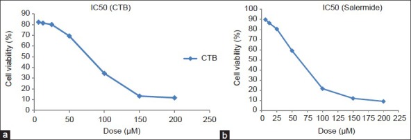
After the treatment of MCF-7 cells with MTT solution in this assay, the dark blue formazan crystals were seen in cells, which indicated their metabolic activity. The reduction in the number of cells was dependent on the cell type as shown by the half-maximal IC50 index. The IC50 values for the CTB and Salermide were established [Figure 1a and b]. The results showed that the essential CTB and Salermide concentration to achieve the IC50 in MCF-7 cells at 24 h respectively were 85.43 and 80.56 μmol/L [Figure 1a and b].
Figure 1.

Inhibitory concentration (IC50) assay for half-maximal IC analysis of cholera toxin B (CTB). (a) And Salermide. (b) In MCF-7 cancer cell lines after 24 h of treatment. Cells were incubated with or without the CTB and Salermide using 0, 6.25, 12.5, 25, 50, 100, 150 and 200 μM doses and the relative amount of viable cells were estimated by measuring the absorbance of the cell suspension after incubation with MTT assay was carried out and a graph of viability versus drug concentration was used to calculate IC50 values for MCF-7cell line
Flow-cytometry
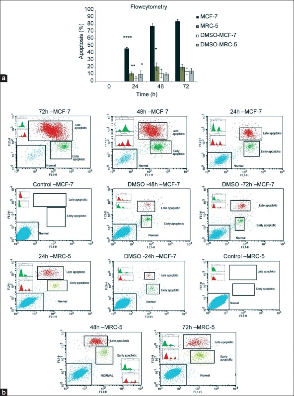
To establish the anti-apoptosis potential of the combination CTB and Salermide, we first investigated the effects of this combination on the proliferation of the breast carcinoma cell line (MCF-7). The flow cytometry results showed that this combination drugs at different times (24, 48 and 72 h) could significantly induce apoptosis in MCF-7 cells and it was increased with ascending time (P < 0.001) [Figure 2a and b]. Combination drugs treatment arrested MCF-7 cell proliferation (≥95% of inhibition) in 72 h, whereas its inhibition on MRC-5 cells proliferation in all different times were negligible; although, different times (24, 48 and 72 h) could significantly induce apoptosis in MRC-5 cells and it was increased with ascending time (P > 0.05) [Figure 2a and b]. MCF-7 apoptotic cells showed a sharp increase at all times in comparison with MRC-5 cells (P < 0.001). DMSO was used in the control sample (vehicle Drugs) a small amount of cell death in both cell lines at different times was observed (P < 0.05) [Figure 2a and b].
Figure 2.

Relative levels of apoptotic cells in Michigan Cancer Foundation (MCF-7) and MRC-5 treated with combination of drugs for different times. Cells incubated with the vehicle dimethyl sulfoxide (DMSO) were used as a control. (a and b) The percentage of apoptotic cells was measured using the AnnexinV FITC (FL1-H) and propidium iodide (PI) (FL2-H) assay. ****P < 0.001 versus all other groups MCF-7 cells treated with combination of cholera toxin B and Salermide. ***P < 0.05 versus all other groups MRC-5 cells. **P < 0.05 versus all other groups MCF-7 cells incubated with the DMSO was used as a control. *P < 0.05 versus 48 and 72 h groups MRC-5 cells incubated with the DMSO were used as control. (b) Cells that are AnnexinV-positive and propidium iodide negative are in early apoptosis as phosphatidyl serine (PS) translocation has occurred; although, the plasma membrane remains intact. Cells that are positive for both AnnexinV and PI either are in the late stages of apoptosis or are already dead as PS translocation has occurred and the loss of plasma membrane integrity is visible
RT-PCR
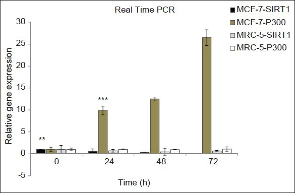
We examined the inhibitory effects of Salermide and the activator effects of CTB in combination of drugs at different times on the mRNA expression of Sirtuin1 and P300 on MCF-7 and MRC-5 cells using RT-PCR. The P300 gene expression was dramatically up-regulated by CTB treatment in combination of drugs with an ascending time in MCF-7 cells, in particular, at 72-h treatment its increased expression was significantly raised [Figure 3, P < 0.001]. The Sirtuin1 gene expression was dramatically down-regulated by Salermide treatment in combination of drugs with time in MCF-7 cells [Figure 3, P < 0.01]. In MRC-5 cells, the expression of P300 was increased 72 h after the CTB treatment (P > 0.05), but it was not statistically significant in different times (P > 0.05) [Figure 3] and the expression of Sirtuin1 was also significantly reduced at all treatment times, but the difference in the expression among different times were not statistically significant (P > 0.05) [Figure 3]. However, in combination of drugs the effect of CTB treatment on up-regulation of P300 and Salermide treatment on down-regulation of Sirtuin1 expression were significantly higher in MCF-7 cells in comparison with MRC-5 cells [Figure 3, P < 0.01].
Figure 3.

Results of real-time quantitative polymerase chain reaction before and after combination of cholera toxin B and Salermide at different times on the P300 and Sirtuin1 messenger ribonucleic acid expression in Michigan Cancer Foundation (MCF-7) and MRC-5 cells. Relative expression levels of each gene were obtained by using the comparative Ct (ΔΔct) method. Histone acetyltransferases activator and Sirtuin1 inhibitor-caused epigenetically activated P300 and inhibited Sirtuin1. Values were the means of triplicate experiments ***P< 0.001 versus and **P < 0.05 versus control (non-treated drugs) and other MCF-7 groups. No significant difference was seen in other groups
Acetylated and total P53 sandwich ELISA
To investigate further distinct effects of combination drugs on cell apoptosis, the ELISA analysis was conducted in MCF-7 (wild-type P53) and MRC-5 cells. The cells were treated at different times (0, 24, 48 and 72 h) with a combination of drugs, to study its effects on the acetylation status of the P53 as targets of P300 and Sirtuin1. The results of ELISA analysis was calculated based on control index. The results showed that combination drugs could induce P53 acetylation in MCF-7 and MRC-5 cells and significantly increase in the total protein levels with ascending time until 48-h treatment in MCF-7 cells, but not in MRC-5 cells (P < 0.05). Interestingly, between 48 and 72 h, decrease of protein levels was observed in MCF-7 cells [Figure 4a]. Notably, after treatment by combination drugs at all different times, the acetylated P53 protein levels in MCF-7 cells was significantly higher than in MRC-5 cells (P < 0.05) [Figure 4a]. Consistently, we also performed the mentioned method to examine the total P53 protein levels in both cell lines. These results were similar to results of acetylated P53 except for an increase in the total P53 protein levels in MRC-5 cells until 48 h after treatment (P < 0.05) [Figure 4b]. In the control samples (using DMSO without combination drugs), there was negligible effect on inducing total and acetylated P53 in both cell lines at the different times of study (P > 0.05) [Figure 4a and b].
Figure 4.

The results of enzyme linked immunosorbent assay analysis based on the control index for acetylated and total P53 protein generated in Michigan Cancer Foundation (MCF-7) and MRC-5 cells were treated with and without drugs at different times. Cells were treated with combination of drugs for 0, 24, 48 and 72 h. Values are mean + standard error of triplicate experiments. (a) ***P < 0.001 versus all other groups in different time. **P < 0.05 versus acetylated P53 in MCF-7 with combination of drugs treatment at 72 h. (b) *P < 0.001 versus all other groups in different time. **P < 0.05 versus total P53 in MRC-5 without drugs treatment and with drugs treatment at time 48 and 72 h. No significant difference was shown in total and acetylated P53 content of other groups
DISCUSSION
The potencies and functional mechanisms of the combination drugs (CTB and Salermide) were studied at the concentration that was confirmed by IC50 on the MCF-7 as breast cancer cell line and MRC-5 as non-tumorigenic control cells. In recent years, a few researchers have described the therapeutic effect of P300 activation and inhibitor of Sirtuin1, but there are no reports of their combination effects on the diverse types of cancerous and non-cancerous cells.[29,40] The role of P300 and Sirtuin1 during stress are complex and their activator and inhibitor effects are probably cell context-specific.[41,42] Until date, no clear explanation exists about molecular mechanisms of the combination of P300 activators (particularly CTB) and Sirtuin1 inhibitors (particularly Salermide) in different cells or about comparing their effects on cancerous and normal cells at different times. It is notable that in this study, the apoptotic potency of combination drugs was examined on the MCF-7 because expression level of P300 and Sirtuin1 are respectively down-regulated and up-regulated, P53 is wild-type and this kind of cancer is the most prevalent malignancy in woman.[43] In this study, treatment by the combination of CTB and Salermide at various time showed a time-dependent increase in apoptotic cell count of the cancerous cells, but not in non-tumorigenic MRC-5 cells as measured by flow-cytometric assay. On the other hand, although CTB and Salermide could effectively induce activation of P300 and inhibition of Sirtuin1 and subsequent cancer cell death it did not have such an effect on fibroblastic cells (poor cell apoptosis). These results revealed that probable apoptotic sensitivity of MRC-5 cells to CTB and Salermide stimulation was negligible, which agreed with the previous findings of Lara et al.[4] showed that Sirtuin inhibitors can induce massive apoptosis in cancerous, but not in non-malignant cells. The severe apoptotic effect in the MCF-7 was observed after 72 h of incubation with CTB and Salermide, so as approximately 4% of MCF-7 cells, were viable while at this time MRC-5 cells showed only a slight increase in level of apoptosis (only about 25%). It seems that this result is because of the presence of the wild-type P53. Our results were supported by the findings of Janknecht[44] which shows modulation of P300 function may consider novel therapies directed against tumors with wild-type P53. Therefore, it was assumed that in MCF-7 cells, up-regulation of Sirtuin1 and degradation of P300 expression promoted cell survival and combination of CTB and Salermide could induce apoptosis in these cancer cells with wild-type P53 in a time-dependent manner. Our results were matched with the findings of Zou et al.[1] They state that trichostatin A (HDAC inhibitor) can induce apoptosis in BGC-823 gastric cell line in a time-dependent manner. In addition, our results were consistent with the findings of Vempati et al.[7] and Molvaersmyr et al.[41] who demonstrated that P300 may be an important regulator of wild-type P53 function and P300 fails to acetylated mutant P53. It was observed that combination of CTB and Salermide was ineffective to promote acetylation of P53 and it seemed that it cannot activate P53 to induce apoptosis in fibroblastic cells. Once it was determined that combination of CTB and Salermide anti-tumor activity was primarily because of the promotion of apoptosis, we decided to study the molecular mechanisms involved in this process. We first studied the expression of P300 and Sirtuin1 in combination of CTB and Salermide-mediated apoptosis by using RT-PCR. The results of RT-PCR assay indicated that Salermide mediated inhibition of Sirtuin1 expression in a time-dependent manner and CTB was responsible for the over-expression of P300 in a time-dependent manner in MCF-7 cells (progressive increase in P300 mRNA levels after 24, 48 and 72 h) by exposure to CTB. This was in accordance with the findings of Mantelingu et al.[27] which showed that incubation of P300 with increasing concentration of CTBP results in a dose-dependent enhancement of P300 HAT activity by HAT assay. It was found that P300 expression level in MRC-5 cells by the treatment of CTB slightly increase in a time-dependent manner only in 72 h. No alteration of P300 expression levels between the other groups of (non-treatment, 24 and 48 h) MRC-5 samples (normal P300 expression) was found. We also found that Sirtuin1 expression levels in MRC-5 cells was low and after treatment with Salermide in combination of drugs slightly decreased in a time dependent manner. Importantly, we found that in the non-cancerous cells, Sirtuin1 silencing due to Salermide was at least equivalent to MCF-7 breast cancer cells. Subsequently, we observed that Sirtuin1 silencing occurred particularly after 48 h of Salermide treatment in MCF-7 cells. More importantly, it was discovered that in the non-cancerous cells, P300 expression may increase because CTB was at least equivalent to MCF-7 breast cancer cells. Subsequently, we observed that strong P300 transcription occurred particularly after 48 h of CTB treatment in MCF-7 cells. However, Sirtuin1 silencing and relative stability of P300 expression until 48 h of treatment and only little enhancement after 72-h incubation was not effective on the cell apoptosis and viability of the non-cancerous cells. Importantly, it seemed that low levels of apoptosis, which was observed in all MRC-5 samples after treatment with a combination of CTB and Salermide, was more significantly relevant to apoptotic effects of DMSO as carrier and solvent of drugs on fibroblastic cells. This was in agreement with the findings of Ikushima and Miyazono[45] that Smad complex and transforming growth factor beta (TGF-β) recruit co-activators such as P300 to induce growth arrest and/or apoptosis through P53 protein interaction in cancerous cells and loss of P300 genes could lead to tumor progression, which in contrast with normal human epithelial cells seemed to be refractory to P300 activation and Sirtuin1 silencing (in the absence of applied stress, Sirtuin1 silencing induces growth arrest and/or apoptosis in human epithelial cancerous. Inversely, these observations disagree with the findings of Bedford et al.[46] that knockdown and lack of P300 gene suppress cell growth and increase apoptosis effect in the cancerous cells. The results indicated that function of P300 and Sirtuin1 were different in MCF-7 and MRC-5 cells and that Sirtuin1 inhibition and P300 activation might enable MCF-7 cancer cell apoptosis, but it seems non-essential for the apoptosis of lung fibroblast cells. These results were also similar to the study of Karamouzis et al.[47] that the down-regulation of P300 expression was observed in breast cancer cells and P300 may provide a tumor suppressor-like function although the tumor-suppressor function of P300 is still unclear. Also, similar to the study of Kojima et al.[48] who noted that the up-regulation of Sirtuin1 expression was observed in breast cancer MCF-7 cells and that treatment with a Sirtuin1 inhibitor, Sirtinol, induced inhibition of Sirtuin1 expression and subsequently inhibited cell growth in human prostate cancer cell lines. These results were dissimilar with the findings of Fermento et al.[24] which showed up-regulation of P300 expression in murine mammary adenocarcinoma LM3 cells. Our results showed that the effect of 24 and 48 h of treatment of MRC-5 cells by CTB in combination of drugs on the expression of P300 mRNA were similar to the non-treatment condition. Subsequently, we used RT-PCR to evaluate P300 and Sirtuin1 expression in both cell lines before treatment. We observed lower expression of P300 and over-expression of Sirtuin1 in MCF-7 cells in comparison to MRC-5 cells, which might be explained by the fact that decrease of P300 and increased level of Sirtuin1 expression in MCF-7 cells leads to inhibited apoptosis and mediated survival in response to stress. Therefore, these results suggest that P300 and Sirtuin1 keep a balance of specific acetylation levels for proper cellular function and mediate survival in normal cells. This finding was in accordance with the previous findings of Peck et al.[25] that indicate that the degree of acetylation is largely mediated by a balance between HATs and HDAC in normal cells. Our findings indicate that stimulation of cell death by CTB and Salermide requires respectively the activation of P300 and Sirtuin1 genes, showing the combination of drugs potential anti-tumor effect. This observation, similar to a recent study by Chen et al.[49] has showed that small molecule activators of P300 may act as anti-cancer agents. To investigate further and determine the total and acetylated status of P53 in response to the combination of CTB and Salermide in the cells, ELISA analysis was performed after ensuring the level of total protein concentration using the Bradford method. We found a remarkable increase in P53 acetylation level in a time-dependent manner until 48 h in MCF-7 but not in MRC-5 cells. Consistently, combination of drugs induced a similar increase of total P53 in both cell lines as proposed earlier. In MRC-5 cells, we saw a little increase of total and acetylated P53 protein levels in a time-dependent manner of combination CTB and Salermide treatment compared with MCF-7 cells, which showed CTB over-expression of P300 and Salermide lower-expression of Sirtuin1 in combination of drugs resulted in up-regulation of acetylated P53 and subsequently P53 activation in MCF-7 cells (based on previous results by flow-cytometry and RT-PCR assay). Different researchers reported various data about how P300 activation and inhibition of Sirtuin1 could induce (Dornan et al.[10] and Lain et al.[50]) or not induce (Vempati et al.[7] and Ota et al.[30]) P53 acetylation in cancer cell lines. A direct correlation between total and acetylated P53 protein levels and combination of CTB and Salermide toxicity in the MCF-7 cell line was discovered. These results suggested that incubation of MCF-7 with a combination of CTB and Salermide might induce hyperacetylation of P53 protein and apoptosis in MCF-7 cells. Our results indicated that slight decrease of total and acetylated P53 was evident at 72-h incubation in MCF-7 cells. We suggested that although an increase in total and acetylated P53 levels in response to P300 activation and Sirtuin1 inhibition at this time was accorded, P53 protein was undetectable by ELISA assay due to the release of proteases and degradation process inside the cancer cells after 48 h of cell death. A decrease in total P53 level in response to drugs incubation after 48 h in the control MRC-5 cells was not observed, which indicated that combination of drugs could not induce significant apoptosis even after 48 h in MRC-5 cells. These observations showed that although in MCF-7 cells, P53 was wild-type it was a target for deacetylation of P300 and acetylation of Sirtuin1 and so could not induce apoptosis due to this aberrant epigenetic event. These data were in accordance with the findings of Iyer et al.[51] that P300 contributes to maintain P53 stability by regulating its ubiquitination and P53 acetylation (activate P53 function) may promote the P53-P300 complex (epigenetic changes). Furthermore in accordance with the findings of Pruitt et al.[52] who reported that Sirtuin1 is a cancer-related gene and inhibits P53 function through epigenetic changes. Our findings suggested that acetylation of wild-type P53 as a tumor suppressor might lead to activation of apoptotic program and was integral to cytotoxic activity of the combination drugs to induce massive apoptosis in less than 24 h of treatment in MCF-7 cells. This finding further highlights our theoretical assumptions, indicating that activation of P300 and inhibition of Sirtuin1 is required for the induction of cell death and P53 acetylation in cancer cells only for wild-type P53 and combination of CTB and Salermide triggers neither cell death nor P53 acetylation in normal cells. Our results are similar to the previous study of Gu and Roeder[53] that indicates a novel pathway for wild-type P53 acetylated by co-activator, P300 to induce apoptosis in some cell types. Kamel et al.[54] suggested that Sirtuin inhibitors are targets for Sirtuin1 and Sirtuin2 and P53 acetylation is important for the cell death induction. We suggested that combination of P300 activators, such as CTB and inhibitors of Sirtuin1 like Salermide, may function through common pathways and mediate their cytotoxic effects through targeting P53 and its acetylation.
CONCLUSIONS
We formulated the hypothesis that dysfunction of P300 and up-regulation of Sirtuin1 could plays crucial roles in inhibiting pro-apoptotic protein expression in cancer cells, so it seems that P53 aberrantly repressed in MCF-7 cells. Thus, the treatment with a combination of CTB and Salermide could predominantly induce apoptosis through enhancing activity of P300 and inhibition of Sirtuin1 and consequent hyperacetylation and reactivation of tumor suppressor P53 to induce cell death in MCF-7 cancer cells. We described that combination of CTB and Salermide as an activator of P300 and inhibitor of Sirtuin1 could make a promising novel class of agents to the future anti-tumorigenic drugs that target acetylation of proteins; thus might be a target for cancer therapy.
ACKNOWLEDGMENTS
We would like to appreciate Dr. Mohammad Kazemi, Dr. Nafiseh Esmaeili, Dr. Marjan Gharagozloo and Dr. Batol Hashemibani for their sincere help without which this study could not be performed.
Footnotes
Source of Support: Nil
Conflict of Interest: None declared
REFERENCES
- 1.Zou XM, Li YL, Wang H, Cui W, Li XL, Fu SB, et al. Gastric cancer cell lines induced by trichostatin A. World J Gastroenterol. 2008;14:4810–5. doi: 10.3748/wjg.14.4810. [DOI] [PMC free article] [PubMed] [Google Scholar]
- 2.Giahuai T, Shundong C, Yuehua M, Richard LP, Delong L. Novel histone deacetylase inhibitors in clinical trials as anti-cancer agents. Hematol Oncol. 2010;3:15–28. doi: 10.1186/1756-8722-3-5. [DOI] [PMC free article] [PubMed] [Google Scholar]
- 3.Rotili D, Tarantino D, Carafa V, Paolini C, Schemies J, Jung M, et al. Benzodeazaoxaflavins as sirtuin inhibitors with antiproliferative properties in cancer stem cells. J Med Chem. 2012;55:8193–7. doi: 10.1021/jm301115r. [DOI] [PubMed] [Google Scholar]
- 4.Lara E, Mai A, Calvanese V, Altucci L, Lopez-Nieva P, Martinez-Chantar ML, et al. Salermide, a Sirtuin inhibitor with a strong cancer-specific proapoptotic effect. Oncogene. 2009;28:781–91. doi: 10.1038/onc.2008.436. [DOI] [PubMed] [Google Scholar]
- 5.Calvanese V, Fraga MF. SirT1 brings stemness closer to cancer and aging. Aging (Albany NY) 2011;3:162–7. doi: 10.18632/aging.100272. [DOI] [PMC free article] [PubMed] [Google Scholar]
- 6.Iglesias-Linares A, Yañez-Vico RM, González-Moles MA. Potential role of HDAC inhibitors in cancer therapy: Insights into oral squamous cell carcinoma. Oral Oncol. 2010;46:323–9. doi: 10.1016/j.oraloncology.2010.01.009. [DOI] [PubMed] [Google Scholar]
- 7.Vempati RK, Jayani RS, Notani D, Sengupta A, Galande S, Haldar D. P300-mediated acetylation of histone H3 lysine 56 functions in DNA damage response in mammals. J Biol Chem. 2010;285:28553–64. doi: 10.1074/jbc.M110.149393. [DOI] [PMC free article] [PubMed] [Google Scholar]
- 8.Lacroix M, Toillon RA, Leclercq G. P53 and breast cancer, an update. Endocr Relat Cancer. 2006;13:293–325. doi: 10.1677/erc.1.01172. [DOI] [PubMed] [Google Scholar]
- 9.Isharwal S, Miller MC, Marlow C, Makarov DV, Partin AW, Veltri RW. P300 (histone acetyltransferase) biomarker predicts prostate cancer biochemical recurrence and correlates with changes in epithelia nuclear size and shape. Prostate. 2008;68:1097–104. doi: 10.1002/pros.20772. [DOI] [PMC free article] [PubMed] [Google Scholar]
- 10.Dornan D, Shimizu H, Perkins ND, Hupp TR. DNA-dependent acetylation of P53 by the transcription coactivator P300. J Biol Chem. 2003;278:13431–41. doi: 10.1074/jbc.M211460200. [DOI] [PubMed] [Google Scholar]
- 11.Emanuele S, Lauricella M, Tesoriere G. Histone deacetylase inhibitors: Apoptotic effects and clinical implications (Review) Int J Oncol. 2008;33:637–46. [PubMed] [Google Scholar]
- 12.Wang TY, Schoene NW, Kim EK, Kim YS. Pleiotropic effects of the sirtuin inhibitor sirtinol involve concentration-dependent modulation of multiple nuclear receptor-mediated pathways in androgen-responsive prostate cancer cell LNCaP. Mol Carcinog. 2012;284:3823–32. doi: 10.1002/mc.21906. [DOI] [PubMed] [Google Scholar]
- 13.Cazzalini O, Perucca P, Savio M, Necchi D, Bianchi L, Stivala LA, et al. Interaction of p21(CDKN1A) with PCNA regulates the histone acetyltransferase activity of P300 in nucleotide excision repair. Nucleic Acids Res. 2008;36:1713–22. doi: 10.1093/nar/gkn014. [DOI] [PMC free article] [PubMed] [Google Scholar]
- 14.Cang S, Ma Y, Liu D. New clinical developments in histone deacetylase inhibitors for epigenetic therapy of cancer. J Hematol Oncol. 2009;2:22. doi: 10.1186/1756-8722-2-22. [DOI] [PMC free article] [PubMed] [Google Scholar]
- 15.Milne JC, Denu JM. The Sirtuin family: Therapeutic targets to treat diseases of aging. Curr Opin Chem Biol. 2008;12:11–7. doi: 10.1016/j.cbpa.2008.01.019. [DOI] [PubMed] [Google Scholar]
- 16.Liu G, Chen X. Regulation of the P53 transcriptional activity. J Cell Biochem. 2006;97:448–58. doi: 10.1002/jcb.20700. [DOI] [PubMed] [Google Scholar]
- 17.Knights CD, Catania J, Di Giovanni S, Muratoglu S, Perez R, Swartzbeck A, et al. Distinct P53 acetylation cassettes differentially influence gene-expression patterns and cell fate. J Cell Biol. 2006;173:533–44. doi: 10.1083/jcb.200512059. [DOI] [PMC free article] [PubMed] [Google Scholar]
- 18.Gregoretti IV, Lee YM, Goodson HV. Molecular evolution of the histone deacetylase family: Functional implications of phylogenetic analysis. J Mol Biol. 2004;338:17–31. doi: 10.1016/j.jmb.2004.02.006. [DOI] [PubMed] [Google Scholar]
- 19.Olmos Y, Brosens JJ, Lam EW. Interplay between SIRT proteins and tumour suppressor transcription factors in chemotherapeutic resistance of cancer. Drug Resist Updat. 2011;14:35–44. doi: 10.1016/j.drup.2010.12.001. [DOI] [PubMed] [Google Scholar]
- 20.Chim CS, Wong AS, Kwong YL. Absence of P300 gene promoter methylation in acute leukemia. Cancer Genet Cytogenet. 2004;150:164–7. doi: 10.1016/j.cancergencyto.2003.09.006. [DOI] [PubMed] [Google Scholar]
- 21.Iwahashi S, Ishibashi H, Utsunomiya T, Morine Y, Ochir TL, Hanaoka J, et al. Effect of histone deacetylase inhibitor in combination with 5-fluorouracil on pancreas cancer and cholangiocarcinoma cell lines. J Med Invest. 2011;58:106–9. doi: 10.2152/jmi.58.106. [DOI] [PubMed] [Google Scholar]
- 22.Escande C, Chini CC, Nin V, Dykhouse KM, Novak CM, Levine J, et al. Deleted in breast cancer-1 regulates SIRT1 activity and contributes to high-fat diet-induced liver steatosis in mice. J Clin Invest. 2010;120:545–58. doi: 10.1172/JCI39319. [DOI] [PMC free article] [PubMed] [Google Scholar]
- 23.Giahuai T, Shundong C, Yuehua M, Richard LP, Delong L. Novel histone deacetylase inhibitors in clinical trials as anti-cancer agents. J Hematol Oncol. 2010;3:15–28. doi: 10.1186/1756-8722-3-5. [DOI] [PMC free article] [PubMed] [Google Scholar]
- 24.Fermento ME, Gandini NA, Lang CA, Perez JE, Maturi HV, Curino AC, et al. Intracellular distribution of P300 and its differential recruitment to aggresomes in breast cancer. Exp Mol Pathol. 2010;88:256–64. doi: 10.1016/j.yexmp.2010.01.007. [DOI] [PubMed] [Google Scholar]
- 25.Peck B, Chen CY, Ho KK, Di Fruscia P, Myatt SS, Coombes RC, et al. SIRT inhibitors induce cell death and P53 acetylation through targeting both SIRT1 and SIRT2. Mol Cancer Ther. 2010;9:844–55. doi: 10.1158/1535-7163.MCT-09-0971. [DOI] [PubMed] [Google Scholar]
- 26.Dworkin AM, Huang TH, Toland AE. Epigenetic alterations in the breast: Implications for breast cancer detection, prognosis and treatment. Semin Cancer Biol. 2009;19:165–71. doi: 10.1016/j.semcancer.2009.02.007. [DOI] [PMC free article] [PubMed] [Google Scholar]
- 27.Mantelingu K, Kishore AH, Balasubramanyam K, Kumar GV, Altaf M, Swamy SN, et al. Activation of P300 histone acetyltransferase by small molecules altering enzyme structure: Probed by surface-enhanced Raman spectroscopy. J Phys Chem B. 2007;111:4527–34. doi: 10.1021/jp067655s. [DOI] [PubMed] [Google Scholar]
- 28.McCarthy AR, Pirrie L, Hollick JJ, Ronseaux S, Campbell J, Higgins M, et al. Synthesis and biological characterisation of sirtuin inhibitors based on the tenovins. Bioorg Med Chem. 2012;20:1779–93. doi: 10.1016/j.bmc.2012.01.001. [DOI] [PubMed] [Google Scholar]
- 29.Devipriya B, Parameswari AR, Rajalakshmi G, Palvannan T, Kumaradhas P. Exploring the binding affinities of P300 enzyme activators CTPB and CTB using docking method. Indian J Biochem Biophys. 2010;47:364–9. [PubMed] [Google Scholar]
- 30.Ota H, Tokunaga E, Chang K, Hikasa M, Iijima K, Eto M, et al. Sirt1 inhibitor, Sirtinol, induces senescence-like growth arrest with attenuated Ras-MAPK signaling in human cancer cells. Oncogene. 2006;25:176–85. doi: 10.1038/sj.onc.1209049. [DOI] [PubMed] [Google Scholar]
- 31.Nikbakht M, Salahshoor MR, Mardani M, Rabbani M, Hashemibeni B, Gharagozloo M, et al. The apoptotic effects of Sirtuin1 inhibitor on MCF-7 and MRC-5 cell lines. Res Pharm Sci. 2013;8:79–89. [PMC free article] [PubMed] [Google Scholar]
- 32.Wong PP, Pickard A, McCance DJ. P300 alters keratinocyte cell growth and differentiation through regulation of p21(Waf1/CIP1) PLoS One. 2010;5:e8369. doi: 10.1371/journal.pone.0008369. [DOI] [PMC free article] [PubMed] [Google Scholar]
- 33.Nikbakht Dastjerdi M, Salahshoor MR, Mardani M, Hashemibeni B, Roshankhah SH. The effect of CTB on P53 protein acetylation and consequence apoptosis on MCF-7 and MRC- cell lines. Advanced Biomedical Research. 2013;2:147–156. doi: 10.4103/2277-9175.108005. [DOI] [PMC free article] [PubMed] [Google Scholar]
- 34.Chen S, Sang N. Histone deacetylase inhibitors: the epigenetic therapeutics that repress hypoxia-inducible factors. BioMed Research International 2011. 2010:197946–197946. doi: 10.1155/2011/197946. [DOI] [PMC free article] [PubMed] [Google Scholar]
- 35.Gallinari P, Di Marco S, Jones P, Pallaoro M, Steinkühler C. HDACs, histone deacetylation and gene transcription: From molecular biology to cancer therapeutics. Cell Res. 2007;17:195–211. doi: 10.1038/sj.cr.7310149. [DOI] [PubMed] [Google Scholar]
- 36.Kim HJ, Bae SC. Histone deacetylase inhibitors: Molecular mechanisms of action and clinical trials as anti-cancer drugs. Am J Transl Res. 2011;3:166–79. [PMC free article] [PubMed] [Google Scholar]
- 37.Yang T, Fu M, Pestell R, Sauve AA. SIRT1 and endocrine signaling. Trends Endocrinol Metab. 2006;17:186–91. doi: 10.1016/j.tem.2006.04.002. [DOI] [PubMed] [Google Scholar]
- 38.Jones PA, Baylin SB. The fundamental role of epigenetic events in cancer. Nat Rev Genet. 2002;3:415–28. doi: 10.1038/nrg816. [DOI] [PubMed] [Google Scholar]
- 39.Heltweg B, Gatbonton T, Schuler AD, Posakony J, Li H, Goehle S, et al. Antitumor activity of a small-molecule inhibitor of human silent information regulator 2 enzymes. Cancer Res. 2006;66:4368–77. doi: 10.1158/0008-5472.CAN-05-3617. [DOI] [PubMed] [Google Scholar]
- 40.Kim JC, Kim DD, Lee YM, Kim TW, Cho DH, Kim MB, et al. Evaluation of novel histone deacetylase inhibitors as therapeutic agents for colorectal adenocarcinomas compared to established regimens with the histoculture drug response assay. Int J Colorectal Dis. 2009;24:209–18. doi: 10.1007/s00384-008-0590-1. [DOI] [PubMed] [Google Scholar]
- 41.Molvaersmyr AK, Saether T, Gilfillan S, Lorenzo PI, Kvaløy H, Matre V, et al. A SUMO-regulated activation function controls synergy of c-Myb through a repressor-activator switch leading to differential P300 recruitment. Nucleic Acids Res. 2010;38:4970–84. doi: 10.1093/nar/gkq245. [DOI] [PMC free article] [PubMed] [Google Scholar]
- 42.Cen Y. Sirtuins inhibitors: The approach to affinity and selectivity. Biochim Biophys Acta. 2010;1804:1635–44. doi: 10.1016/j.bbapap.2009.11.010. [DOI] [PubMed] [Google Scholar]
- 43.Autier P, Boniol M, Gavin A, Vatten LJ. Breast cancer mortality in neighbouring European countries with different levels of screening but similar access to treatment: Trend analysis of WHO mortality database. BMJ. 2011;343:d4411. doi: 10.1136/bmj.d4411. [DOI] [PMC free article] [PubMed] [Google Scholar]
- 44.Janknecht R. The versatile functions of the transcriptional coactivators P300 and CBP and their roles in disease. Histol Histopathol. 2002;17:657–68. doi: 10.14670/HH-17.657. [DOI] [PubMed] [Google Scholar]
- 45.Ikushima H, Miyazono K. TGFbeta signalling: A complex web in cancer progression. Nat Rev Cancer. 2010;10:415–24. doi: 10.1038/nrc2853. [DOI] [PubMed] [Google Scholar]
- 46.Bedford DC, Kasper LH, Fukuyama T, Brindle PK. Target gene context influences the transcriptional requirement for the KAT3 family of CBP and P300 histone acetyltransferases. Epigenetics. 2010;5:9–15. doi: 10.4161/epi.5.1.10449. [DOI] [PMC free article] [PubMed] [Google Scholar]
- 47.Karamouzis MV, Konstantinopoulos PA, Papavassiliou AG. Roles of CREB-binding protein (CBP)/P300 in respiratory epithelium tumorigenesis. Cell Res. 2007;17:324–32. doi: 10.1038/cr.2007.10. [DOI] [PubMed] [Google Scholar]
- 48.Kojima K, Ohhashi R, Fujita Y, Hamada N, Akao Y, Nozawa Y, et al. A role for SIRT1 in cell growth and chemoresistance in prostate cancer PC3 and DU145 cells. Biochem Biophys Res Commun. 2008;373:423–8. doi: 10.1016/j.bbrc.2008.06.045. [DOI] [PubMed] [Google Scholar]
- 49.Chen S, Feng B, George B, Chakrabarti R, Chen M, Chakrabarti S. Transcriptional coactivator P300 regulates glucose-induced gene expression in endothelial cells. Am J Physiol Endocrinol Metab. 2010;298:E127–37. doi: 10.1152/ajpendo.00432.2009. [DOI] [PubMed] [Google Scholar]
- 50.Lain S, Hollick JJ, Campbell J, Staples OD, Higgins M, Aoubala M, et al. Discovery, in vivo activity, and mechanism of action of a small-molecule P53 activator. Cancer Cell. 2008;13:454–63. doi: 10.1016/j.ccr.2008.03.004. [DOI] [PMC free article] [PubMed] [Google Scholar]
- 51.Iyer NG, Ozdag H, Caldas C. P300/CBP and cancer. Oncogene. 2004;23:4225–31. doi: 10.1038/sj.onc.1207118. [DOI] [PubMed] [Google Scholar]
- 52.Pruitt K, Zinn RL, Ohm JE, McGarvey KM, Kang SH, Watkins DN, et al. Inhibition of SIRT1 reactivates silenced cancer genes without loss of promoter DNA hypermethylation. PLoS Genet. 2006;2:e40. doi: 10.1371/journal.pgen.0020040. [DOI] [PMC free article] [PubMed] [Google Scholar]
- 53.Gu W, Roeder RG. Activation of P53 sequence-specific DNA binding by acetylation of the P53 C-terminal domain. Cell. 1997;90:595–606. doi: 10.1016/s0092-8674(00)80521-8. [DOI] [PubMed] [Google Scholar]
- 54.Kamel C, Abrol M, Jardine K, He X, McBurney MW. SirT1 fails to affect P53-mediated biological functions. Aging Cell. 2006;5:81–8. doi: 10.1111/j.1474-9726.2006.00191.x. [DOI] [PubMed] [Google Scholar]


