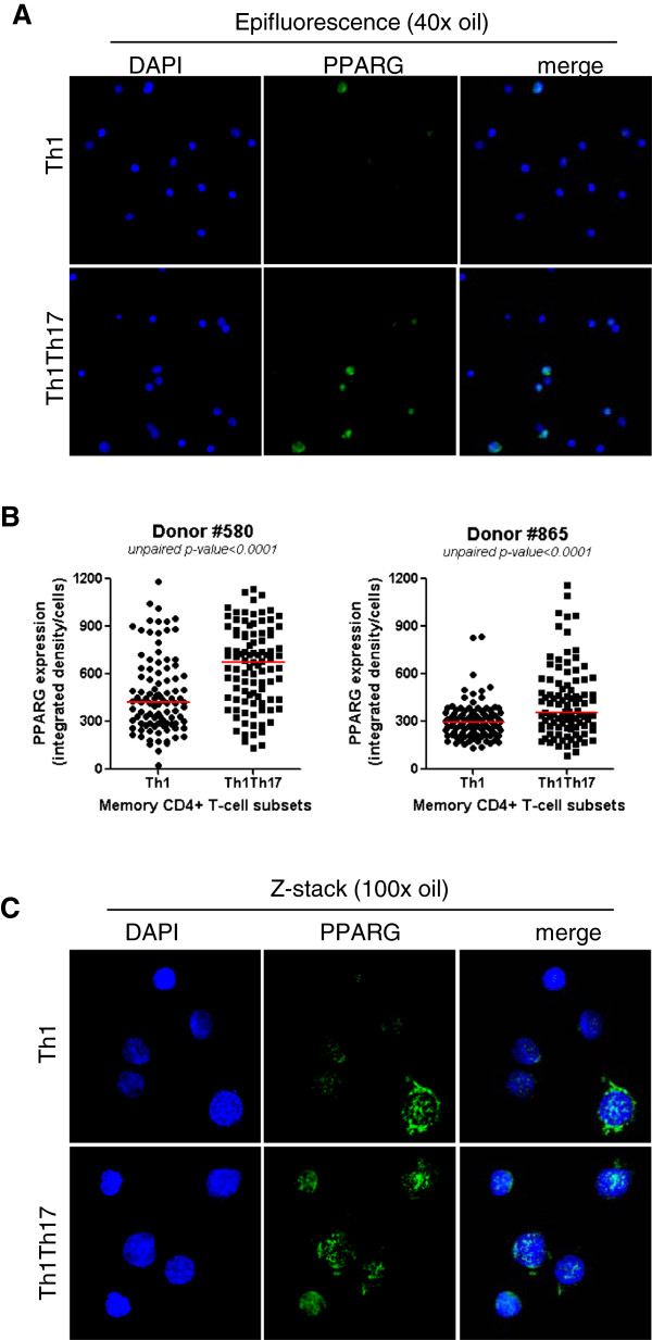
Figure 5.
Confocal microscopy quantification of PPARγ expression in Th1Th17 vs. Th1 cells. Matched Th1Th17 and Th1 cells were stimulated via CD3/CD28 for 3 days. Cells were fixed on poly-L-lysine coated slides and intracellular staining was performed with rabbit anti-human PPARγ Abs and then AlexaFluor 488 goat anti-rabbit Abs. Slides were mounted with the ProLong Gold Antifade reagent containing the nuclear dye DAPI. Slides were observed by fluorescence microscopy. (A-B) PPARγ expression was observed by epifluorescence at 40x magnification. (A) Shown are fields of Th1 and Th1Th17 cells from one donor representative of two donors. (B) Shown is statistical analysis of PPARγ expression in Th1 vs. Th1Th17 cells in two different donors (n = 100 cells per subsets per donor). Horizontal red lines indicate median values. Unpaired p-values are indicated on the figures. (C) The intracellular localization of PPARγ was observed by maximum intensity z-projection of z-stack from representative field of each subset observed with a 100x oil objective in a spinning-disc confocal mode system. Shown images are representative of observations made with Th1 and Th1Th17 from two different donors.

