Abstract
Paracoccidioidomycosis presenting as a sarcoid-like plaque may be misdiagnosed as leprosy, especially when shared endemic areas are concerned. We report the case of a Brazilian male patient presenting with an ulcerated plaque on his left ear and neighboring areas. The plaque simulated tuberculoid leprosy type 1 reaction, both clinically and histopathologically. A perineural granuloma with no organisms detected by routine and Fite-Faraco staining reinforced that diagnosis. Paracoccidioidomycosis was confirmed only after a second biopsy, taken from the ulcerated area.
Keywords: Adult, Granuloma, Neuritis, Paracoccidioidomycosis
Abstract
Paracoccidioidomicose expressando-se como placa sarcoídica pode ser confundida com hanseníase, especialmente em zonas endêmicas comuns às duas condições. Apresentamos um paciente brasileiro, com placa ulcerada na orelha direita e em áreas vizinhas que simulava, clinica e histopatologicamente, hanseníase tuberculoide em reação tipo 1. O encontro de granuloma perineural, sem parasitas detectáveis às colorações de rotina e Fite-Faraco, reforçou a hipótese de hanseníase. Apenas com uma nova biópsia, desta vez da área ulcerada, a paracoccidioidomicose pôde ser confirmada.
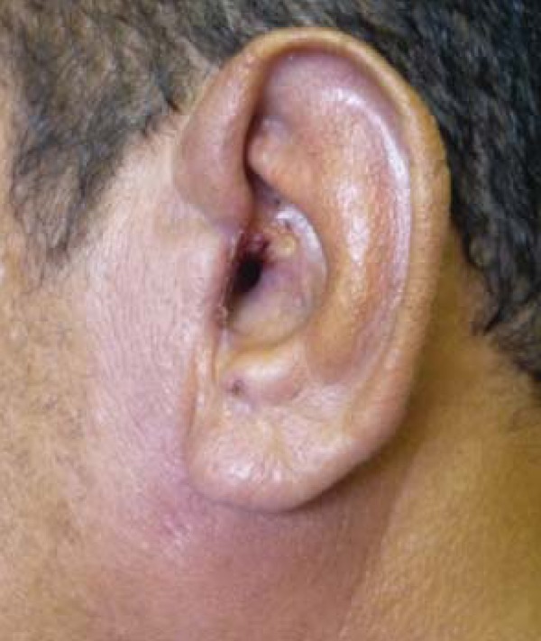
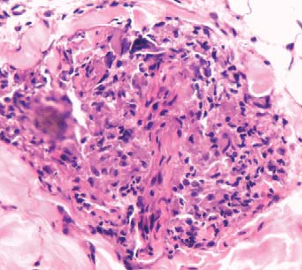
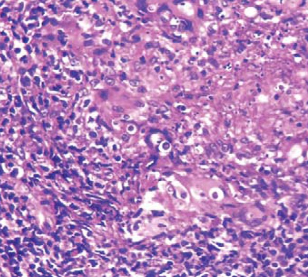
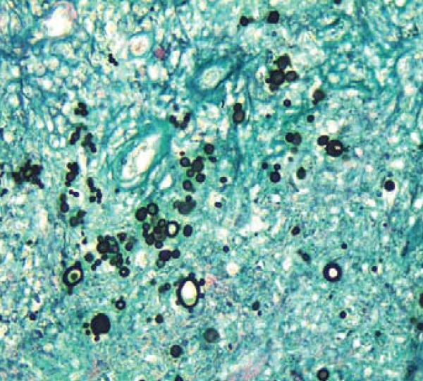
A 47-year-old male complained of a pruritic lesion on his left ear, which had been present for 6 years. He worked as a bricklayer and an iron miner in the Brazilian States of Amapá and Pará and in French Guiana. Following a positive intradermal leishmanin test, he had been given 220 meglumine and five pentamidine injections in another facility, obtaining poor results. On admission, he presented with an infiltrated, partly ulcerated plaque involving the left ear and adjacent areas (Figure 1). Blood cell counts, FAN, VDRL, FTA-Abs, anti-HIV and anti-HTLV yielded normal/negative results. A biopsy from the pre-auricular area showed a tuberculoid granulomatous infiltrate of epithelioid and multinucleated histiocytes, lymphocytes, and plasmocytes involving vessels, adnexa, and a nerve (Figure 2). No organisms were observed with H-E and Fite-Faraco stains. Tuberculoid leprosy type 1 reaction was considered, but there was no improvement after a two-month treatment with dapsone 100 mg/day, rifampicin 600 mg/month, and prednisone 60 mg/day. Paracoccidioidomycosis (PCM) was confirmed in a second biopsy taken from the ulcerated area (Figures 3 and 4).
FIGURE 1.

Violaceous, infiltrated plaque on the left ear and adjacent areas. Note central ulceration
FIGURE 2.

First biopsy: A nerve twig surrounded by a compact aggregate of epithelioid histiocytes (HE, 400x)
FIGURE 3.

Second biopsy: Numerous pleomorphic yeast organisms within a granuloma (H-E, 400x)
FIGURE 4.

Typical budding forms of Paracoccidioides brasiliensis (Grocott stain, 400x)
PCM is a fungal infection of the skin, mucous membranes, and internal organs caused by Paracoccidioides brasiliensis. Skin lesions are mostly papules, papulopustules, vegetations, and ulcers.1 Sarcoid-like plaques are occasionally seen and may lead to misdiagnosis, being misinterpreted as other granulomatous conditions such as tuberculoid leprosy, leishmaniasis, and sarcoidosis.1-3 The perineural granuloma and clinical presentation strongly suggested leprosy in our case; however, perineural inflammation may also occur in other infections such as syphilis and herpesvirus infections.4 We could not find reports on granulomatous perineural infiltrates in PCM in the literature.
Footnotes
Study conducted at the Dermatology Service, Institute of Health Sciences (Instituto de Ciências da Saúde), School of Medicine, Federal University of Pará (Universidade Federal do Pará-UFPA) - Belém (PA), Brazil.
Financial Support: None
Conflict of Interests: None.
REFERENCES
- 1.Sampaio SAP, Rivitti EA. Dermatologia. 3 ed. São Paulo: Artes Médicas; 2008. pp. 723–733. [Google Scholar]
- 2.Marques SA, Lastória JC, Putinatti MS, Camargo RM, Marques ME. Paracoccidioidomycosis: infiltrated, sarcoid-like cutaneous lesions misinterpreted as tuberculoid leprosy. Rev Inst Med Trop São Paulo. 2008;50:47–50. doi: 10.1590/s0036-46652008000100010. [DOI] [PubMed] [Google Scholar]
- 3.Nascimento CR, Delanina WF, Soares CT. Paracoccidioidomycosis: sarcoid-like form in childhood. An Bras Dermatol. 2012;87:486–487. doi: 10.1590/s0365-05962012000300025. [DOI] [PubMed] [Google Scholar]
- 4.Abbas O, Bhawan J. Cutaneous perineural inflammation: a review. J Cutan Pathol. 2010;37:1200–1211. doi: 10.1111/j.1600-0560.2010.01614.x. [DOI] [PubMed] [Google Scholar]


