Abstract
In contrast to most other sensory systems, hardly anything is known about the neuroanatomical development of central projections of primary vestibular neurons and how their second order target neurons develop. Recent data suggest that afferent projections may develop not unlike other sensory systems, forming first the overall projection by molecular means followed by an as yet unspecified phase of activity mediated refinement. The latter aspect has not been tested critically and most molecules that guide the initial projection are unknown.
The molecular and topological origin of the vestibular and cochlear nucleus neurons is also only partially understood. Auditory and vestibular nuclei form from several rhombomeres and a given rhombomere can contribute to two or more auditory or vestibular nuclei. Rhombomere compartments develop as functional subdivisions from a single column that extends from the hindbrain to the spinal cord. Suggestions are provided for the molecular origin of these columns but data on specific mutants testing these proposals are not yet available. Overall, the functional significance of both overlapping and segregated projections are not yet fully experimentally explored in mammals. Such lack of details of the adult organization compromises future developmental analysis.
Keywords: Vestibular, Hindbrain, Sensory system
1. Introduction
The primary vestibular and auditory neurons delaminate from the ventromedial aspect of the otocyst, and migrate away to the presumptive vestibular (Scarpa’s) and auditory (Spiral) ganglion between the ear and hindbrain. Here, these neuroblasts differentiate into bipolar neurons which project peripheral dendrites and central axons to connect with peripheral sensory endorgans and central targets, respectively [39]. Concomitantly, an intricate mosaic of gene expression drives patterning, cell fate specification, growth, and differentiation [30]. These local molecular events, along with global interaction between the epithelium of the otocyst and the surrounding mesoderm finally convert the simple otocyst into a three-dimensionally complicated series of interconnected sacs and tubes, housing five vestibular sensory patches and the cochlea in mammals and up to nine sensory patches in other vertebrates [30,101]. Each vestibular sensory patch and the cochlea, by virtue of their morphology, spatial orientation, and unique central projection, are specialized to convey specific information and bring about a specific sensory response.
In the last two decades, researchers have identified many molecules underlying different developmental events for different cell populations. Although, a rudimentary knowledge of genetic and molecular pathways controlling different aspects of growth and differentiation is now emerging, several outstanding problems at the basic anatomical and developmental levels remain unsolved. For example, we know that the determination and differentiation of vestibular ganglion neurons are the work of genes such as ngn1, NeuroD, and Brn3a ([39] and this issue). However, we do not know how the pan-neuronal nature of these vestibular ganglion cells is sorted out during development into endorgan-specific subtypes. Neither do we know what the precise connectivity pattern of these neurons with their central targets determines, nor do we know the developmental mechanisms setting up this pattern. The question of whether the vestibular system employs mechanisms or molecules similar to those regulating the visual system connectivity, or has evolved novel sets of mechanisms or molecules to fulfill unique functional requirements is still open.
Even less clear are the details of second order auditory and vestibular neuron development. Comparative anatomy has long recognized that spinal pathways extend into the hindbrain. For example, the continuity of the descending tract of the trigeminal nerve with Lissauer’s tract of the spinal cord has been established since over 100 years. Likewise, there is little doubt that the spinal somatic motor column extends as the hypoglossal and abducens motoneurons into the hindbrain [76]. Thus, continuity exists between general somatic aspects of both the motor and sensory system throughout the hindbrain and spinal cord. And yet, the hindbrain has components such as the branchial motor neurons which are both molecularly and evolutionary distinct from the spinal cord [11,42]. Those motoneurons appear to be a novelty of the hindbrain and may be derived from spinal motoneurons through the evolution of a single gene, the paralogous homeodomain gene Phox2b. Such suggestions derive from the fact that ectopic expression of Phox2b in the spinal cord results in a branchial motoneuron-like phenotype [11]. Other genes appear also to be uniquely associated with either branchial/visceral motoneurons of the hindbrain or somatic motoneurons [11,103]. Likewise, another apparent novelty of the hindbrain, the viscerosensory solitary nucleus, seems to depend on a set of genes for its formation, some of which are also expressed in the spinal cord [11,79]. This suggests either complete continuity or transformational continuity of specific cell groups of the hindbrain and the spinal cord.
Unfortunately, the molecular identity acquisition during development as well as the evolutionary history of another, apparently novel group of neurons of the hindbrain, the second order neurons of the vestibular and auditory nuclei, is much less clear. While numerous suggestions have been raised for the auditory nuclei to be evolutionary derived from lateral line or electroreceptive nuclei [33,43,60], those suggestions do not address the deeper, underlying problem: even if this transformation could be proven, we still would not know where the precursors of either the vestibular or lateral line nuclei are coming from. This ignorance is well reflected in the use of the term ‘special somatic afferent nuclei’, used in medical text books without any explanation as to what is so special about these nuclei and how they relate to somatic sensory nuclei.
The formation of second order neurons of the vestibular and auditory systems, but also the development of the central projections of the primary vestibular and auditory neurons is essential for the establishment of a functioning sensory-neural circuit. Understanding the developmental mechanisms, at the molecular and physical levels, which underlies the establishment of proper connections between primary and secondary auditory and vestibular neurons is a paramount goal in developmental neurobiology.
In this review we will establish some of the basic principles of development of the afferent projections of the inner ear and relate this to the growing information of molecular and topologic aspects of vestibular and auditory nuclei development. We hope to establish a framework that can guide future studies in this largely unexplored field. This review will also address some of the outstanding issues and questions concerning how the primary vestibular sensory neurons achieve their connectivity pattern during development. We will propose models of how these neurons acquire their target specificity and connect precisely with their targets, based on the current data available on the vestibular system, and the insights gained on research on other, more thoroughly studied systems, such as visual and auditory systems. The development of spiral neurons and their central connections was recently reviewed [85] and will not be dealt with here.
2. Acquisition of target specificity of the primary vestibular neurons: place versus time as a determining factor
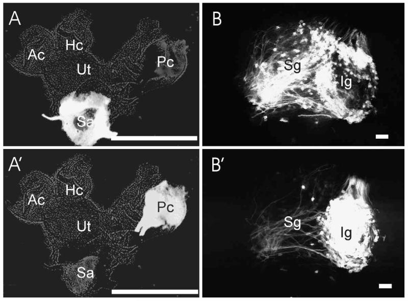
Neuroanatomists have investigated the topographical organization of the vestibular ganglion neurons with respect to their endorgans since 100 years in an attempt to delineate the anatomical substrate of specific function. A common notion has emerged that implies a clear-cut segregation of the vestibular ganglion neurons with respect to their endorgans and the sensation they convey [14,16,19,59,63], essentially suggesting a scheme similar to the retinotopic or tonotopic map in the visual and auditory systems, respectively [61,77,84,97]. In the last two decades, several reports contradicted that traditional notion [58,67]. These reports concluded that the vestibular ganglion neurons are not exclusively organized in an endorgan-specific fashion: they are only partially segregated with some random distribution and the neurons from different endorgans that convey different information show an overlapping distribution (Fig. 1).
Fig. 1.
These images are from a single case in which two different colored lipophilic dyes were injected into the saccule (A) and posterior crista (A′). The vestibular ganglion of this case was prepared and the distribution of primary vestibular neurons was investigated using a confocal microscope (B, B′). Note that the posterior crista primary neurons are almost all in the inferior vestibular ganglion (Ig, B′) with fairly few outliers in the superior vestibular ganglion (SG, B′). The fibers in the superior vestibular ganglion represent efferent fibers to other vestibular end organs. In contrast, the primary saccular neurons are widely dispersed, with a tendency to be near the caudal end of the superior and rostral end of the inferior vestibular ganglion. Ac, anterior crista; Hc, horizontal crista; Ig, inferior vestibular ganglion; Pc, posterior crista; Sa, saccule; Sg, superior vestibular ganglion; Ut, utricle. Bar indicates 1 mm (A, A′) and 100 μm (B, B′); anterior is to the left.
The original view seems intuitively plausible since one would assume that different sensory information should be well compartmentalized to evoke selective response. However, the refined, more recent view does not run counterintuitive or impose any conceptual challenge with respect to the functioning of the vestibular system. Indeed, the location and distribution of primary vestibular neurons should not be relevant for evoking a specific response as long as primary vestibular neurons sort out their peripheral projections to topographically, and functionally distinct receptor sites and project centrally to a functionally organized output of second order neurons. This sorting of vestibular afferent input to second order vestibular neurons has been shown by Straka et al. [93] despite the fact that afferent fibers emanating from different endorgans have an overlapping distribution [9]. Such a scheme of distribution is similar to dorsal root ganglion neurons whose different subpopulations, conveying different sensory information, are overlapping at the level of the ganglion [65]. However, these neurons sort out very sharply into modality-specific laminar projections into the spinal cord such as the Lissauer’s tract and dorsal columns [27].
These two conflicting traditional and modern views on vestibular ganglion organization represent a formidable obstacle to our understanding of the mechanism(s) underlying the development of the target specificity in the primary vestibular neurons. Such conflicting views imply different sets of mechanisms for target specificity, one that happens at the level of ganglion and one that happens independently of the ganglion. Clarifying these two issues will consequently help direct our research strategy for finding the molecules employed by each mechanism.
2.1. Do primary vestibular neurons follow similar developmental principles employed to set up target specificity of the visual and auditory system in which the location is the determining factor?
Irrespective of the particular weakness or strength of either the traditional view (clustered distribution of primary neurons) or the revised view (limited or absence of clustering of primary neurons projecting to a given sensory epithelium), we will assume here, for the sake of argument, that both views are correct and we will discuss the developmental implications of both views.
The presence of clear-cut topographical organization of the vestibular ganglion neurons (traditional view) implies similar developmental mechanisms to those setting up the retinal ganglion cells, spiral ganglion cells, and the efferent motor neurons projections. In this model, the final locations of these neurons and the gene expression specific to this location specify their targets. In the visual system, a gradient of molecules distributed in a spatial fashion does or can confer differential identity of these neurons according to their relative position with respect to different threshold concentrations along the gradient of a diffusible molecule. This position-based identity label match a complementary set of address label at the target terrain [89,96]. Here we refer to this model as “the spatial model of target specificity.”
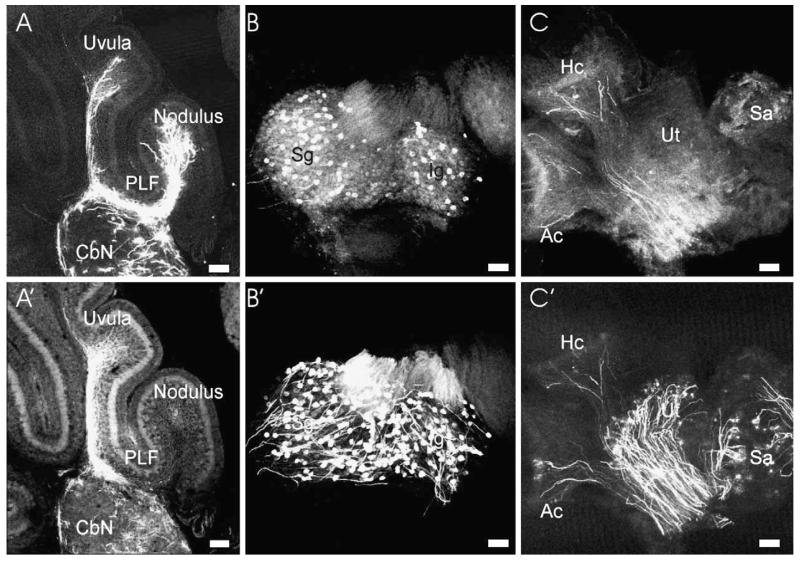
In contrast to the expectations of this model, the partial stochastic distribution of the vestibular ganglion cells with respect to their target found in more recent work can not be satisfactorily accounted for based on a spatial molecular gradient. If the primary vestibular neurons acquire their target specificity in a spatial fashion inside the vestibular ganglion, one might ask how can two adjacent cells, within a similar threshold of concentration, project differently if they do not obtain a different competence in the first place (Fig. 2)? This distributed assignment of the neurons to their targets would rather imply that target recognition is acquired, at least in part, prior to the arrival of sensory neurons at their final position in the vestibular ganglion [39]. Thus far, we do not know of any molecules that are asymmetrically distributed among the vestibular ganglion neurons that might play a role in this fate assignment. The known neuronal determination (ngn1), and differentiation (NeuroD and Brn3a) genes are common among the different populations and lack of their function leads to partial or complete absence of the entire population ([39] and this issue).
Fig. 2.
The distribution of afferents to and from the cerebellum is shown. Labeling the posterior crista (A) and the saccule (A′) results in distinct, partially overlapping projections to the cerebellum in this 7-day-old mouse. Retrograde labeling from the nodule (B) and uvula (B′) shows overlapping distribution of primary vestibular neurons in both the superior (Sg) and inferior (Ig) vestibular ganglion. Investigation of the peripheral organs shows that nodular injections (C) result in many more fibers to the anterior and horizontal crista (Ac, Hc) than to the utricle (Ut) and saccule (Sa). In contrast, fibers from the uvula (C′) end prominently in the utricle and saccule and fewer fibers reach the canal cristae. Bar indicates 100 μm.
In the olfactory system with equally dispersed rather than clustered sensory neurons, each olfactory neuron uses the odorant specific molecular identity to navigate toward the same olfactory glomerulus as all other receptors of this specific identity [100]. Such molecular fate determination could play also a role in the development of the vestibular sensory projection and specify, for example, primary neurons to the canal organs different from those to the gravistatic sensory epithelia. This raises the question at what level along the road of differentiation do different vestibular sensory neurons diverge from commonality to the point of molecular distinction, and differential fate acquisition? One may argue that the presence of a common marker, such as ngn1, NeuroD or Brn3a, for all sensory neuron populations does not necessarily mean that all populations are homogenous. There may be other, as yet unidentified marker(s) that are differentially distributed, conferring as yet undetected differential competence or identity, much like in the CNS [79,80].
2.2. How is such cellular heterogeneity or differential cell fate acquired?
Studies on other systems such as neural crest may provide some insights of how cell fate can be specified. In the neural crest [4,5,10], regional derivation of some populations may alter their developmental potential by endowing different competence for subsequent environmental interactions (cell-cell or cell-matrix interactions). In the otocyst, a similar mechanism may be used to establish different competence prior to or among the migrating neuroblasts. It has become clear that the otocyst is already a mosaic of gene expression domains at the time the neuroblasts start to migrate from various parts of the otocyst. Different domains may thus contribute neuroblasts which are molecularly distinct and it has been speculated that the neuroblasts may acquire their molecular heterogeneity as a function of their regional derivation [39]. This mechanism of fate assignment implies a topological effect. However, in contrast to the retinotopic fate assignment, it is the position where these neurons are generated, rather than the place where they finally settle, that is the key determinant factor for fate assignment; here we refer to it as “predetermination model of target recognition.”
2.3. Is it only the position?
The answer may be no. Not only the position, but also the time may be a mechanism to assign cell fate, like in other systems. In the cerebral cortex, there is inside-out temporal gradient of neuronal birth dates [2,71], it is the time of birth, not the place that orchestrates the laminar organization of the cerebral cortex which eventually determines the connectivity pattern of each layer. In some retinal ganglion cells, the date at which the neurons are born can instruct the neurons as to whether or not to cross the optic chiasm [81]. In the otocyst, the neuroblasts may leave the wall of the otocyst in different chronological contingents and may be sorted out for their targets through differential birth dates [87]. This may involve asymmetric distribution of fate determinant molecules in a cell cycle dependent fashion [2,71], here we refer to it as the “temporal gradient model for target specificity.”
Closely associated with the temporal gradient model and the predetermination model is another mechanism that may be used to specify target for vestibular ganglion neurons: the possible clonal relationship between neuronal cells and their targets [38]. In the ototcyst, the primary vestibular neuron precursors and the sensory epithelia are derived from the same or closely adjacent precursor populations and a specific population of sensory neurons and their sensory targets may be the descendent of the same clone. However, this mechanism is not supported by data on the central nervous system, as clonally related cells were found dispersed in different cortical layers and functional regions, having different fates and connections [78,99]. Also, in the dorsal root ganglion there is evidence for clonal dispersion [32] and clonal dispersion is well known for the hindbrain [52]. How far clonal fate determination will apply to the ear remains to be determined.
2.4. Do these mechanisms work in a mutually exclusive fashion?
In the visual system, the central nervous system, and the neural crest cells, variable combinations of time of birth, place of origin, and spatial placement of cells in their final environment can work in concert to dictate as well as modify fate of the different populations [65]. A detailed mapping study of the vestibular ganglion cell locations [67] concluded that the distribution of the vestibular ganglion cells for a given endorgan, is not entirely random. Each ganglion population, projecting to specific endorgans, has an area of main preference where the main population of cells are tightly packed, surrounded with an area of sparse distribution, shared with other primary neurons projecting to other endorgans (Figs. 1 and 2). This distribution pattern implies heterogeneity in the cellular behavior among the primary neurons of the same endorgan, suggesting different developmental mechanisms in target selection for the same peripheral target.
3. Establishing central connections
Having migrated to their definitive location, vestibular ganglion cells send axons to second order neurons in the vestibular nuclei and the cerebellum. It is generally agreed that, in their central representation, each endorgans has a domain of almost exclusive projection and a domain of sparse projection overlapping with other endorgans, reproducing the peripheral distribution pattern at the level of the ganglion [16,26,59,68,75]. Such a partial functional as well as anatomical overlap is best known for the frog [9,92,93]. This central overlapping pattern may be a universal functional feature of the vestibular system to converge monosynaptically afferent canal and otolith inputs [93] to integrate multiple inputs, and yet to respond in precise and finely graded fashion to unique signals (Newlands and Perachio, this volume).
Compared to other sensory systems, such as the visual and the auditory system, the lack of information as to when and how this pattern emerges during development is astounding. In the visual and auditory systems, there is a two-stage model for setting up the retinotopic and cochleotopic map of projection, respectively [85,96].
In the visual system, an initial phase is genetically determined, and functions via expression of sets of axon guidance molecules that work to establish coarse-grained patterns of connections. This phase is followed by a second phase in which physical cues in the form of spontaneous activity of the projecting axons or physiological stimuli drive refinement of the initial pattern into a fine-grained, highly tuned pattern of connection. More recent work suggests that the retinotopic map may initially start as precise as it will ever be at the connectional level [21,22]. In the auditory system[61,84], the two stage model may not be quite faithfully reproduced by the spiral neurons to establish the tonotopic map. The auditory neurons achieve a full-scale precision a long time before the functional maturity to perceive auditory stimuli has occurred, and do so in the absence of a significant amount of spontaneous activity to account for this precise projection as functionally driven. Indeed, some topologically refined projection can be obtained in mouse mutants without formation of hair cells [102]. Thus, absence of any discharge activity driven by hair cells appears to be compatible with the formation of a crude topological projection.
However, more detailed analyses are needed to verify how much of the complicated terminal arbor of individual fibers (Ryugo and Parks, this volume) forms in the absence of hair cells or hair cell mediated activity. In conclusion, the two-stage model of development of connection in the visual system can not be uniformly applied to other systems even if they share a very precise topologic organization such as the auditory system [84].
In the vestibular system, no clear prediction on the developmental organization principals can be derived from the adult pattern of extensively overlapping or partially segregated central projections or from the function of the vestibular system. Clearly not enough is understood at the level of individual terminals of a given fiber to precisely predict the function of each terminal in the context of ear function as a spatial analyzer decoding three-dimensional head movement to affect three-dimensional eye, neck, body movements through a three-leg reflex arc. Given that the detailed information processing from primary afferents to vestibular nuclei is only partially understood in mammals[75,98], the target for developmental processes operating in the vestibular system to form connections must be somewhat speculative. Selective ablations of function in one or more developing vestibular endorgan, while not as easy as lid suture, is now possible using specific mutations [44] and may help untangling molecular guidance and functional verification of connection formation.
A framework for neonatal reorganizations was recently established for two endorgans representing two different vestibular information, the posterior vertical canal as a representative of the angular acceleration, and saccule as a representative of gravistatic and linear acceleration [68]. In this kind of analysis of the differential nature of the projection from the different endorgans, neuroanatomists are trying to cope with three technical problems.
Nonspecific labeling, due to diffusion of the tracer to other endorgans, gives a false positive overlap.
Incomplete labeling, spatially or temporally as in degeneration techniques, gives false positive segregation.
Failure to compare equivalent sections due to difficulty to reproduce exactly the same section level in different animals for comparison of different endorgan projections.
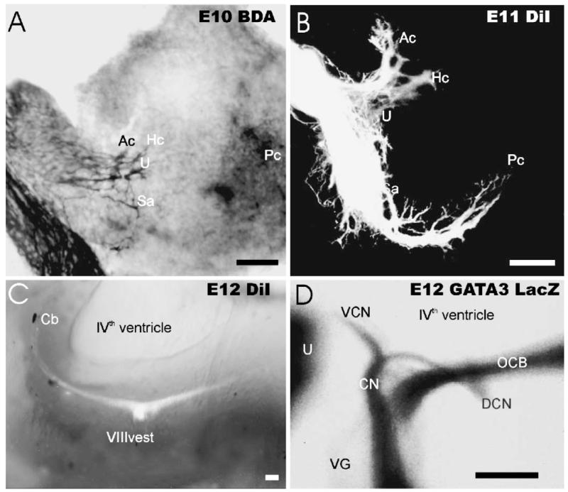
These technical difficulties have generated inconsistency in the results among different studies. Such inconsistencies increase the difficulty to establish a uniform rule for modeling the parameters of vestibular projection development. The simultaneous two-color labeling of two endorgans with low molecular weight dextran amines [34] or lipophilic dyes [68] avoids almost all of these technical difficulties. Using these approaches as well as transgenic approaches [57], the segregation of vestibular and cochlear central projections can be delineated in the mouse between embryonic day 12 and E13 (Fig. 3). Following up on this initial segregation of afferents between the auditory and the vestibular system, axons of the primary vestibular neurons from the posterior canal and the saccule form a remarkably detailed scaffold of projection in the vestibular nuclear complex at the earliest stage examined [68]. The growing axons made no gross errors such as penetrating nonvestibular targets. The extravestibular projections were to functionally related nuclei, such as prepositus hypoglossi and external cuneate, and persisted to at least postnatal day 7. At the embryonic ages in which the fibers sort the innervation to these central targets, there is no clear evidence of sensory transduction for the external vestibular stimuli nor for existence of spontaneous discharge at the primary vestibular neurons to provide cues to drive that phase of projection. It seems thus reasonable to conclude that this embryonic phase is dominated by molecular cues, and is activity independent. This phase, as in other systems, may regulate axon guidance through expression of molecules in a complimentary fashion in the projecting axons and their central targets [53].
Fig. 3.
The development of the peripheral and central projection of the vestibular (A-C) and auditory system (D) is shown. Already at E10.5, biotinylated dextran amine shows fibers to the antero-ventral aspect of the developing ear. These fibers show already some split into bundles that extend toward specific endorgans. One day later (B) DiI labeling shows that all endorgans receive afferent fibers, except for the cochlea (B). Injecting DiI selectively into the vestibular system labels only vestibular fibers which extend in a single fascicle from the forming cerebellum (Cb) toward the obex region. Using GATA3 TauLacZ as a molecular marker (D), spiral afferents can be visualized as early as E12 and project into a discrete fascicle which extends from the forming antero-ventral cochlear nucleus (AVCN) to the forming dorsal cochlear nucleus (DCN). Ac, anterior crista; Cb, cerebellum; DCN, dorsal cochlear nucleus; Hc, horizontal crista; Pc, posterior crista; Sa, saccule; U, utricle; VCN, ventral cochlear nucleus; VG, vestibular ganglion. Bar indicates 100 μm.
Molecules such as Eph receptors and their ligands, the ephrins, are known to pattern the connectivity of the retinotopic projection [96]. These receptors and ligands are widely expressed in the primary vestibular afferents and their peripheral and probably also their central targets [8]. Interestingly, mice mutant for EphB2 receptors exhibit circling behavior. Analysis of these mutant demonstrated defective formation of the dark cells in the stria vascularis with subsequent defective ionic homeostasis and production of the endolymph [18]. Despite the fact that this phenotype could account for the circling behavior, the primary afferent projections in these mutants need to be examined to exclude possible disoriented connections. Such disoriented connections could add to the circling behavior of this mutant.
In general, the embryonic development of central vestibular projections is characterized by two main features:
Parts of the different vestibular nuclei are devoid of projections during embryonic life.
The projections from a gravistatic and angular acceleration organ are indistinguishable from each other based on their pattern: it is only the simultaneous imaging with the help of the color of the tracer that allows distinguishing the two projections.
The latter feature implies that in this early genetically determined and molecularly guided phase, the pan-vestibular label is strong as evident in the correct homing only to the vestibular nuclei, and to the functionally related cell groups. Such homing even works in tissue culture [95], strongly supporting our notion.
Unlike the auditory system, which is immature at birth, the vestibular system displays some degrees of functional maturity and ability to transduce external vestibular stimuli and display proper vestibular-evoked righting reflex [41,83]. Consistent with this onset of rudimentary behavior is the emergence of the differential, endorgan-specific projection through elaboration of exuberant branches that fill almost all regions of the vestibular nuclei in a fashion characteristic for each endorgan. The coincidence of these dramatic changes in the pattern of connectivity, i.e. the emergence of endorgan based individuality, with the commencement of function, provides at least circumstantial evidence for the role of physiological stimuli to drive some remodeling of the initial projections. Studying these developmental events in experimental alteration of the basal level of activity in mutants or in microgravity/hypergravity should provide evidence for the existence of such putative role for activity.
It has been reported that exposure of the animal to altered gravitational states produce a wide range of morphological and behavioral effects on the vestibular system[17,23,31,41,56,83]. These behavioral changes, which imply underlying structural changes such as altered membrane excitability in the neural circuits, were either temporary or permanent, depending on the time of exposure. This invokes the idea of the presence of a critical developmental window during the development of the neural circuits [54,55]. Indeed, one report [41,83] suggests that exposure of rat embryos to microgravity from E9 to E19, results in decrease in the branching pattern in the medial vestibular nucleus from a gravity sensing endorgan, the saccule. Unfortunately, no postnatal stages could be examined and require in fact a different approach such as the use of mutants without otoconia.
These data suggest that the vestibular system may follow the two-stage paradigm of other sensory systems in establishing the connectivity pattern. However, in the vestibular system, the genetically programmed molecular phase generates a scaffold of pan-vestibular projection with a limited definition of distinct projections of the different endorgans. The second, activity-driven phase may reinforce the discrete nature of the endorgan specific projection through exuberant tertiary and quaternary sets of branches of a given endorgan at the domains of their correct synaptic targets. This suggestion implies some role for physiological stimuli which transform a naïve vestibular projection into an endorgan-specific pattern of precise connections. A test of this model can now be conducted in animals that lack gravistatic input via otoconia and are thus vestibular deprived. Altering the specific density of otoconia (e.g. strontium instead of calcium) could provide increased inertia mediated relative movement, thus allowing for overstimulation of gravistatic epithelia. Last, but not least, animals devoid of all acellular vestibular covering structures exist, owing to the discovery of the gene otogelin [28,88]. The available null mutants allow testing the absence of specific input to all sensory epithelia.
4. Development of vestibular and auditory nuclei: longitudinal columns and their transverse boundaries
The developing hindbrain of vertebrates generates two or more continuous longitudinal columns of neurons that receive the afferent fibers from the vestibular part of the ear, from the auditory part of the ear, and, if present, from the mechanosensory lateral line and electroreceptive system[36,72]. In the last 10 years we have come to understand two principle features of the development of these sensory columns. One feature is the molecular continuity with gene expression domains of the spinal cord (longitudinal columns). The second feature is the relationship of subdivisions of these columns that become functionally specified within the hindbrain rhombomeres (transverse boundaries). We will first relate the mammalian data that indicate the longitudinal continuity of the vestibular and cochlear nuclei with the spinal cord, suggesting that the evolutionary novelty of those nuclei is only a problem of appearance, not of essence. In the second part of this paper we will relate how the spacemap of vestibular nuclei projections appears to relate to the rhombomeric spacemap. Those comparisons will also be attempted for the auditory nuclei, but the data are even less clear.
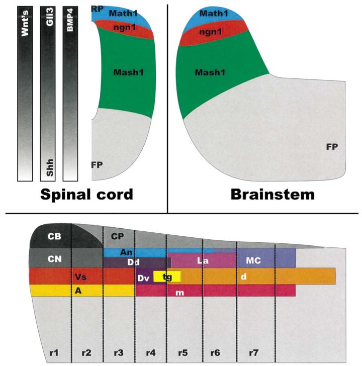
The pioneering work of Ma et al. [66] indicated that expression of specific basic Helix-Loop-Helix (bHLH) genes forms unique dorso-ventral strata in the developing hindbrain and spinal cord. Later work of others have clearly demonstrated that the two most dorsally expressed bHLH genes (ngn2/1; Math1) are expressed in essentially similar pattern throughout the spinal cord [50]. Thus, both the hindbrain and the spinal cord have a dorsalmost ventricular zone of the bHLH gene Math1. Adjacent and more ventral is the zone of expression of ngn2/1, which appear to be temporarily segregated expression of closely related, unique progenitors defining bHLH genes [65]. Below this zone is a large zone expressing Mash1. The latter zone extends throughout the rest of the alar plate [50,66]. A major difference between the spinal cord and the hindbrain seems to be the relative size of the Math1 and ngn1/2 expressing ventricular zone. Concomitant with this size difference we can expect that fewer (spinal cord) or more (hindbrain) neurons will derive from the otherwise continuous ventricular zone. The apparent stability of the dorso-ventral patterning is related to at least three parallel signaling systems, the sonic hedgehog/gli3 system [62], the BMP-4 system that may directly control the expression of bHLH genes in the ventricular zone [50], and the effect of Wnt genes [74]. The latter system is apparently able to signal dorso-ventral patterning independent of BMP-4, whereas elimination of both Shh and Gli3 unmasks a Shh independent dorso-ventral patterning system (Fig. 4).
Fig. 4.
This drawing shows the distribution of three BHLH genes and the gradients of three potential regulators of expression, Wnt’s, Shh-GLi3 and BMP4 in the hindbrain and the spinal cord (Top). The bottom shows the distribution of auditory and vestibular columns according to rhombomeric origin. All vestibular and cochlear nuclei derive from one or more rhombomere(s) and several rhombomeres contribute to more than one nucleus in a given column. Abbreviations: A, vestibular group A; An, angular nucleus; CB, cerebellum; CN, cerebellar nuclei; CP, choroid plexus; d, descending vestibular nucleus; Dd, dorsal Deiters nucleus; Dv, ventral Deiters nucleus; FP, floor plate; La, laminar nucleus; m, medial vestibular nucleus; MC, magnocellular nucleus; RP, roof plate; tg, tangential nucleus; Vs, superior vestibular nucleus. Modified according to Refs. [15,50,65,66,74].
In summary, there is some overall continuity of initial regulation of neuronal formation between the spinal cord and the hindbrain as indicated by the longitudinal continuity of bHLH expression domains. Those longitudinal zones that are related to the formation of vestibular and cochlear nuclei show an increase in ventricular zone bHLH expression domain likely correlated with the production of more neurons in the hindbrain compared to the spinal cord.
The next step in cell fate acquisition is more complex and we are just beginning to understand it in the spinal cord. For example, Math1 establishes in the spinal cord projection interneurons that form the dorsal and ventral spinocerebellar tract [7]. However, in more rostral parts of the hindbrain Math1 dependent precursors form pontine nuclei, parts of the auditory nuclei and of the cerebellum. Unfortunately, what neurons depend on Math1 in the cochlear nuclei has not yet been worked out. Based on the loss of granule cells in the cerebellum, it can be expected that they will be among the granule cells of the cochlear nuclei (Ryugo and Parks, this volume). A detailed analysis of Math1-LacZ reporter positive neurons and their phenotype in Math1 heterozygotic animals should help to clarify this issue. Such studies are currently under way in our laboratory.
Much more complex is the formation of neurons that derive from the lowest bHLH gene expressing zone, the Mash1 positive zone. In the spinal cord, this zone gives rise through an intricate overlap of several genes to three distinct classes of neurons [51]. Much of the downstream genes and the specification of neurons generated by the Mash1 positive zone in the hindbrain is still unclear, but at least the solitary nucleus has recently been identified as being derived from the most dorsal part of the Mash1 expressing zone in the hindbrain [79] and is specified to do so by the homeobox genes Rnx/Tlx-1 [80].
Based on this topology, it appears that the vestibular and cochlear nuclei may be derived from the ngn1/2 expression zone that is wedged between the Math1 and the Mash1 positive zone. As suggested by the spinal cord data, one would expect that genes related to Foxd3, Lhx1/5, Brn3a, Pax3 and Pax6 should be expressed in vestibular and cochlear nuclei precursor cells. If that pattern of expression differs significantly from the pattern in the spinal cord, the differences could potentially be related to the specific vestibular and cochlear cellular phenotype(s) (Cant, this volume; Barmack, this volume) and could thus help establish how neuronal diversification of an ancestral longitudinal column is modified through increased proliferation and subsequent changes in downstream gene expression patterns. Such a scheme has recently been suggested as a general principle of brain evolution [37].
While these expression data are interesting and offer some, admittedly speculative insights, other ways of obtaining data on this subject are needed that critically test this idea and its underlying assumptions. Such data could be generated through the use of specific null mutations and the analysis of the deficits obtained in these loss or gain of function mutations. We have started such an analysis in ngn1 and Math1 null mutations for the auditory and vestibular nuclei, but have not yet obtained conclusive data. Nevertheless, a start is made to derive the ‘special somatic sensory columns’ through altered gene expression out of a continuous hindbrain-to-spinal-cord precursor pool.
It has recently been suggested that ear evolution might represent a transformation of an ancestral mechanosensory system [38]. The latter suggestion is in line with the hypothesis we propose here. Ultimate proof that those ‘special nuclei’ are indeed transformed spinal sensory columns that have acquired a novel input of a uniquely derived sensory system clearly requires more detailed follow up studies.
5. Rhombomeric origin of vestibular and cochlear nuclei
Within recent years, the original concept of rhombomeric boundaries in the hindbrain, long recognized for its possible significance as well as its limits [76] has been revived based on matching gene expression [46,64]. Such gene expression, in particular of homeobox domain containing genes, appears to specify in a partly overlapping fashion the identity of hindbrain rhombomeres [73]. Two basic attempts have been made in chicken to relate rhombomeres to vestibular and cochlear nuclei.
One attempt is trying to establish contributions of various hindbrain rhombomeres to cochlear and vestibular nuclei using grafting or dye injection as a means to trace the future fate. Cramer et al. [20] used rhombomere specific dye injections to trace the latter distribution of auditory nuclei. Based on these data they suggested that the chicken auditory nucleus laminaris derives from rhombomere 5 with a few cells from rhombomere 6, nucleus magnocellularis from rhombomeres 5 to 7 and the nucleus angularis from rhombomeres 4 and 5. Largely identical results were reported by Puelles and co-workers [15,69] who used the transplantation of quail rhombomeres into the chicken hindbrain. The latter authors independently found that nucleus laminaris derives from r5 to r6, nucleus magnocellularis predominantly from r7 to r8 with a small addition from r6 and nucleus angularis derives from r3 to r4 with a small addition from r5 and perhaps r6. These combined data suggest thus a selective formation of only one auditory nucleus out of rhombomeres 3 + 4, and 7 + 8. In contrast, rhombomeres 5 and 6 produce neurons that will later be situated in at least two different auditory nuclei. Indeed, cells for up to four auditory nuclei seem to derive from rhombomere 4 (nucleus angularis, nucleus magnocellularis, nucleus laminaris, and superior olivary nucleus). These data suggest that there is only limited auditory nuclei specificity provided to the future fate of cells originating in a given or multiple rhombomeres. In essence, the longitudinal origin in a specific longitudinal area of ventricle (possibly an ngn and/or Math1 dependent zone) rather than the rhombomeric origin (Hox-code dependent rhombomeres) may specify much of the future fate of auditory nuclei and the combination of both longitudinal and transverse divisions can in essence establish a precise three-dimensional map of cell fate assignment.
No comparable data for nuclear origin exists for any other vertebrate species. However, using a different approach for transgenic misexpression of homeobox genes, Studer et al. [94] reported an apparent reduction of the antero-ventral auditory nucleus in an animal in which rhombomere 2 has been transformed into rhombomere 4 through misexpression of the HoxB2 homeobox gene. These preliminary data suggest that at least some of the auditory nuclei in a mammal derive from rhombomeres that do not produce auditory nuclei in chicken as Cambronero and Puelles [15] claim that no auditory, but cerebellar nuclei derive from r2 in chicken. Clearly, our tracing data suggest that the auditory afferents extend in mice from the glossopharyngeal nerve to the trigeminal nerve root (Fig. 3), suggesting that mammalian auditory nuclei derive from rhombomeres 2 to 6. Unfortunately, no such specific basilar papilla data on initial projections into rhombomeres exist in birds in which only the entire ear projections have been mapped during early development when they can be related to rhombomeres [40].
If further studies can confirm this finding and can extend the rhombomeric auditory nuclei origin to other vertebrates such as amphibians we could at the very least establish a spacemap of auditory nuclei generating rhombomeres that would allow a more detailed comparison of auditory nuclei across vertebrates. Such data are essential as many neurons derived from the rhombic lip can migrate rather long distances, e.g. the cerebellar pontine nuclei which migrate from r6 to r3 [7]. However, given the limited specificity that is apparently provided by the rhombomeric origin to the auditory nuclei as presented above, it might not be too surprising that different rhombomeres give rise to functionally equivalent auditory nuclei in various vertebrates.
Four experimental studies for the rhombomeric origin of vestibular neurons exist, and all are in chicken. Puelles and co-workers [15,24,69] proposed that the superior vestibular nucleus derives from r1 to r3, Deiters (or lateral) vestibular nucleus derives from r4 with a small contribution of r3, tangential nucleus derives from both r4 and r5, the medial vestibular nucleus derives from r4 to r8 and the descending vestibular nucleus derives from r5 to r9. The data of Cramer et al. [20] using dye injections largely agree, but cover only the area from r4 to r7. They show that the lateral and tangential vestibular nuclei derive from r4, the medial vestibular nucleus derives from r4 to r7 and the descending vestibular nucleus derives from r5 to r6. As was above described for the auditory nuclei, there is a topologically restricted origin from a given rhombomere or rhombomeres in some but not in all vestibular nuclei. As in the auditory nuclei, this suggests that longitudinal origin imposes most of the cell fate specification with the rhombomeric origin providing only some limited rostro-caudal specification, with the possible exception of rhombomere 4 (see below). No experimental data exist on the origin of vestibular nuclei of other vertebrates thus precluding any direct comparison of the stability of the rhombomere specific vestibular nuclei formation and the stability of their phenotype across phyla.
A second attempt to compare topological origin with adult nuclei is the embryonic and adult tracing of connections of these nuclei. The idea basically is to establish continuity between output neurons of a given rhombomere and relate those populations of output neurons to the adult pattern of connections. No such data exist as yet for auditory nuclei thus precluding addressing precisely the connectional similarities between auditory and vestibular nuclei development.
A growing literature exists for this kind of connectional analysis for the vestibular nuclei of chicken [48], frogs[90,91], bonyfish [6] and lamprey [35]. The following populations of neurons have been analyzed: ipsi- and contralateral projecting vestibulo-ocular neurons, ipsi- and contralateral projecting vestibulo-spinal neurons and, to some extend, ipsi- and contralateral projecting vestibulo-cerebellar neurons. Based on this analysis, a common scheme appears to be present among vertebrates that suggest a high degree of evolutionary conservation.
Most rostral is an ipsilateral and a contralateral projecting vestibulo-ocular group derived from several rhombomers (probably from r2 to r3/4 in chicken and lampreys[35,47,48]). Judging from the tracings published in various species, the distribution seems to be minimally different. The contralateral group appears to be more rostral and less extensive in birds, lampreys and bonyfish [6,35,48]. In contrast, the ipsilateral population appears to be in more rostral rhombomeres in frogs [91]. The contralateral projecting vestibulo-ocular group consists in lampreys of the large celled anterior octavo-motor group [35]. Both excitatory and inhibitory components exist in these populations in some of the vertebrates tested, some of which are uniquely associated in r3 with the horizontal canal system in at least frogs [91] and bonyfish [6]. Such components are unlikely to exist in lampreys as neither the horizontal canal nor the medial rectus muscle exists in these species [35,44].
An ipsilateral projecting vestibulo-ocular population exists rostral to r4 (characterized by the Mauthner cell in lampreys, bonyfish and frogs) whose input into the oculomotor nuclei has not yet been characterized. It might be excitatory as is the ipsilateral input from the horizontal canal and also inhibitory on vertical and oblique extraocular motoneurons [6,91]. Interestingly, the central projection of the dorsal macula of lampreys, the proposed homologue of the horizontal crista [44], is most extensive to what appears to be r2/3 vestibular neurons [35]. It remains to be determined whether the vestibulo-ocular neurons that receive input from the dorsal macula excite specific subpopulations of the ocular motoneurons.
Between the rostral and caudal vestibulo-ocular population is an ipsilateral projecting vestibulo-spinal population. This population appears to be the medial octavo-motoneurons in lampreys [42] and may be derived from r4. In virtually all vertebrates, this population of lateral or Deiters vestibular nucleus appears to be the most constant output of the vestibular nuclear complex and seems to exist even in hagfish, an animal with no known vestibulo-ocular connections [82].
Caudal to the lateral (or Deiters) vestibulo-spinal projection neurons are two populations of ipsi- and contralateral projecting vestibulo-ocular neurons. In chicken, the ipsilateral vestibulo-ocular group may derive from r5 to r6 whereas the larger contralateral vestibulo-ocular group may derive from r4 to r6 [24,25,48]. In the frog, similar excitatory and inhibitory canal neurons of the medial and descending vestibular nuclei seem to derive from r5 to r6, while ipsilateral projecting neurons appear to be restricted to parts of r6 [91]. Only contralateral projecting vestibulo-ocular neurons have been reported in lampreys[35,49] suggesting that the ipsilateral projecting group that relates horizontal canal information [6,24,91] is either very small in lampreys or does not exist.
Only the projection of vestibulo-spinal neurons to rhombomeres has been established thus far in embryonic mammals [3]. However, judging from the projection patterns analyzed in both bulk and single neuron fillings of adult mammals, it appears that largely comparable ipsi- and contralateral projecting populations of vestibular neurons exist in mammals [12,13]. Overall, the wiring of the vestibular-ocular and vestibular-spinal projections seems to be extraordinarily conserved among vertebrates. However, some additional new tracts may have formed in jawed vertebrates and elasmobranches may have a reduction of second order projections owing to the simpler wiring diagram of vestibulo-ocular interactions [29,45,49].
In summary, the tracing studies show some specification of neuronal populations with rhombomeres but also show that specificity of vestibular populations can vary somewhat between taxa. Combining more sophisticated tracing techniques with cell fate labeling is needed to fully establish the range of variation in taxa like birds and mammals. Most importantly, the rhombomeric origin of traditionally recognized vestibular nucleus groups is less than clear [24,91] and requires the above outlined approach.
6. Time of origin of auditory and vestibular nuclei neurons
The origin of auditory and vestibular nuclei neurons has been studied with thymidine radiography in mice, rats and chicken [1,70,86]. Altman and Bayer [1] described an orderly progression of birth dates for vestibular and cochlear nuclei in rats. The lateral vestibular nucleus is generated first (peak around E12) followed by the superior (E13), inferior or descending (E13-14) and medial vestibular nucleus (E13-14), with a broad overlap around E12-14. This suggests a lateral to medial as well as a superior to inferior gradient, essentially generating the large output neurons (the vestibulo-spinal or Deiters neurons) first and the small interneurons last. In contrast, terminal mitosis of the cells for the three auditory nuclear complexes of the rat peaks on the same day (E15) with the broadest generation time (range E12-P2) to be found in the dorsal cochlear nucleus. Overall, the neurons are generated in the usual sequence: large output neurons first followed by small interneurons with production of granule cells in the dorsal cochlear nucleus lasting into neonatal stages. It is likely that the granule cells of the dorsal cochlear nucleus share a common anlage with the granule cells of the cerebellum [1] and may both depend on Math1 for their generation [7].
In contrast to the absence of a clear cytogenic gradient in the auditory nuclei, the superior olivary complex has such cytogenic gradients [1]. Specifically, the lateral trapezoid nucleus peaks at E12, the medial superior olivary nucleus peaks at E13, the trapezoid nucleus peaks at E15 and the lateral superior olivary nucleus peaks at E16. This order of terminal mitosis results in a medial to lateral gradient in the more dorsal, and a lateral to medial gradient in the more ventral compartment of this nuclear complex. This countergradient is reminiscent to that found in the cochlea hair cells and spiral sensory neurons [85,87].
A gradient of medial to lateral was observed in the formation of the chicken auditory nuclei. Specifically, the study of Rubel et al. [86] found that the magnocellular nuclei had their terminal mitosis between E2.5 and 3 whereas the laminar nuclei were generated between E3.5 and 4. Comparable to mammals, the vestibular nuclei are generated somewhat earlier, around E2 [70]. No such data exist for any other vertebrate to date, but would be interesting with respect to formation of vestibular neurons prior to auditory neurons in a given rhombomere. In other words, a combination of labeling for terminal mitosis with lineage tracing and connectivity tracing is needed to move the field forward in the analysis of specific neuronal types and their spatial and temporal origin.
Acknowledgements
This work was supported by grants from NIH (RO1 DC005590; 2PO1 DC00215) and NASA (NAG2-1353). We wish to express our gratitude to numerous collaborators who helped with this review, in particular Drs. K.W. Beisel, D. Nichols, N. Bermingham, H. Zoghbi, and J.F. Brunet. We wish to express our gratitude to three reviewers who provided detailed suggestions for improvements on an earlier version of this manuscript: Drs. Q. Ma, J. Glover, and H. Straka.
References
- [1].Altman J, Bayer SA. Development of the brain stem in the rat. III. Thymidine-radiographic study of the time of origin of neurons of the vestibular and auditory nuclei of the upper medulla. J. Comp. Neurol. 1980;194:877–904. doi: 10.1002/cne.901940410. [DOI] [PubMed] [Google Scholar]
- [2].Anderson SA, Kaznowski CE, Horn C, Rubenstein JL, McConnell SK. Distinct origins of neocortical projection neurons and interneurons in vivo. Cereb. Cortex. 2002;12:702–709. doi: 10.1093/cercor/12.7.702. [DOI] [PubMed] [Google Scholar]
- [3].Auclair F, Marchand R, Glover JC. Regional patterning of reticulospinal and vestibulospinal neurons in the hindbrain of mouse and rat embryos. J. Comp. Neurol. 1999;411:288–300. doi: 10.1002/(sici)1096-9861(19990823)411:2<288::aid-cne9>3.0.co;2-u. [DOI] [PubMed] [Google Scholar]
- [4].Baker CV, Bronner-Fraser M. The origins of the neural crest. Part II. An evolutionary perspective. Mech. Dev. 1997;69:13–29. doi: 10.1016/s0925-4773(97)00129-9. [DOI] [PubMed] [Google Scholar]
- [5].Baker CV, Bronner-Fraser M. The origins of the neural crest. Part I. Embryonic induction. Mech. Dev. 1997;69:3–11. doi: 10.1016/s0925-4773(97)00132-9. [DOI] [PubMed] [Google Scholar]
- [6].Baker R. From genes to behavior in the vestibular system. Otolaryngol Head Neck Surg. 1998;119:263–275. doi: 10.1016/S0194-5998(98)70061-0. [DOI] [PubMed] [Google Scholar]
- [7].Bermingham NA, Hassan BA, Wang VY, Fernandez M, Banfi S, Bellen HJ, Fritzsch B, Zoghbi HY. Proprioceptor pathway development is dependent on Math1. Neuron. 2001;30:411–422. doi: 10.1016/s0896-6273(01)00305-1. [DOI] [PubMed] [Google Scholar]
- [8].Bianchi LM, Liu H. Comparison of ephrin-A ligand and EphA receptor distribution in the developing inner ear. Anat. Rec. 1999;254:127–134. doi: 10.1002/(SICI)1097-0185(19990101)254:1<127::AID-AR16>3.0.CO;2-Q. [DOI] [PubMed] [Google Scholar]
- [9].Birinyi A, Straka H, Matesz C, Dieringer N. Location of dye-coupled second order and of efferent vestibular neurons labeled from individual semicircular canal or otolith organs in the frog. Brain Res. 2001;921:44–59. doi: 10.1016/s0006-8993(01)03075-x. [DOI] [PubMed] [Google Scholar]
- [10].Bronner-Fraser M, Sternberg PW. Pattern formation and developmental mechanisms. The cell biological basis of inductive signaling. Curr. Opin. Genet. Dev. 2000;10:347–349. doi: 10.1016/s0959-437x(00)00094-0. [DOI] [PubMed] [Google Scholar]
- [11].Brunet JF, Pattyn A. Phox2 genes—from patterning to connectivity. Curr. Opin. Genet. Dev. 2002;12:435–440. doi: 10.1016/s0959-437x(02)00322-2. [DOI] [PubMed] [Google Scholar]
- [12].Buttner-Ennever JA. Patterns of connectivity in the vestibular nuclei. Ann. N.Y. Acad. Sci. 1992;656:363–378. doi: 10.1111/j.1749-6632.1992.tb25222.x. [DOI] [PubMed] [Google Scholar]
- [13].Buttner-Ennever JA. A review of otolith pathways to brainstem and cerebellum. Ann. N.Y. Acad. Sci. 1999;871:51–64. doi: 10.1111/j.1749-6632.1999.tb09175.x. [DOI] [PubMed] [Google Scholar]
- [14].Cajal SR. Accion neurotropica de los epitelios. Trab. del Lab. de Invest. Biol. 1919;17:1–153. [Google Scholar]
- [15].Cambronero F, Puelles L. Rostrocaudal nuclear relationships in the avian medulla oblongata: a fate map with quail chick chimeras. J. Comp. Neurol. 2000;427:522–545. doi: 10.1002/1096-9861(20001127)427:4<522::aid-cne3>3.0.co;2-y. [DOI] [PubMed] [Google Scholar]
- [16].Carpenter MB, Stein BM, Peter P. Primary vestibulocerebellar fibers in the monkey: distribution of fibers arising from distinctive cell groups of the vestibular ganglia. Am. J. Anat. 1972;135:221–249. doi: 10.1002/aja.1001350209. [DOI] [PubMed] [Google Scholar]
- [17].Cohen B, Wearne S, Dai M, Raphan T. Spatial orientation of the angular vestibulo-ocular reflex. J. Vestib. Res. 1999;9:163–172. [PubMed] [Google Scholar]
- [18].Cowan CA, Yokoyama N, Bianchi LM, Henkemeyer M, Fritzsch B. EphB2 guides axons at the midline and is necessary for normal vestibular function. Neuron. 2000;26:417–430. doi: 10.1016/s0896-6273(00)81174-5. [DOI] [PubMed] [Google Scholar]
- [19].Cox RG, Peusner KD. Horseradish peroxidase labeling of the central pathways in the medulla of the ampullary nerves in the chicken, Gallus gallus. J. Comp. Neurol. 1990;297:564–581. doi: 10.1002/cne.902970409. [DOI] [PubMed] [Google Scholar]
- [20].Cramer KS, Fraser SE, Rubel EW. Embryonic origins of auditory brain-stem nuclei in the chick hindbrain. Dev. Biol. 2000;224:138–151. doi: 10.1006/dbio.2000.9779. [DOI] [PubMed] [Google Scholar]
- [21].Crowley JC, Katz LC. Early development of ocular dominance columns. Science. 2000;290:1321–1324. doi: 10.1126/science.290.5495.1321. [DOI] [PubMed] [Google Scholar]
- [22].Crowley JC, Katz LC. Ocular dominance development revisited. Curr. Opin. Neurobiol. 2002;12:104–109. doi: 10.1016/s0959-4388(02)00297-0. [DOI] [PubMed] [Google Scholar]
- [23].Dai M, Raphan T, Kozlovskaya I, Cohen B. Vestibular adaptation to space in monkeys. Otolaryngol. Head Neck Surg. 1998;119:65–77. doi: 10.1016/S0194-5998(98)70175-5. [DOI] [PubMed] [Google Scholar]
- [24].Diaz C, Glover JC. Comparative aspects of the hodological organization of the vestibular nuclear complex and related neuron populations. Brain Res. Bull. 2002;57:307–312. doi: 10.1016/s0361-9230(01)00673-6. [DOI] [PubMed] [Google Scholar]
- [25].Diaz C, Puelles L, Marin F, Glover JC. The relationship between rhombomeres and vestibular neuron populations as assessed in quail-chicken chimeras. Dev. Biol. 1998;202:14–28. doi: 10.1006/dbio.1998.8986. [DOI] [PubMed] [Google Scholar]
- [26].Dickman JD, Fang Q. Differential central projections of vestibular afferents in pigeons. J. Comp. Neurol. 1996;367:110–131. doi: 10.1002/(SICI)1096-9861(19960325)367:1<110::AID-CNE8>3.0.CO;2-6. [DOI] [PubMed] [Google Scholar]
- [27].Eide AL, Glover JC. Developmental dynamics of functionally specific primary sensory afferent projections in the chicken embryo. Anat. Embryol. Berl. 1997;195:237–250. doi: 10.1007/s004290050043. [DOI] [PubMed] [Google Scholar]
- [28].El-Amraoui A, Cohen-Salmon M, Petit C, Simmler MC. Spatiotemporal expression of otogelin in the developing and adult mouse inner ear. Hear. Res. 2001;158:151–159. doi: 10.1016/s0378-5955(01)00312-4. [DOI] [PubMed] [Google Scholar]
- [29].Ezure K, Graf W. A quantitative analysis of the spatial organization of the vestibulo-ocular reflexes in lateral- and frontal-eyed animals—II. Neuronal networks underlying vestibulo-oculomotor coordination. Neuroscience. 1984;12:95–109. doi: 10.1016/0306-4522(84)90141-6. [DOI] [PubMed] [Google Scholar]
- [30].Fekete DM, Wu DK. Revisiting cell fate specification in the inner ear. Curr. Opin. Neurobiol. 2002;12:35–42. doi: 10.1016/s0959-4388(02)00287-8. [DOI] [PubMed] [Google Scholar]
- [31].Fermin CD, Martin D, Jones T, Vellinger J, Deuser M, Hester P, Hullinger R. Microgravity in the STS-29 space shuttle discovery affected the vestibular system of chick embryos. Histol. Histopathol. 1996;11:407–426. [PubMed] [Google Scholar]
- [32].Frank E, Sanes JR. Lineage of neurons and glia in chick dorsal root ganglia: analysis in vivo with a recombinant retrovirus. Development. 1991;111:895–908. doi: 10.1242/dev.111.4.895. [DOI] [PubMed] [Google Scholar]
- [33].Fritzsch B. On the coincidence of loss of electroreception and reorganization of brain stem nuclei. In: Finlay B, Innocenti GM, Scheich H, editors. The Neocortex: Ontogeny and Phylogeny. Plenum Press; London: 1990. pp. 103–109. [Google Scholar]
- [34].Fritzsch B. Fast axonal diffusion of 3000 molecular weight dextran amines. J. Neurosci. Methods. 1993;50:95–103. doi: 10.1016/0165-0270(93)90060-5. [DOI] [PubMed] [Google Scholar]
- [35].Fritzsch B. Evolution of the vestibulo-ocular system. Otolaryngol. Head Neck Surg. 1998;119:182–192. doi: 10.1016/S0194-5998(98)70053-1. [DOI] [PubMed] [Google Scholar]
- [36].Fritzsch B. Hearing in two worlds: theoretical and realistic adaptive changes of the aquatic and terrestrial ear for sound reception. In: Fay RR, Popper AN, editors. Comparative Hearing: Fish and Amphibians. Springer-Verlag; New York: 1999. pp. 15–42. [Google Scholar]
- [37].Fritzsch B. Evolution of the ancestral vertebrate brain. In: Arbib MA, editor. The Handbook of Brain Theory and Neural Networks. MIT Press; Cambridge: 2002. in press. [Google Scholar]
- [38].Fritzsch B, Beisel KW. Evolution and development of the vertebrate ear. Brain Res. Bull. 2001;55:711–721. doi: 10.1016/s0361-9230(01)00558-5. [DOI] [PubMed] [Google Scholar]
- [39].Fritzsch B, Beisel KW, Jones K, Farinas I, Maklad A, Lee J, Reichardt LF. Development and evolution of inner ear sensory epithelia and their innervation. J. Neurobiol. 2002;53:143–156. doi: 10.1002/neu.10098. [DOI] [PMC free article] [PubMed] [Google Scholar]
- [40].Fritzsch B, Christensen MA, Nichols DH. Fiber pathways and positional changes in efferent perikarya of 2.5- to 7-day chick embryos as revealed with DiI and dextran amines. J. Neurobiol. 1993;24:1481–1499. doi: 10.1002/neu.480241104. [DOI] [PubMed] [Google Scholar]
- [41].Fritzsch B, Maklad A, Bruce LL, Crapon de Caprona MD. Development of the ear and of connections between the ear and the brain: is there a role for gravity? Adv. Space Res. 2001;28:595–600. doi: 10.1016/s0273-1177(01)00387-8. [DOI] [PubMed] [Google Scholar]
- [42].Fritzsch B, Northcutt RG. Cranial and spinal nerve organization in amphioxus and lampreys: evidence for an ancestral craniate pattern. Acta Anat. 1993;148:96–109. doi: 10.1159/000147529. [DOI] [PubMed] [Google Scholar]
- [43].Fritzsch B, Ryan M, Wilczynski W, Hetherington T, Walkowiak W. The Evolution of the Amphibian Auditory System. Wiley; New York: 1988. [Google Scholar]
- [44].Fritzsch B, Signore M, Simeone A. Otx1 null mutant mice show partial segregation of sensory epithelia comparable to lamprey ears. Dev. Genes Evol. 2001;211:388–396. doi: 10.1007/s004270100166. [DOI] [PubMed] [Google Scholar]
- [45].Fritzsch B, Sonntag R, Dubuc R, Ohta Y, Grillner S. Organization of the six motor nuclei innervating the ocular muscles in lamprey. J. Comp. Neurol. 1990;294:491–506. doi: 10.1002/cne.902940402. [DOI] [PubMed] [Google Scholar]
- [46].Gavalas A, Studer M, Lumsden A, Rijli FM, Krumlauf R, Chambon P. Hoxa1 and Hoxb1 synergize in patterning the hindbrain cranial nerves and second pharyngeal arch. Development. 1998;125:1123–1136. doi: 10.1242/dev.125.6.1123. [DOI] [PubMed] [Google Scholar]
- [47].Glover JC. Development of specific connectivity between premotor neurons and motoneurons in the brain stem and spinal cord. Physiol. Rev. 2000;80:615–647. doi: 10.1152/physrev.2000.80.2.615. [DOI] [PubMed] [Google Scholar]
- [48].Glover JC. Neuroepithelial ‘compartments’ and the specification of vestibular projections. Prog. Brain Res. 2000;124:3–21. doi: 10.1016/S0079-6123(00)24004-1. [DOI] [PubMed] [Google Scholar]
- [49].Gonzalez MJ, Pombal MA, Rodicio MC, Anadon R. Internuclear neurons of the ocular motor system of the larval sea lamprey. J. Comp. Neurol. 1998;401:1–15. doi: 10.1002/(sici)1096-9861(19981109)401:1<1::aid-cne1>3.0.co;2-q. [DOI] [PubMed] [Google Scholar]
- [50].Gowan K, Helms AW, Hunsaker TL, Collisson T, Ebert PJ, Odom R, Johnson JE. Crossinhibitory activities of Ngn1 and Math1 allow specification of distinct dorsal interneurons. Neuron. 2001;31:219–232. doi: 10.1016/s0896-6273(01)00367-1. [DOI] [PubMed] [Google Scholar]
- [51].Gross MK, Dottori M, Goulding M. Lbx1 specifies somatosensory association interneurons in the dorsal spinal cord. Neuron. 2002;34:535–549. doi: 10.1016/s0896-6273(02)00690-6. [DOI] [PubMed] [Google Scholar]
- [52].Hemond SG, Glover JC. Clonal patterns of cell proliferation migration and dispersal in the brainstem of the chicken embryo. J. Neurosci. 1993;13:1387–1402. doi: 10.1523/JNEUROSCI.13-04-01387.1993. [DOI] [PMC free article] [PubMed] [Google Scholar]
- [53].Huang X, Cheng HJ, Tessier-Lavigne M, Jin Y. MAX-1, a novel PH/MyTH4/FERM domain cytoplasmic protein implicated in netrin-mediated axon repulsion. Neuron. 2002;34:563–576. doi: 10.1016/s0896-6273(02)00672-4. [DOI] [PubMed] [Google Scholar]
- [54].Hubel DH, Wiesel TN. Stereoscopic vision in macaque monkey. Cells sensitive to binocular depth in area 18 of the macaque monkey cortex. Nature. 1970;225:41–42. doi: 10.1038/225041a0. [DOI] [PubMed] [Google Scholar]
- [55].Hubel DH, Wiesel TN. Early exploration of the visual cortex. Neuron. 1998;20:401–412. doi: 10.1016/s0896-6273(00)80984-8. [DOI] [PubMed] [Google Scholar]
- [56].Johnson JE, Jr., Mehler WR, Oyama J. The effects of centrifugation on the morphology of the lateral vestibular nucleus in the rat: a light and electron microscopic study. Brain Res. 1976;106:205–221. doi: 10.1016/0006-8993(76)91021-0. [DOI] [PubMed] [Google Scholar]
- [57].Karis A, Pata I, van Doorninck JH, Grosveld F, de Zeeuw CI, de Caprona D, Fritzsch B. Transcription factor GATA-3 alters pathway selection of olivocochlear neurons and affects morphogenesis of the ear. J. Comp. Neurol. 2001;429:615–630. doi: 10.1002/1096-9861(20010122)429:4<615::aid-cne8>3.0.co;2-f. [DOI] [PubMed] [Google Scholar]
- [58].Kevetter GA, Perachio AA. Central projections of first order vestibular neurons innervating the sacculus and posterior canal in the gerbil. Prog. Clin. Biol. Res. 1985;176:279–291. [PubMed] [Google Scholar]
- [59].Kevetter GA, Perachio AA. Distribution of vestibular afferents that innervate the sacculus and posterior canal in the gerbil. J. Comp. Neurol. 1986;254:410–424. doi: 10.1002/cne.902540312. [DOI] [PubMed] [Google Scholar]
- [60].Larsell O. The Comparative Anatomy and Histology of the Cerebellum from Myxinoids Through Birds. The University of Minnesota Press; Minneapolis: 1967. [Google Scholar]
- [61].Leake PA, Snyder RL, Hradek GT. Postnatal refinement of auditory nerve projections to the cochlear nucleus in cats. J. Comp. Neurol. 2002;448:6–27. doi: 10.1002/cne.10176. [DOI] [PMC free article] [PubMed] [Google Scholar]
- [62].Litingtung Y, Chiang C. Specification of ventral neuron types is mediated by an antagonistic interaction between Shh and Gli3. Nat. Neurosci. 2000;3:979–985. doi: 10.1038/79916. [DOI] [PubMed] [Google Scholar]
- [63].Lorente de No R. Anatomy of the eighth nerve: the central projections of the nerve endings of the internal ear. Laryngoscope. 1933;43:1–38. doi: 10.1097/00005537-199605000-00004. [DOI] [PubMed] [Google Scholar]
- [64].Lumsden A, Krumlauf R. Patterning the vertebrate neuraxis. Science. 1996;274:1109–1115. doi: 10.1126/science.274.5290.1109. [DOI] [PubMed] [Google Scholar]
- [65].Ma Q, Fode C, Guillemot F, Anderson DJ. Neurogenin1 and neurogenin2 control two distinct waves of neurogenesis in developing dorsal root ganglia. Genes Dev. 1999;13:1717–1728. doi: 10.1101/gad.13.13.1717. [DOI] [PMC free article] [PubMed] [Google Scholar]
- [66].Ma Q, Sommer L, Cserjesi P, Anderson DJ. Mash1 and neurogenin1 expression patterns define complementary domains of neuroepithelium in the developing CNS and are correlated with regions expressing notch ligands. J. Neurosci. 1997;17:3644–3652. doi: 10.1523/JNEUROSCI.17-10-03644.1997. [DOI] [PMC free article] [PubMed] [Google Scholar]
- [67].Maklad A, Fritzsch B. Incomplete segregation of endorgan-specific vestibular ganglion cells in mice and rats. J. Vestib. Res. 1999;9:387–399. [PubMed] [Google Scholar]
- [68].Maklad A, Fritzsch B. The developmental segregation of posterior crista and saccular vestibular fibers in mice: a carbocyanine tracer study using confocal microscopy. Dev. Brain Res. 2002;135:1–17. doi: 10.1016/s0165-3806(01)00327-3. [DOI] [PubMed] [Google Scholar]
- [69].Marin F, Puelles L. Morphological fate of rhombomeres in quail/chick chimeras: a segmental analysis of hindbrain nuclei. Eur. J. Neurosci. 1995;7:1714–1738. doi: 10.1111/j.1460-9568.1995.tb00693.x. [DOI] [PubMed] [Google Scholar]
- [70].McConnell JA, Sechrist JW. Identification of early neurons in the brainstem and spinal cord. I. An autoradiographic study in the chick. J. Comp. Neurol. 1980;192:769–783. doi: 10.1002/cne.901920410. [DOI] [PubMed] [Google Scholar]
- [71].McConnell SK, Kaznowski CE. Cell cycle dependence of laminar determination in developing neocortex. Science. 1991;254:282–285. doi: 10.1126/science.254.5029.282. [DOI] [PubMed] [Google Scholar]
- [72].McCormick CA. Anatomy of the central auditory pathways of fish and amphibians. In: Fay RR, Popper AN, editors. Comparative Hearing: Fish and Amphibians. Springer-Verlag; New York: 1999. pp. 155–217. [Google Scholar]
- [73].Moens CB, Prince VE. Constructing the hindbrain: insights from the zebrafish. Dev. Dyn. 2002;224:1–17. doi: 10.1002/dvdy.10086. [DOI] [PubMed] [Google Scholar]
- [74].Muroyama Y, Fujihara M, Ikeya M, Kondoh H, Takada S. Wnt signaling plays an essential role in neuronal specification of the dorsal spinal cord. Genes Dev. 2002;16:548–553. doi: 10.1101/gad.937102. [DOI] [PMC free article] [PubMed] [Google Scholar]
- [75].Newlands SD, Purcell IM, Kevetter GA, Perachio AA. Central projections of the utricular nerve in the gerbil. J. Comp. Neurol. 2002;452:11–23. doi: 10.1002/cne.10350. [DOI] [PubMed] [Google Scholar]
- [76].Nieuwenhuys R, Donkelaar HJT, Nicholson C. The Central Nervous System of Vertebrates. Vol. 3. Springer; Berlin, New York: 1998. p. 2219. [Google Scholar]
- [77].O’Leary DD, Wilkinson DG. Eph receptors and ephrins in neural development. Curr. Opin. Neurobiol. 1999;9:65–73. doi: 10.1016/s0959-4388(99)80008-7. [DOI] [PubMed] [Google Scholar]
- [78].O’Rourke NA, Sullivan DP, Kaznowski CE, Jacobs AA, McConnell SK. Tangential migration of neurons in the developing cerebral cortex. Development. 1995;121:2165–2176. doi: 10.1242/dev.121.7.2165. [DOI] [PubMed] [Google Scholar]
- [79].Qian Y, Fritzsch B, Shirasawa S, Chen CL, Choi Y, Ma Q. Formation of brainstem (nor)adrenergic centers and first-order relay visceral sensory neurons is dependent on homeodomain protein Rnx/Tlx3. Genes Dev. 2001;15:2533–2545. doi: 10.1101/gad.921501. [DOI] [PMC free article] [PubMed] [Google Scholar]
- [80].Qian Y, Shirasawa S, Chen CL, Cheng L, Ma Q. Proper development of relay somatic sensory neurons and D2/D4 interneurons requires homeobox genes Rnx/Tlx-3 and Tlx-1. Genes Dev. 2002;16:1220–1233. doi: 10.1101/gad.982802. [DOI] [PMC free article] [PubMed] [Google Scholar]
- [81].Reese BE, Guillery RW, Mallarino C. Time of ganglion cell genesis in relation to the chiasmatic pathway choice of retinofugal axons. J. Comp. Neurol. 1992;324:336–342. doi: 10.1002/cne.903240304. [DOI] [PubMed] [Google Scholar]
- [82].Ronan M, Northcutt RG. Projections ascending from the spinal cord to the brain in petromyzontid and myxinoid agnathans. J. Comp. Neurol. 1990;291:491–508. doi: 10.1002/cne.902910402. [DOI] [PubMed] [Google Scholar]
- [83].Ronca AE, Fritzsch B, Alberts J, Bruce LL. Effects of microgravity on vestibular development and function in rats: genetics and environment. Korean J. Biol. Sci. 2000;4:215–221. doi: 10.1080/12265071.2000.9647547. [DOI] [PubMed] [Google Scholar]
- [84].Rubel EW, Cramer KS. Choosing axonal real estate: location, location, location. J. Comp. Neurol. 2002;448:1–5. doi: 10.1002/cne.10255. [DOI] [PubMed] [Google Scholar]
- [85].Rubel EW, Fritzsch B. Auditory system development: primary auditory neurons and their targets. Annu. Rev. Neurosci. 2002;25:51–101. doi: 10.1146/annurev.neuro.25.112701.142849. [DOI] [PubMed] [Google Scholar]
- [86].Rubel EW, Smith DJ, Miller LC. Organization and development of brain stem auditory nuclei of the chicken: ontogeny of n. magnocellularis and n. laminaris. J. Comp. Neurol. 1976;166:469–489. doi: 10.1002/cne.901660408. [DOI] [PubMed] [Google Scholar]
- [87].Ruben RJ. Development of the inner ear of the mouse: a radioautographic study of terminal mitoses. Acta Otolaryngol. Suppl. 1967;220:221–244. [PubMed] [Google Scholar]
- [88].Simmler MC, Cohen-Salmon M, El-Amraoui A, Guillaud L, Benichou JC, Petit C, Panthier JJ. Targeted disruption of otog results in deafness and severe imbalance. Nat. Genet. 2000;24:139–143. doi: 10.1038/72793. [DOI] [PubMed] [Google Scholar]
- [89].Sperry RW. Chemoaffinity in the orderly growth of nerve fiber patterns and connections. Proc. Natl. Acad. Sci. U.S.A. 1963;50:703–710. doi: 10.1073/pnas.50.4.703. [DOI] [PMC free article] [PubMed] [Google Scholar]
- [90].Straka H, Baker R, Gilland E. Rhombomeric organization of vestibular pathways in larval frogs. J. Comp. Neurol. 2001;437:42–55. doi: 10.1002/cne.1268. [DOI] [PubMed] [Google Scholar]
- [91].Straka H, Baker R, Gilland E. The frog as a unique vertebrate model for studying the rhombomeric organization of functionally identified hindbrain neurons. Brain Res. Bull. 2002;57:301–305. doi: 10.1016/s0361-9230(01)00670-0. [DOI] [PubMed] [Google Scholar]
- [92].Straka H, Biesdorf S, Dieringer N. Spatial distribution of semicircular canal nerve evoked monosynaptic response components in frog vestibular nuclei. Brain Res. 2000;880:70–83. doi: 10.1016/s0006-8993(00)02768-2. [DOI] [PubMed] [Google Scholar]
- [93].Straka H, Holler S, Goto F. Patterns of canal and otolith afferent input convergence in frog second-order vestibular neurons. J. Neurophysiol. 2002;88:2287–2301. doi: 10.1152/jn.00370.2002. [DOI] [PubMed] [Google Scholar]
- [94].Studer M, Gavalas A, Marshall H, Ariza-McNaughton L, Rijli FM, Chambon P, Krumlauf R. Genetic interactions between Hoxa1 and Hoxb1 reveal new roles in regulation of early hindbrain patterning. Development. 1998;125:1025–1036. doi: 10.1242/dev.125.6.1025. [DOI] [PubMed] [Google Scholar]
- [95].Tashiro Y, Miyahara M, Shirasaki R, Okabe M, Heizmann CW, Murakami F. Local nonpermissive and oriented permissive cues guide vestibular axons to the cerebellum. Development. 2001;128:973–981. doi: 10.1242/dev.128.6.973. [DOI] [PubMed] [Google Scholar]
- [96].Tessier-Lavigne M, Goodman CS. The molecular biology of axon guidance. Science. 1996;274:1123–1133. doi: 10.1126/science.274.5290.1123. [DOI] [PubMed] [Google Scholar]
- [97].Thanos S, Mey J. Development of the visual system of the chick. II. Mechanisms of axonal guidance. Brain Res. Brain Res. Rev. 2001;35:205–245. doi: 10.1016/s0165-0173(01)00049-2. [DOI] [PubMed] [Google Scholar]
- [98].Uchino Y, Sato H, Suwa H. Excitatory and inhibitory inputs from saccular afferents to single vestibular neurons in the cat. J. Neurophysiol. 1997;78:2186–2192. doi: 10.1152/jn.1997.78.4.2186. [DOI] [PubMed] [Google Scholar]
- [99].Walsh C, Cepko CL. Widespread dispersion of neuronal clones across functional regions of the cerebral cortex. Science. 1992;255:434–440. doi: 10.1126/science.1734520. [DOI] [PubMed] [Google Scholar]
- [100].Wang F, Nemes A, Mendelsohn M, Axel R. Odorant receptors govern the formation of a precise topographic map. Cell. 1998;93:47–60. doi: 10.1016/s0092-8674(00)81145-9. [DOI] [PubMed] [Google Scholar]
- [101].Whitfield TT, Riley BB, Chiang MY, Phillips B. Development of the zebrafish inner ear. Dev. Dyn. 2002;223:427–458. doi: 10.1002/dvdy.10073. [DOI] [PubMed] [Google Scholar]
- [102].Xiang M, Maklad A, Pirvola U, Fritzsch B. Brn3c null mutant mice show long-term, incomplete retention of some afferent inner ear innervation. Developmental Biomed Central. 2003 doi: 10.1186/1471-2202-4-2. submitted. [DOI] [PMC free article] [PubMed] [Google Scholar]
- [103].Zhou Q, Anderson DJ. The bHLH transcription factors OLIG2 and OLIG1 couple neuronal and glial subtype specification. Cell. 2002;109:61–73. doi: 10.1016/s0092-8674(02)00677-3. [DOI] [PubMed] [Google Scholar]


