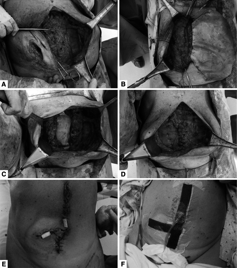
Fig. 2.
Intraoperative photographs: a components separation; b PADM underlay using transfascial sutures; c completion of the underlay PADM placement; d completion of midline fascial closure; e “French fry” technique with 2 portals with white foam; f black foam placed over entire incision line; PADM non-crosslinked intact porcine-derived acellular dermal matrix

