Description
Tuberculosis (TB) is a multisystem disease, 90% of which locates primarily in lungs, unlike isolated splenic tuberculosis, a rare form of extrapulmonary TB.1 Patients with AIDS or who are immunocompetent have been reported to be at a high risk for splenic TB.2 Some scholars insist that all patients with splenic TB are secondary to the previous infection of tubercle bacillus in other organs.3
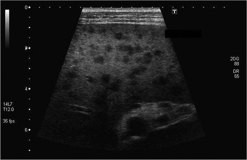
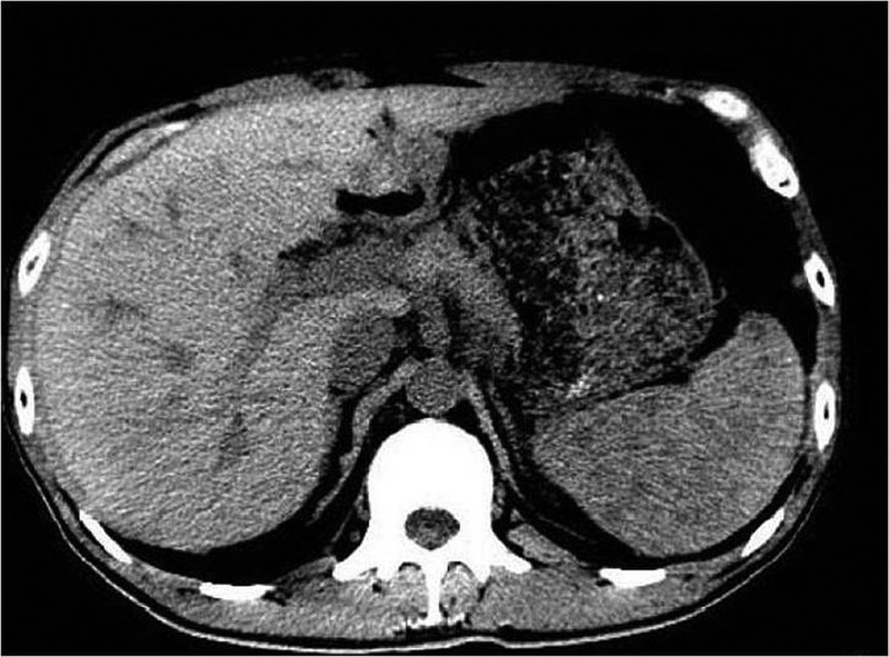
A 53-year-old male patient reported of intermittent left hypochondrium aching pain, occurring after vomiting since past 2 months. He did not report fever, cough or any other respiratory problems. He was diagnosed to have type 2 diabetes mellitus 1 year ago. On examination, it was found that there was no organomegaly and no other abnormalities were detected. No primary focus was found. X-ray findings were normal. On further investigations, ultrasonography of the abdomen and pelvis was performed, which showed splenomegaly with multiple hypoechoic areas, suggestive of granuloma (figure 1), and a CT scan revealed hypodense area in the spleen (figure 2). Para-aortic lymphadenopathy and bilateral renal parenchymal changes were detected in both. Also, fine-needle aspiration cytology was reported to have granulomatous inflammation. Urine protein was present in traces. Serum uric acid (8.4 mg/dL) and calcium were high. Liver function tests were normal. These investigations suggested the patient of having splenic TB. The person was advised not to go for splenectomy as he was diabetic and removal of the spleen would increase chances of infections. The patient was started on antitubercular treatment which showed a rapid improvement in his condition.
Figure 1.

Ultrasonography of the abdomen and pelvis showing: splenomegaly with multiple hypoechoic areas suggestive of granuloma.
Figure 2.

CT scan revealing hypointensities in the spleen.
Learning points.
Spleen is the third most common organ becoming involved in military tuberculosis (TB). Virtually all organ systems may be affected. Owing to haematogenous dissemination in HIV-infected individuals, extrapulmonary tuberculosis seems to be more common today than in the past.
There are many modalities of investigations, yet the diagnosis is usually delayed. Ultrasonography, CT scan and MRI are sensitive investigations but CT scan is preferred for the abdomen. The characteristic CT features of splenic tuberculosis include solitary/multiple nodular or saccular foci or hypodense areas in the spleen.
Splenic TB has a lot of differential diagnosis because of which diagnosis is often delayed. Moreover, typical nodules on the splenic capsule are usually too small to be detected. In differential diagnosis of CT findings, lymphoma, hydatid disease and metastases must be considered.
Acknowledgments
The authors wish to thank the entire department of radiology of Teerthanker Mahaveer Medical Hospital and Research Centre for cooperating and guiding us.
Footnotes
Competing interests: None.
Patient consent: Obtained.
Provenance and peer review: Not commissioned; externally peer reviewed.
References
- 1.Chakraborty AK. Epidemiology of tuberculosis: current status in India. Indian J Med Res 2004;120:248–76 [PubMed] [Google Scholar]
- 2.Gotor MA, Mur M, Guerrero L, et al. Tuberculous splenic abscess in an immunocompetent patient. Gastroenterol Hepatol 1995;18:15–17 [PubMed] [Google Scholar]
- 3.Zhan F, Wang CJ, Lin JZ, et al. Isolated splenic tuberculosis: a case report. World J Gastrointest Pathophysiol 2010;1:109–11 [DOI] [PMC free article] [PubMed] [Google Scholar]


