Abstract
The intermolecular interaction between mefenamic acid (MFA), a poorly water-soluble non-steroidal anti-inflammatory drug, and Eudragit® EPO (EPO), a water-soluble polymer, is investigated in their supersaturated solution using high-resolution magic-angle spinning (HRMAS) nuclear magnetic resonance (NMR) spectroscopy. The stable supersaturated solution with a high MFA concentration of 3.0 mg/mL is prepared by dispersing the amorphous solid dispersion into a d-acetate buffer at pH 5.5 and 37 °C. By virtue of MAS at 2.7 kHz, the extremely broad and unresolved 1H resonances of MFA in one-dimensional 1H NMR spectrum of the supersaturated solution are well resolved, thus enabling the complete assignment of MFA 1H resonances in the aqueous solution. Two-dimensional (2D) 1H/1H nuclear Overhauser effect spectroscopy (NOESY) and radio frequency-driven recoupling (RFDR) under MAS conditions reveal the interaction of MFA with EPO in the supersaturated solution at an atomic level. The strong cross-correlations observed in the 2D 1H/1H NMR spectra indicate a hydrophobic interaction between the aromatic group of MFA and the backbone of EPO. Furthermore, the aminoalkyl group in the side chain of EPO forms a hydrophilic interaction, which can be either electrostatic or hydrogen bonding, with the carboxyl group of MFA. We believe these hydrophobic and hydrophilic interactions between MFA and EPO molecules play a key role in the formation of this extremely stable supersaturated solution. In addition, 2D 1H/1H RFDR demonstrates that the molecular MFA-EPO interaction is quite flexible and dynamic.
Keywords: Eudragit® EPO, supersaturated solution, 1H NMR, NOESY, RFDR, intermolecular interaction
INTRODUCTION
Strategies for pharmaceutical formulations must take into account the various characteristics of active pharmaceutical ingredients (APIs) such as their taste, smell, color, stability, solubility, and bioavailability.1 In particular, enhancing the solubility of APIs is one of the most challenging tasks in the development of new solid oral formulations because most drug candidates are poor water-soluble substances.2 In this regard, several methods have been applied to increase drug solubility such as designing its crystal form,2, 3 complexation,4 amorphous solid dispersion,5-8 and nanoparticle formation.9 Amorphous solid dispersion has particularly been attracting a great deal of attention as a practical and useful technique in the field of pharmaceutical sciences.10 When amorphous solid dispersion is dispersed into water, a temporary enhancement of drug solubility can be generally achieved; this temporary supersaturated state with drug concentration higher than drug solubility can contribute to a significant enhancement of drug absorption into the body.11 Recently, solid dispersions producing highly stable supersaturated solutions have been studied in order to obtain further drug absorption using efficient polymers such as hydroxypropyl methylcellulose acetate succinate6 and methacrylate copolymers (Eudragit®).5, 7 These polymers possess a strong inhibitory effect on drug recrystallization in solutions, although the corresponding stabilization mechanisms are yet to be understood. Eudragit® and its derivatives have been extensively used as pharmaceutical excipients for a wide range of purposes.12
Mefenamic acid (MFA; Figure 1), which has a prevalent usage in high-dose drug formulations, is a non-steroidal anti-inflammatory drug used for pain relief in menstrual disorders.13 According to the United States Food and Drug Administration, MFA is categorized as a class II drug by the biopharmaceutical classification system since it shows high permeability and low water solubility.14, 15 Recently, we have demonstrated that the solid dispersion of acidic drugs with aminoalkyl methacrylate copolymer E, Eudragit® EPO (EPO; Figure 1) produces a stable supersaturated solution with high drug concentration.5 When the supersaturated solution of MFA with EPO is orally administered to rats in vivo, an intense increase in bioavailability of MFA is also observed.5 These results strongly support the promising applicability of supersaturated solutions using acidic drug/EPO for the purpose of enhancing the oral absorption.
Figure 1.
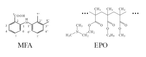
Molecular structures of mefenamic acid (MFA) and Eudragit® EPO (EPO). Proton numbering of MFA represents peak assignment in 1H NMR spectra.
In order to unravel the underlying stabilization mechanism of the supersaturated solution and for a more efficient design of new drug formulations, it is of paramount significance to identify the intermolecular interactions between the drug and the polymer in such solutions. Although several attempts have been made to investigate the interactions in a solid dispersion,8, 16 there are still only few studies that investigate the intermolecular interactions in supersaturated solutions. Indeed, the intermolecular interaction site between MFA and EPO in the solid dispersion by infra-red spectroscopy, 13C NMR and 13C spin-lattice relaxation time (13C T1) NMR measurements was reported.5 However, no useful information about the interaction site in the supersaturated solution was obtained from the one-dimensional (1D) 1H NMR experiments due to its colossal molecular size and dynamics.5
Magic-angle spinning (MAS) solid-state NMR spectroscopy is a powerful tool to acquire well-resolved solution-like spectra by averaging anisotropic interactions in a variety of samples including rigid solids, semi-solids, and viscous samples. High-resolution MAS technique has been widely used for various types of samples such as biomolecules,17, 18 food materials19 and lipid membranes,20 to name a few. Recently, this NMR approach has been applied in pharmaceutical formulations to detect APIs in the formulation of creams, gels, and pastes.21 However, to the best of our knowledge, the application of this technique to investigate the intermolecular interactions between APIs and their excipients has not been reported to date. In order to gain insight into the mechanism of pharmaceutical formulation for further improvement of drug efficacy, it is essential to obtain the atomic-level information of molecular interactions of formulations in aqueous solutions. For this purpose, it is indispensable to employ multidimensional NMR methodologies such as 2D 1H/1H nuclear Overhauser effect spectroscopy (NOESY)22 and radio-frequency driven recoupling (RFDR)23 experiments.18 2D 1H/1H NOESY measurements have been generally utilized to determine the intermolecular interactions in drug/cyclodextrin and drug/surfactant systems.24 Through the cross-peak intensities that arise in the NOESY spectrum, the spatial location of protons can be determined within a distance of ~ 5 Å.24 In this study, we report the results of our investigation of the intermolecular interactions that exist between MFA and EPO in their supersaturated solution using 2D 1H/1H NOESY and RFDR NMR techniques under MAS conditions. Based on the NMR results obtained, we disclose the model molecular structure of a MFA/EPO supersaturated solution in an attempt to rationalize the intermolecular interactions.
EXPERIMENTAL SECTION
Materials
Mefenamic acid (MFA; MW:241.29) was purchased from Wako Pure Chemical Industries, Ltd. (Osaka, Japan). Aminoalkyl methacrylate copolymer E, Eudragit® EPO (EPO; mean MW:135,000) was kindly provided by Evonik Degussa Japan Co. Ltd. (Tokyo, Japan). All chemicals were of chemical grade and were used as received. Chemical structures of MFA and EPO are shown in Figure 1. Proton numbering of MFA represents peak assignment in 1H NMR spectra.
Preparation of Amorphous Solid Dispersion and Supersaturated Solution of MFA and EPO for NMR Measurements
Sample preparation processes in this study are represented in Scheme 1. The preparation of the amorphous solid dispersion of MFA and EPO was reported previously.5 MFA and EPO were mixed at a weight ratio of 24/76 to obtain a physical mixture. The physical mixture was ground in a TI-500ET vibrational rod mill (CMT Co. Ltd., Fukushima, Japan) at −180 °C for 90 min to prepare the amorphous solid dispersion. The formation of amorphous solid dispersion by cryogenic grinding was confirmed by powder X-ray diffractometry using a Miniflex II X-ray diffractometer (Rigaku, Tokyo, Japan) with the temperature at 25 °C, voltage at 30 kV, current at 15 mA, scanning speed at 4°/min, and CuKα radiation source with a Ni filter.
The amorphous solid dispersion of MFA and EPO was dispersed into 0.1 M d-acetate buffer at pH 5.5 and 37 °C with the MFA concentration of 3.0 mg/mL. The dispersion was stirred at 37 °C until the transparent supersaturated solution was obtained. The MFA/EPO supersaturated solution was transparent before and after NMR measurements.
One-dimensional 1H NMR Measurement under Static Conditions
One-dimensional 1H NMR spectrum under static conditions was acquired at a magnetic field of 14.1 T on a JNM-ECA600 NMR spectrometer using an 1H/X probe (JEOL RESONANCE, Tokyo, Japan). The supersaturated solution of MFA and EPO was transferred into a 5 mm NMR sample tube. The 1D 1H NMR spectrum was obtained at 37 °C using 128 scans with a recycle delay of 5 s and a 1H 45° pulse of 7.75 μs. The signal of HDO at 4.67 ppm was used for 1H chemical shift referencing. The NMR data was processed using Delta® software (JEOL RESONANCE, Tokyo, Japan).
MAS NMR Experiments
MAS NMR measurements were conducted at 14.1 T on an Agilent/Varian VNMRS 600 MHz solid-state NMR spectrometer using a 4 mm 1H/X double-resonance nanoprobe (Agilent, CA, USA). The MFA/EPO supersaturated solution of 20 μL was placed into a glass rotor for MAS measurements. All spectra were acquired at 37 °C with a MAS rate of 2.7 kHz. The samples were equilibrated at 37 °C for about 30 minutes prior to NMR measurements. The 1D 1H NMR spectrum was obtained using 24 scans with a recycle delay of 1.5 s and 1H 90° pulse of 5 μs. The signal of HDO at 4.67 ppm was used for 1H chemical shift referencing. The 2D 1H/1H NOESY and RFDR experiments were performed using a mixing time of 50 ms and an acquisition time of 0.5 s. The 2D 1H/1H NOESY and RFDR spectra were recorded using 200 t1 increments and 8446 t2 complex points. The experimental data sets were zero-filled in both the t1 and t2 dimensions to form a 2048 × 2048 data matrix. Phase-shifted sine bell multiplication was applied in both dimensions prior to Fourier transformation. NMR data were processed using TopSpin 2.1 (Bruker, MA, USA). Assignment of proton resonances was completed based on a previous report14 and 2D 1H/1H NOESY spectrum.
RESULTS AND DISCUSSION
Effect of MAS on 1D 1H NMR Spectrum of MFA/EPO Supersaturated Solution
It is well known that MAS NMR spectroscopy has the advantage of significantly narrowing the spectral lines in the NMR spectrum by averaging out the anisotropic (orientation-dependent) interactions, namely the chemical shift anisotropy and the internuclear dipolar couplings. Therefore, we chose to investigate the effect of MAS on the MFA/EPO supersaturated solution, in an attempt to understand the binding mechanism underlying the intermolecular interaction between the drug and the polymer species. Figure 2 shows the static (Figure 2a) and the 2.7 kHz MAS (Figure 2b) 1D 1H NMR spectra of MFA/EPO supersaturated solution in a d-acetate buffer (pH 5.5, 37 °C). Although MFA is a small molecule that can undergo fast tumbling in solutions, it is obvious that the MFA peaks in the static spectrum suffer from significant line broadening caused by the strong 1H-1H homonuclear dipolar couplings. Such dipolar couplings are not averaged completely in this case because the small MFA molecule strongly binds to the large EPO polymer in the supersaturated solution, thus reducing the overall molecular mobility in MFA.5 These severe line broadenings make it impossible to resolve the overlapped peaks of H4-H6 and H4`-H6` in MFA. However, the application of modest spinning speed (2.7 kHz) successfully enables the resolution of the MFA peaks (Figure 2b), thus allowing for the unambiguous assignment of MFA resonances in the solution. To the best of our knowledge, this is the first report in which MAS NMR technique is applied to characterize drug-polymer interactions in a supersaturated solution at an atomic level. Our results indicate that MAS NMR spectroscopy can be a powerful tool for efficient characterization of the molecular states in supersaturated solutions where small drug molecules display effective binding activity to large polymeric species.
Figure 2.
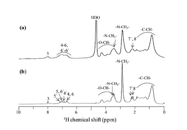
One-dimensional 1H NMR spectra of MFA/EPO supersaturated solution in d-acetate buffer (pH 5.5, 37 °C) under (a) static and (b) 2.7 kHz magic angle spinning conditions.
Two-Dimensional 1H/1H NOESY and RFDR Measurements under MAS
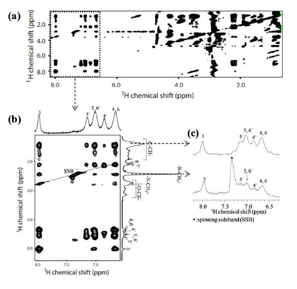
In order to observe the long-range intermolecular interaction between MFA and EPO directly, 2D 1H/1H NOESY experiment under MAS conditions was performed, and the obtained spectrum is shown in Figure 3a. Clearly, NOE correlations are observed between the peaks of MFA around 6.5–8.0 ppm and those of EPO around 0.5–5.0 ppm as shown in Figure 3b. The difference NOE experiments showed only less specific NOE cross-peaks throughout the spectrum,5, 26 which gave minimal information about the MFA-EPO interaction site. Here, the specific interactions between MFA and EPO are clearly observed due to the suppression of the residual dipolar interaction by MAS. The C-CH3 peaks of EPO at 0.82 ppm, corresponding to the backbone of EPO, exhibits strong correlation with the aromatic peaks of MFA (H3, H4-H6, and H4`-H6`). The protons near the aminoalkyl group in the side chain of EPO (N-CH3 at 2.86 ppm and N-CH2 at 3.55 ppm) show NOE correlations with the ring protons (H3-H6) of MFA. On the other hand, the cross-peaks between O-CH protons (at 3.3-4.3 ppm) in the side chain of EPO and the protons in MFA are too small to be detected in the 2D NOESY spectrum, which suggests that the O-methyl group in the side chain of EPO side chain does not participate in the interaction with MFA.
Figure 3.
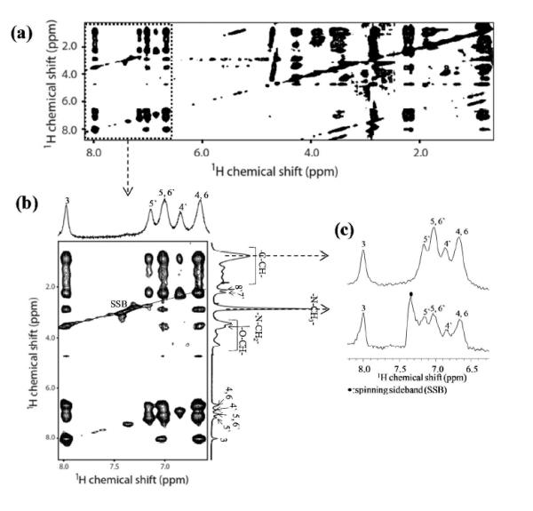
Two-dimensional 1H/1H NOESY NMR spectrum of MFA/EPO supersaturated solution in d-acetate buffer (pH 5.5, 37 °C) under 2.7 kHz MAS. (b) Expanded region of the spectrum in (a). (c) 1D 1H slices extracted from the 2D NOESY spectrum along 0.82 and 2.86 ppm.
To gain a detailed high-resolution insight into the interaction between MFA and EPO in the supersaturated solution, the intensities of NOE correlation peaks were compared. Figure 3c shows the 1D 1H spectra obtained by slicing the 2D 1H/1H NOESY spectrum along 0.82 and 2.86 ppm corresponding to C-CH3 and N-CH3 resonances of EPO, respectively. The relative intensities of MFA protons are similar between the 1D 1H NOESY spectrum sliced along C-CH3 peaks (Figure 3c) and 1D 1H NMR spectrum (t2 projection in Figure 3b). This indicates that C-CH3 protons in the backbone of EPO could lie close to all of the aromatic protons (H3, H4-H6 and H4`-H6`) of MFA. In contrast, the relative intensity of the H3 peak to each of the H4-H6 and H4`-H6` peaks is much higher in the 1D 1H spectrum sliced along the N-CH3 peak (Figure 3c). It follows that N-CH3 protons in the side chain of EPO are in close proximity to H3 of MFA.
In order to possibly quantify dipolar couplings and measure proton–proton distances, 2D 1H/1H RFDR experiment under MAS was performed on the MFA-EPO in supersaturated solution. In the 2D RFDR experiment, the 1H-1H dipolar couplings, which are averaged out under MAS, are reintroduced by applying a series of rotor-synchronized 180° pulses during the mixing time.23 Hence, the cross-peaks in the RFDR spectrum originate from the combined effect of NOE cross-relaxation and spin diffusion through homonuclear dipolar coupling among protons.18 As shown in Figure 4, the 2D 1H/1H RFDR spectrum produces similar information about cross-correlation peaks as the NOESY spectrum in Figure 3, albeit with lower cross-peak intensities. This can be attributed to the fact that the MFO-EPO interaction is quite flexible and dynamic, thus rendering the reintroduction of the dipolar coupling ineffective in the RFDR experiment. This result suggests that the solvent plays an important role in the flexibility of molecular constituents. It is worth noting that the 2D 1H/1H RFDR experiments can be successfully implemented for distance measurements on rigid samples, or on mobile samples at low temperatures, for which the molecular mobility is restricted. Such experiments performed at ultra-high magnetic fields will provide even more piercing insights into the molecular interactions, which can further be utilized to optimize the formulation of drugs.
Figure 4.
Two-dimensional 1H/1H RFDR NMR spectrum of MFA/EPO supersaturated solution in d-acetate buffer (pH 5.5, 37 °C) under 2.7 kHz MAS. (b) Expanded region of the spectrum in (a). (c) 1D 1H slices extracted from the 2D RFDR spectrum along 0.82 and 2.86 ppm.
Intermolecular Interaction of MFA with EPO in the Supersaturated Solution
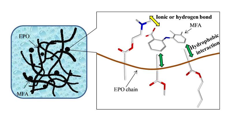
In light of the aforementioned discussion about the plausible intermolecular interactions between MFA and EPO, a possible model structure of MFA dispersed in EPO polymer matrices in the supersaturated solution is proposed in Figure 5. It is possible that MFA recrystallization was inhibited due to its binding with EPO, which stabilizes the supersaturated solution for a longer time. At the atomic level, MFA and EPO can interact with each other mainly via hydrophobic and hydrophilic interactions. Two aromatic groups of MFA interact with the backbone of EPO by hydrophobic interaction. In addition, hydrophilic interaction such as ionic or hydrogen bonding is formed between the carboxyl group of MFA and the aminoalkyl group of the EPO side chain. Indeed, this hydrophilic interaction is supported by results from the previous study, in which stable supersaturated solutions were effectively obtained using the solid dispersion of EPO with acidic drugs such as indomethacin and piroxicam.5 Furthermore, the previous report demonstrated that the carboxyl group of MFA interacts with the aminoalkyl group of the EPO side chain through the hydrophilic interaction in the solid dispersion.5 The same hydrophilic interaction is observed in the present study for the supersaturated solution, thus indicating that the hydrophilic interaction formed in solid state is potentially conserved upon dispersion into solution. Indeed, this hydrophilic interaction in the solid state underlies the enhanced MFA solubility in the amorphous solid dispersion over that in the MFA/EPO physical mixture.
Figure 5.
A model molecular structure of MFA/EPO supersaturated solution and intermolecular interaction between MFA and EPO.
CONCLUSIONS
In summary, we have demonstrated that the intermolecular interaction between MFA and EPO in the supersaturated solution can be clearly observed by 2D 1H/1H NOESY and RFDR MAS NMR experiments. MAS NMR is a powerful technique for analyzing and characterizing the molecular structure and dynamics of supersaturated solutions. Our results reveal that both hydrophobic and hydrophilic intermolecular interactions contribute to the stabilization of the supersaturated solution. Hydrophobic interaction is formed between the aromatic groups of MFA and the backbone of EPO, while the carboxyl group of MFA interacts with the aminoalkyl group of the EPO side chain via hydrophilic interaction. On the other hand, the O-methyl groups in the side chain of EPO do not exhibit any interaction with MFA. To our knowledge, this is the first report that determines an atomic-level drug/polymer interaction in a supersaturated solution. The obtained results provide better understanding of the formation and stabilization mechanisms of supersaturated solutions, and open up further opportunities for designing new pharmaceutical formulations. We believe that multidimensional high-resolution MAS NMR spectroscopy can be widely implemented for pharmaceutical formulations in solution state as well as in semi-solid state, and will be of ample applicability in this field for the characterization of molecular interactions.
Scheme 1.

A schematic representation of sample preparation of MFA/EPO supersaturated solution for high-resolution magic-angle spinning NMR measurements.
ACKNOWLEDGMENT
This study was supported by NIH (GM095640 to A.R.) and partly by the Japan Health Sciences Foundation for Public-private sector joint research on Publicly Essential Drugs, by the Institutional Program for Young Researcher Overseas Visits (JSPS) and by Grants-in-Aid for Young Scientist (B) (JSPS, 24790041) from the Japan Society for the Promotion of Sciences. We also thank Evonik Degussa Japan Co. Ltd (Tokyo, Japan) for gifting EPO.
REFERENCES
- 1.Rowland M, Noe CR, Smith DA, Tucker GT, Crommelin DJA, Peck CC, Rocci ML, Jr, Besançon L, Shah VP. Impact of the pharmaceutical sciences on health care: A reflection over the past 50 years. J. Pharm .Sci. 2012;101(11):4075–4099. doi: 10.1002/jps.23295. [DOI] [PubMed] [Google Scholar]
- 2.Kawakami K. Modification of physicochemical characteristics of active pharmaceutical ingredients and application of supersaturatable dosage forms for improving bioavailability of poorly absorbed drugs. Adv. Drug Deliv. Rev. 2012;64(6):480–495. doi: 10.1016/j.addr.2011.10.009. [DOI] [PubMed] [Google Scholar]
- 3.Singhal D, Curatolo W. Drug polymorphism and dosage form design: a practical perspective. Adv. Drug Deliv. Rev. 2004;56(3):335–347. doi: 10.1016/j.addr.2003.10.008. [DOI] [PubMed] [Google Scholar]; Dempah KE, Barich DH, Kaushal AM, Zong Z, Desai SD, Suryanarayanan R, Kirsch L, Munson EJ. Investigating gabapentin polymorphism using solid-state NMR spectroscopy. AAPS PharmSciTech. 2013;14(1):19–28. doi: 10.1208/s12249-012-9879-z. [DOI] [PMC free article] [PubMed] [Google Scholar]
- 4.Childs SL, Kandi P, Lingireddy SR. Formulation of a danazol cocrystal with controlled supersaturation plays an essential role in improving bioavailability. Mol. Pharm. 2013;10(8):3112–3127. doi: 10.1021/mp400176y. [DOI] [PubMed] [Google Scholar]; Higashi K, Ideura S, Waraya H, Moribe K, Yamamoto K. Incorporation of salicylic acid molecules into the intermolecular spaces of γ-cyclodextrin-polypseudorotaxane. Cryst. Growth Des. 2009;9(10):4243–4246. [Google Scholar]; Alhalaweh A, Roy L, Rodríguez-Hornedo N, Velaga SP. pH-Dependent solubility of indomethacin–saccharin and carbamazepine–saccharin cocrystals in aqueous media. Mol. Pharm. 2012;9(9):2605–2612. doi: 10.1021/mp300189b. [DOI] [PubMed] [Google Scholar]; Chattah AK, Mroue KH, Pfund LY, Ramamoorthy A, Longhi MR, Garnero C. Insights into novel supramolecular complexes of two solid forms of norfloxacin and β-cyclodextrin. J. Pharm .Sci. 2013;102(10):3717–3724. doi: 10.1002/jps.23683. [DOI] [PubMed] [Google Scholar]
- 5.Kojima T, Higashi K, Suzuki T, Tomono K, Moribe K, Yamamoto K. Stabilization of a supersaturated solution of mefenamic acid from a solid dispersion with EUDRAGIT® EPO. Pharm. Res. 2012;29(10):2777–2791. doi: 10.1007/s11095-011-0655-7. [DOI] [PubMed] [Google Scholar]
- 6.Ueda K, Higashi K, Limwikrant W, Sekine S, Horie T, Yamamoto K, Moribe K. Mechanistic differences in permeation behavior of supersaturated and solubilized solutions of carbamazepine revealed by nuclear magnetic resonance measurements. Mol. Pharm. 2012;9(11):3023–3033. doi: 10.1021/mp300083e. [DOI] [PubMed] [Google Scholar]; Friesen DT, Shanker R, Crew M, Smithey DT, Curatolo WJ, Nightingale JAS. Hydroxypropyl methylcellulose acetate succinate-based spray-dried dispersions: an overview. Mol. Pharm. 2008;5(6):1003–1019. doi: 10.1021/mp8000793. [DOI] [PubMed] [Google Scholar]; Miller JM, Beig A, Carr RA, Spence JK, Dahan A. A win–win solution in oral delivery of lipophilic drugs: supersaturation via amorphous solid dispersions increases apparent solubility without sacrifice of intestinal membrane permeability. Mol. Pharm. 2012;9(7):2009–2016. doi: 10.1021/mp300104s. [DOI] [PubMed] [Google Scholar]
- 7.Guzmán ML, Manzo RH, Olivera ME. Eudragit E100 as a drug carrier: the remarkable affinity of phosphate ester for dimethylamine. Mol. Pharm. 2012;9(9):2424–2433. doi: 10.1021/mp300282f. [DOI] [PubMed] [Google Scholar]
- 8.Calahan JL, Zanon RL, Alvarez-Nunez F, Munson EJ. Isothermal microcalorimetry to investigate the phase separation for amorphous solid dispersions of AMG 517 with HPMC-AS. Mol. Pharm. 2013;10(5):1949–1957. doi: 10.1021/mp300714g. [DOI] [PubMed] [Google Scholar]
- 9.Fukami T, Ishii T, Io T, Suzuki N, Suzuki T, Yamamoto K, Xu J, Ramamoorthy A, Tomono K. Nanoparticle processing in the solid state dramatically increases the cell membrane permeation of a cholesterol-lowering drug, probucol. Mol. Pharm. 2009;6(3):1029–1035. doi: 10.1021/mp9000487. [DOI] [PubMed] [Google Scholar]; Io T, Fukami T, Yamamoto K, Suzuki T, Xu J, Tomono K, Ramamoorthy A. Homogeneous nanoparticles to enhance the efficiency of a hydrophobic drug, antihyperlipidemic probucol, characterized by solid-state NMR. Mol. Pharm. 2009;7(1):299–305. doi: 10.1021/mp900254y. [DOI] [PMC free article] [PubMed] [Google Scholar]; Li Y, Sun S, Chang Q, Zhang L, Wang G, Chen W, Miao X, Zheng Y. A strategy for the improvement of the bioavailability and antiosteoporosis activity of BCS IV flavonoid glycosides through the formulation of their lipophilic aglycone into nanocrystals. Mol. Pharm. 2013;10(7):2534–2542. doi: 10.1021/mp300688t. [DOI] [PubMed] [Google Scholar]
- 10.Kawakami K. Current status of amorphous formulation and other special dosage forms as formulations for early clinical phases. J. Pharm .Sci. 2009;98(9):2875–2885. doi: 10.1002/jps.21816. [DOI] [PubMed] [Google Scholar]
- 11.Takano R, Takata N, Saito R, Furumoto K, Higo S, Hayashi Y, Machida M, Aso Y, Yamashita S. Quantitative analysis of the effect of supersaturation on in vivo drug absorption. Mol. Pharm. 2010;7(5):1431–1440. doi: 10.1021/mp100109a. [DOI] [PubMed] [Google Scholar]
- 12.Čalija B, Cekić N, Savić S, Daniels R, Marković B, Milić J. pH-Sensitive microparticles for oral drug delivery based on alginate/oligochitosan/Eudragit® L100-55 "sandwich" polyelectrolyte complex. Colloid Surf. B. 2013;110:395–402. doi: 10.1016/j.colsurfb.2013.05.016. [DOI] [PubMed] [Google Scholar]; Piao H, Liu S, Li X, Cui F. Development of an osmotically-driven pellet coated with acrylic copolymers (Eudragit® RS 30 D) for the sustained release of oxymatrine, a freely water soluble drug used to treat stress ulcers (I): In vitro and in vivo evaluation in rabbits. Drug. Develop. Ind. Pharm. 2013;39(8):1230–1237. doi: 10.3109/03639045.2012.707206. [DOI] [PubMed] [Google Scholar]; Tan Q, Zhang L, Liu G, He D, Yin H, Wang H, Wu J, Liao H, Zhang J. Novel taste-masked orally disintegrating tablets for a highly soluble drug with an extremely bitter taste: Design rationale and evaluation. Drug. Develop. Ind. Pharm. 2013;39(9):1364–1371. doi: 10.3109/03639045.2012.718784. [DOI] [PubMed] [Google Scholar]
- 13.van Eijkeren MA, Christiaens GCML, Geuze HJ, Haspels AA, Sixma JJ. Effects of mefenamic acid on menstrual hemostasis in essential menorrhagia. Am. J. Obst. Gynecol. 1992;166(5):1419–1428. doi: 10.1016/0002-9378(92)91614-g. [DOI] [PubMed] [Google Scholar]
- 14.Munro SLA, Craik DJ. NMR conformational studies of fenamate non-steroidal anti-inflammatory drugs. Mag. Reson. Chem. 1994;32(6):335–342. [Google Scholar]
- 15.SeethaLekshmi S, Guru Row TN. Conformational polymorphism in a non-steroidal anti-inflammatory drug, mefenamic acid. Cryst. Growth. Des. 2012;12(8):4283–4289. [Google Scholar]; Yazdanian M, Briggs K, Jankovsky C, Hawi A. The “high solubility” definition of the current FDA guidance on biopharmaceutical classification system may be too strict for acidic drugs. Pharm. Res. 2004;21(2):293–299. doi: 10.1023/b:pham.0000016242.48642.71. [DOI] [PubMed] [Google Scholar]
- 16.Pham TN, Watson SA, Edwards AJ, Chavda M, Clawson JS, Strohmeier M, Vogt FG. Analysis of amorphous solid dispersions using 2D solid-state NMR and 1H T1 relaxation measurements. Mol. Pharm. 2010;7(5):1667–1691. doi: 10.1021/mp100205g. [DOI] [PubMed] [Google Scholar]; Tatton AS, Pham TN, Vogt FG, Iuga D, Edwards AJ, Brown SP. Probing hydrogen bonding in cocrystals and amorphous dispersions using 14N–1H HMQC solid-state NMR. Mol. Pharm. 2013;10(3):999–1007. doi: 10.1021/mp300423r. [DOI] [PubMed] [Google Scholar]; Munson E, Endowed PD. Applications of solid-state NMR spectroscopy to pharmaceuticals. Euro. Pharm. Rev. 2013;18(1) [Google Scholar]
- 17.Wong A, Li X, Sakellariou D. Refined magic-angle coil spinning resonator for nanoliter NMR spectroscopy: Enhanced spectral resolution. Anal. Chem. 2013;85(4):2021–2026. doi: 10.1021/ac400188b. [DOI] [PubMed] [Google Scholar]; Beckonert O, Coen M, Keun HC, Wang Y, Ebbels TMD, Holmes E, Lindon JC, Nicholson JK. High-resolution magic-angle-spinning NMR spectroscopy for metabolic profiling of intact tissues. Nat. Protoc. 2010;5(6):1019–1032. doi: 10.1038/nprot.2010.45. [DOI] [PubMed] [Google Scholar]
- 18.Ramamoorthy A, Xu J. 2D 1H/1H RFDR and NOESY NMR experiments on a membrane-bound antimicrobial peptide under magic angle spinning. J. Phys. Chem. B. 2013;117(22):6693–6700. doi: 10.1021/jp4034003. [DOI] [PubMed] [Google Scholar]
- 19.Mucci A, Parenti F, Righi V, Schenetti L. Citron and lemon under the lens of HR-MAS NMR spectroscopy. Food Chem. 2013;141(3):3167–3176. doi: 10.1016/j.foodchem.2013.05.151. [DOI] [PubMed] [Google Scholar]
- 20.Aucoin D, Camenares D, Zhao X, Jung J, Sato T, Smith SO. High-resolution 1H MAS RFDR NMR of biological membranes. J. Magn. Reson. 2009;197(1):77–86. doi: 10.1016/j.jmr.2008.12.009. [DOI] [PMC free article] [PubMed] [Google Scholar]; Smith PES, Brender JR, Dürr UHN, Xu J, Mullen DG, Banaszak Holl MM, Ramamoorthy A. Solid-State NMR reveals the hydrophobic-core location of poly(amidoamine) dendrimers in biomembranes. J. Am. Chem. Soc. 2010;132(23):8087–8097. doi: 10.1021/ja101524z. [DOI] [PMC free article] [PubMed] [Google Scholar]
- 21.Marzorati M, Bigler P, Plattner M, Vermathen M. Feasibility of 1H-high resolution-magic angle spinning NMR spectroscopy in the analysis of viscous cosmetic and pharmaceutical formulations. Anal. Chem. 2013;85(8):3822–3827. doi: 10.1021/ac400559z. [DOI] [PubMed] [Google Scholar]
- 22.Kumar A, Wagner G, Ernst RR, Wuethrich K. Buildup rates of the nuclear Overhauser effect measured by two-dimensional proton magnetic resonance spectroscopy: implications for studies of protein conformation. J. Am. Chem. Soc. 1981;103(13):3654–3658. [Google Scholar]
- 23.Bennett AE, Ok JH, Griffin RG, Vega S. Chemical shift correlation spectroscopy in rotating solids: Radio frequency-driven dipolar recoupling and longitudinal exchange. J. Chem. Phys. 1992;96(11):8624–8627. [Google Scholar]
- 24.Murray NJ, Williamson MP, Lilley TH, Haslam E. Study of the interaction between salivary proline-rich proteins and a polyphenol by 1H-NMR spectroscopy. Eur. J. Biochem. 1994;219(3):923–935. doi: 10.1111/j.1432-1033.1994.tb18574.x. [DOI] [PubMed] [Google Scholar]; Tezuka Y. Nuclear Overhauser effect spectroscopy (NOESY) detection of the specific interaction between substituents in cellulose and amylose triacetates. Biopolymers. 1994;34(11):1477–1481. doi: 10.1002/bip.360341105. [DOI] [PubMed] [Google Scholar]
- 25.Macura S, Ernst RR. Elucidation of cross relaxation in liquids by two-dimensional N.M.R. spectroscopy. Mol. Phys. 1980;41(1):95–117. [Google Scholar]
- 26.Kalk A, Berendsen HJC. Proton magnetic relaxation and spin diffusion in proteins. J. Magn. Reson. 1976;24(3):343–366. [Google Scholar]


