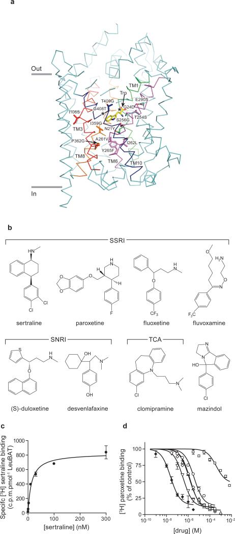
Fig. 1. LeuBAT design and pharmacology.
(a) The representation of mutation positions around the primary binding pocket in wild-type LeuT-Trp structure (PDB 3F3A). Bound tryptophan (yellow) and the mutated residues are in sticks. The transmembrane helices TM1, TM3, TM6, TM8 and TM10 around the pocket are highlighted as green, red, purple, orange and blue, respectively. Asterisks depict the glycine residue positions. (b) Chemical structures of four SSRIs, two SNRIs, one tricyclic antidepressant (clomipramine) and one stimulant (mazindol); (c) Measurement of [3H] sertraline binding (filled circles) to Δ13 LeuBAT; (d) Dose-response curves for inhibition of [3H] paroxetine binding to Δ13 LeuBAT by sertraline (filled diamonds), fluvoxamine (empty circles), fluoxetine (empty diamonds), duloxetine (empty inverted triangles), clomipramine (empty triangles), desvenlafaxine (empty squares). Error bars, s.e.m, n = 3.

