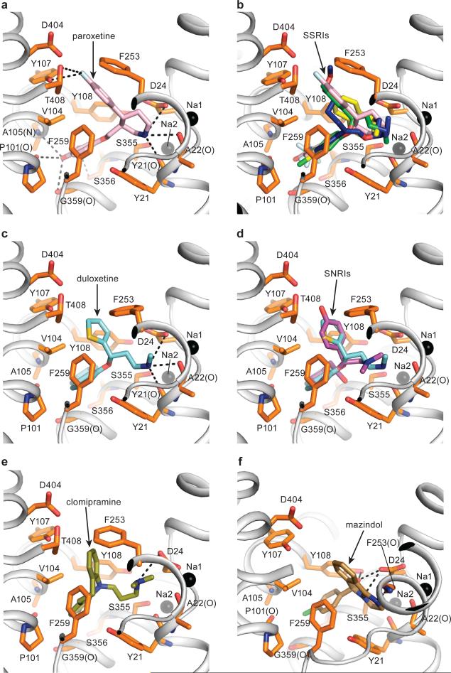
Fig. 3. SSRIs, SNRIs, TCA and mazindol share similar binding features.
(a) Paroxetine binding site in Δ13 LeuBAT mutant viewed within the membrane plane with paroxetine shown as pink sticks. Sodium ions and key residues are shown as black spheres and orange sticks, respectively. Hydrogen bonds, salt bridges and polar interactions are in dashed lines. (b) Superimposition of paroxetine (pink), (R)-fluoxetine (green), fluvoxamine (blue) and sertraline (yellow) in the primary binding pocket of the Δ13 LeuBAT; (c) (S)-Duloxetine binding site in the Δ13 LeuBAT with duloxetine shown as cyan sticks; (d) Superimposition of (S)-duloxetine (cyan) and (S)-desvenlafaxine (magenta) in the primary drug-binding pocket; (e) Clomipramine (CMI) binding site in Δ13 LeuBAT with CMI shown as olive sticks; (f) Mazindol binding site in LeuBAT Δ6 variant with mazindol molecule shown as sand-colored sticks.

